Fusion of the thumb MCP joint for painful arthritis, chronic instability, post-traumatic destruction or rheumatoid Z-collapse | advanced
Surgical Imaging
The trap: Fusing the thumb MCP in too much extension (straight or hyperextended) eliminates effective pulp-to-pulp pinch because the thumb tip cannot reach the index finger pad. Fusing in excessive flexion (greater than 20 degrees) shortens the pinch distance and weakens grip.
The fix: Position the MCP at 10-15 degrees of flexion with slight pronation. Use a temporary K-wire to hold the position, then check pinch with the contralateral hand before committing to definitive fixation. Intraoperative fluoroscopy confirms the angle.
Location: The radial digital nerve of the thumb runs in the volar subcutaneous tissue, crossing into the dorsal wound in the proximal portion of the incision. It lies superficial to the extensor tendons.
Risk: During the dorsal approach between EPB and EPL, the radial digital nerve is vulnerable as the skin flap is elevated. Identify it in the proximal wound before deepening between the tendon intervals. A transection produces a painful neuroma and loss of thumb pulp sensibility.
Why it happens: Inadequate removal of sclerotic subchondral bone leaves two hard, avascular surfaces that cannot consolidate. Poor fixation allowing micro-motion compounds the problem.
Prevention: Resect the articular surfaces down to bleeding cancellous bone on both the metacarpal head and the proximal phalanx base. Use cup-and-cone or flat surfaces with maximal contact area. Apply stable compression fixation (headless screw, plate, or tension band) and protect for 4-6 weeks in a thumb spica.
The mechanism: After MCP fusion the thumb loses its mid-column flexion arc. The IP joint compensates with increased flexion during pinch and grip, accelerating degenerative change.
Prevention: Screen for pre-existing IP joint arthritis before MCP fusion — if the IP joint is symptomatic, consider combined procedures or IP fusion alone. Post-operatively, avoid forceful tip-pinch loading in the early rehabilitation phase. Monitor the IP joint at follow-up.
The trap: Isolating the MCP fusion without addressing symptomatic CMC joint arthritis leaves the patient with persistent basal thumb pain — the CMC is the most common site of thumb arthritis and may coexist.
The fix: Examine the CMC joint pre-operatively (grind test, Kapandji score, AP and lateral radiographs). If the CMC is symptomatic, consider simultaneous CMC arthroplasty (trapeziectomy with ligament reconstruction) or staging the procedures. The CMC arthroplasty should usually precede the MCP fusion in the staged approach.
Plate: A dorsal mini-plate is the most secure fixation but is closest to the skin and tendon. Prominence requiring removal is reported in 10-20% of cases. Use low-profile plates and position the plate ulnar to the EPL to reduce tendon irritation.
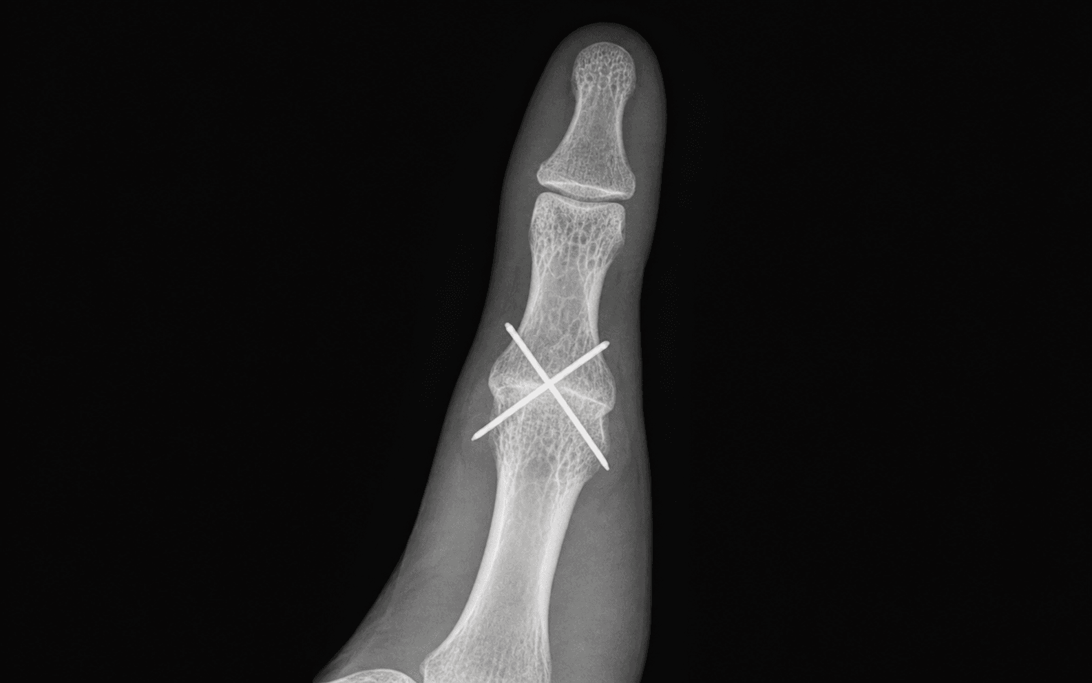
K-wires: Tension band K-wires left protruding are at risk of infection and migration. Bend the ends, bury beneath skin, or use a buried wire technique. Plan for removal at 4-6 weeks if not fully buried.
F.U.S.I.O.NFUSION — Indications for Thumb MCP Arthrodesis
P.O.S.I.T.I.O.NPOSITION — Fusion Angle and Key Technical Points
Surgical Indications
Absolute Indications
- Chronic ulnar collateral ligament insufficiency not amenable to reconstruction — gross instability with volar subluxation during pinch, failed prior UCL repair or graft reconstruction
- Post-traumatic MCP joint destruction with combined arthritis and instability where motion-preserving procedures (arthroplasty, osteotomy) are not viable
- Rheumatoid Z-collapse deformity with painful MCP volar subluxation as part of the zigzag collapse pattern — fusion stabilises the middle link of the collapse
Relative Indications
- Primary MCP osteoarthritis (uncommon in isolation — more often part of pan-trapezial arthritis) causing pain and functional loss refractory to non-operative treatment for greater than 6 months
- MCP instability after infection or tumour resection where joint reconstruction is not possible
- Failed MCP arthroplasty (silicone or pyrocarbon) with painful instability or loosening — revision to fusion
- Salvage after failed external fixation or failed dynamic external fixation for complex MCP peri-articular fractures
Contraindications
Absolute:
- Active infection at or adjacent to the MCP joint
- Uncorrectable soft-tissue deficit precluding wound closure over fixation hardware
Relative:
- Symptomatic CMC arthritis that should be addressed first (either staged before MCP fusion or simultaneously)
- Pre-existing IP joint arthritis — fusion at MCP may overload the IP; consider whether IP fusion alone would suffice
- Patient whose occupation requires full MCP flexion arc (some musicians, specialised manual workers) — counsel thoroughly
Evidence for Non-Operative Treatment
Splinting and Activity Modification
- A resting thumb spica splint reduces MCP loading in arthritic patients and provides symptomatic relief in early disease
- Static splinting at 10-15 degrees of MCP flexion can reduce pain during activities of daily living
- Evidence is largely from case series and expert opinion — no RCTs specifically compare splinting versus surgery for MCP arthritis
- Non-operative treatment is reasonable as a first-line measure in mild disease and when the patient is not yet ready for surgery
Corticosteroid Injection
- Intra-articular corticosteroid injection into the MCP joint provides temporary symptom relief (weeks to months) in inflammatory arthritis
- Limited role in primary OA — the MCP joint has a small capsule and injection provides inconsistent relief
- May be used as a diagnostic tool (symptom relief after injection confirms the MCP as the pain source before committing to surgery)
Evidence for Arthroplasty Versus Arthrodesis
- MCP arthroplasty (silicone, pyrocarbon, or CMC-type interposition) is an option for patients who wish to retain MCP motion
- Silicone arthroplasty has a long track record but is associated with silicone synovitis, implant fracture, and subsidence over time — outcomes are less predictable than fusion
- Pyrocarbon and other resurfacing implants have shorter-term follow-up and higher revision rates than fusion in the available literature
- Arthrodesis provides a predictable, permanent, and painless solution at the cost of MCP motion — most evidence supports fusion as the gold standard for young, active, or high-demand patients
Evidence for Arthrodesis
Functional Outcomes
- Thumb MCP arthrodesis reliably eliminates MCP pain and provides a stable platform for pinch and grip
- Loss of the small MCP flexion arc is well compensated by preserved CMC opposition and IP flexion — most patients report no subjective loss of overall thumb function
- Pinch and grip strength are maintained or improved post-operatively in the majority of series, particularly where instability was the pre-operative problem
- Patient satisfaction rates range from 80% to 95% across published series
Nonunion
- Nonunion is the most commonly reported complication, with rates between 5% and 15% depending on fixation method, bone quality and patient factors
- Compression fixation (headless screw or plate) provides more consistent union than K-wire-only fixation
- Smoking, diabetes, and rheumatoid osteopenia are the most consistently reported risk factors for nonunion
- Re-operation for nonunion involves revision fixation with bone grafting — union rates after revision are lower than primary fusion
Fixation Method Comparison
Fixation Methods for Thumb MCP Arthrodesis
Key Decision Points
- Young, active patient with good bone stock and UCL insufficiency: headless compression screw provides the best balance of compression, low profile and no hardware removal
- Rheumatoid patient with osteoporotic bone: dorsal mini-plate provides the most rigid fixation in poor-quality bone; accept the higher rate of hardware removal
- Low-resource setting or cost-sensitive: tension band wiring with K-wires is reliable and inexpensive; plan for hardware removal at 4-6 weeks
- Concomitant CMC disease: address CMC first (trapeziectomy and LRTI), then reassess the MCP — some patients no longer need MCP fusion after CMC stabilisation
- IP joint arthritis present: if IP disease is mild, proceed with MCP fusion and monitor the IP; if IP disease is severe, consider IP fusion alone (which eliminates tip pinch) or staged procedures
Clinical Decision Scenarios
Practise clinical reasoning and management decisions out loud
“A 58-year-old manual labourer presents with a painful, unstable right thumb MCP joint. He sustained a gamekeeper's thumb injury 3 years ago, had a UCL reconstruction using a palmaris longus graft 2 years ago, and now has recurrent valgus instability with pain during pinch grip. Radiographs show MCP joint space narrowing and volar subluxation of the proximal phalanx. How do you proceed?”
“A 52-year-old woman with longstanding rheumatoid arthritis presents with a Z-collapse deformity of the right thumb. She has dorsal subluxation at the CMC joint, volar subluxation and adduction at the MCP joint, and a mild hyperextension deformity at the IP joint. Her main complaints are pain and inability to grip objects. Describe your surgical plan.”
“You are performing a thumb MCP arthrodesis with a headless compression screw on a 42-year-old carpenter with post-traumatic MCP arthritis. Intraoperatively, after resecting the articular surfaces and positioning the temporary K-wire, you notice that the fusion surfaces are not perfectly congruent — there is a small gap on the ulnar side of the resected surfaces. What do you do?”