Internal (Limb-Sparing) vs External (Hindquarter Amputation) | Enneking-Dunham Classification | Vascular Control | Reconstruction
- Enneking-Dunham classification defines pelvic resection zones (I-IV) - essential for surgical planning
- Internal hemipelvectomy is limb-sparing; external (hindquarter) involves complete limb removal
- Vascular control of common iliac vessels is critical before resection
- Sciatic nerve sacrifice often required for posterior tumours - counsel patient preoperatively
- Reconstruction options include endoprosthesis, allograft, saddle prosthesis, or no reconstruction
- “Type I resection (iliac wing) often requires no formal reconstruction
- “Type II resection (periacetabular) is most challenging - requires acetabular reconstruction
- “Posterior flap (gluteal) is preferred for external hemipelvectomy coverage
- “Mortality rate 1-5% - major blood loss is the primary intraoperative risk
Type I = Iliac wing (above acetabulum). Type II = Periacetabular (most complex). Type III = Pubic rami/ischium. Type IV = Sacral ala. Know the zones and their reconstruction implications.
Common iliac vessels must be controlled early in the procedure. Internal iliac artery is typically ligated. External iliac vessels are preserved in internal hemipelvectomy. Massive blood loss (5-15 units) is expected.
Sciatic nerve sacrifice is often required for posterior tumours. Femoral nerve must be identified and protected anteriorly. Lumbosacral plexus involvement may preclude limb salvage.
Type I: Often no reconstruction needed. Type II: Saddle prosthesis, allograft-prosthetic composite, or custom endoprosthesis. Type III: May not require reconstruction. Flail hip (excision arthroplasty) is an alternative.
- Internal Hemipelvectomy
- Pelvic bone resection with limb preservation
- External Hemipelvectomy
- Complete hindquarter amputation
- Internal Hemipelvectomy
- Bone tumour without neurovascular bundle involvement
- External Hemipelvectomy
- Tumour involving sciatic nerve or major vessels
- Internal Hemipelvectomy
- Preserved external iliac vessels and femoral nerve
- External Hemipelvectomy
- Vessels and nerves sacrificed with specimen
- Internal Hemipelvectomy
- Ambulatory with or without aids depending on reconstruction
- External Hemipelvectomy
- Wheelchair or prosthetic (limited use)
- Internal Hemipelvectomy
- Required for Type II (acetabular) resections
- External Hemipelvectomy
- Flap coverage for wound closure
- Internal Hemipelvectomy
- 1-3%
- External Hemipelvectomy
- 3-5%
- Internal Hemipelvectomy
- 30-50% (infection, flap necrosis, dislocation)
- External Hemipelvectomy
- 20-40% (wound complications, phantom pain)
RECONRECON - Type II Reconstruction Options
Hook:RECON for Type II periacetabular defects - multiple options from no reconstruction to custom prosthesis
Overview and Epidemiology
Definition
Hemipelvectomy refers to surgical resection of part or all of one hemipelvis, performed primarily for malignant bone and soft tissue tumours. The procedure is classified as:
- Internal hemipelvectomy: Resection of pelvic bone with preservation of the ipsilateral lower limb (limb-sparing surgery)
- External hemipelvectomy: Complete amputation through the hemipelvis, removing the entire lower extremity (hindquarter amputation)
Epidemiology
- Primary bone sarcomas: Chondrosarcoma (most common), osteosarcoma, Ewing sarcoma
- Soft tissue sarcomas extending to bone
- Metastatic disease (rarely, for isolated metastasis with curative intent)
- Aggressive benign tumours: Giant cell tumour, chordoma
- Bimodal age distribution: adolescents/young adults (Ewing, osteosarcoma) and older adults (chondrosarcoma)
- Slight male predominance
- Pelvis accounts for 10-15% of primary bone sarcomas
Historical Context
- First hindquarter amputation: Billroth (1891)
- Development of limb-sparing techniques: 1970s-1980s with advances in chemotherapy and imaging
- Current limb salvage rate: 60-70% for pelvic tumours at specialised centres
Clinical Presentation, Indications and Contraindications
Indications for Hemipelvectomy
- Primary bone sarcoma without neurovascular bundle involvement
- Adequate surgical margins achievable (greater than 1cm or good response to chemotherapy)
- Preserved external iliac vessels
- Functional femoral and/or sciatic nerve
- Tumour involving major neurovascular bundle (sciatic nerve, external iliac vessels)
- Extensive soft tissue involvement precluding limb salvage
- Failed limb salvage with local recurrence
- Severe pathological fracture with contaminated field
- Infected tumour or uncontrolled sepsis
Contraindications
- Unresectable tumour (sacral body involvement, bilateral pelvic disease)
- Distant metastatic disease (except for palliation)
- Medical unfitness for major surgery
- Patient refusal after informed consent
- Contralateral limb dysfunction (amputation would leave patient non-ambulatory)
- Advanced age with significant comorbidities
- Poor response to neoadjuvant chemotherapy (for osteosarcoma/Ewing)
- Tumour crossing sacroiliac joint extensively
Tumour-Specific Considerations
Chondrosarcoma
- Most common primary pelvic bone sarcoma
- Chemotherapy and radiation resistant
- Wide surgical margins essential
- Aim for wide margins (greater than 1cm)
- Dedifferentiated chondrosarcoma has worse prognosis
- No role for adjuvant chemotherapy (except dedifferentiated)
- Grade I: 90% 5-year survival
- Grade II: 70% 5-year survival
- Grade III: 30% 5-year survival
Anatomy - Critical Vascular and Neural Structures
Critical Vascular Anatomy
- Common iliac artery bifurcates into external and internal iliac
- Internal iliac artery supplies pelvic viscera and gluteal region - often ligated
- External iliac artery continues as femoral artery - MUST be preserved for limb salvage
- Presacral venous plexus - major source of intraoperative bleeding
- Internal iliac veins - can be ligated
- External iliac vein - must be preserved
- Lumbar arteries provide collateral to gluteal region
- Inferior epigastric provides pelvic wall collateral
- Profunda femoris provides thigh collateral
Classification Systems - Enneking-Dunham Zones
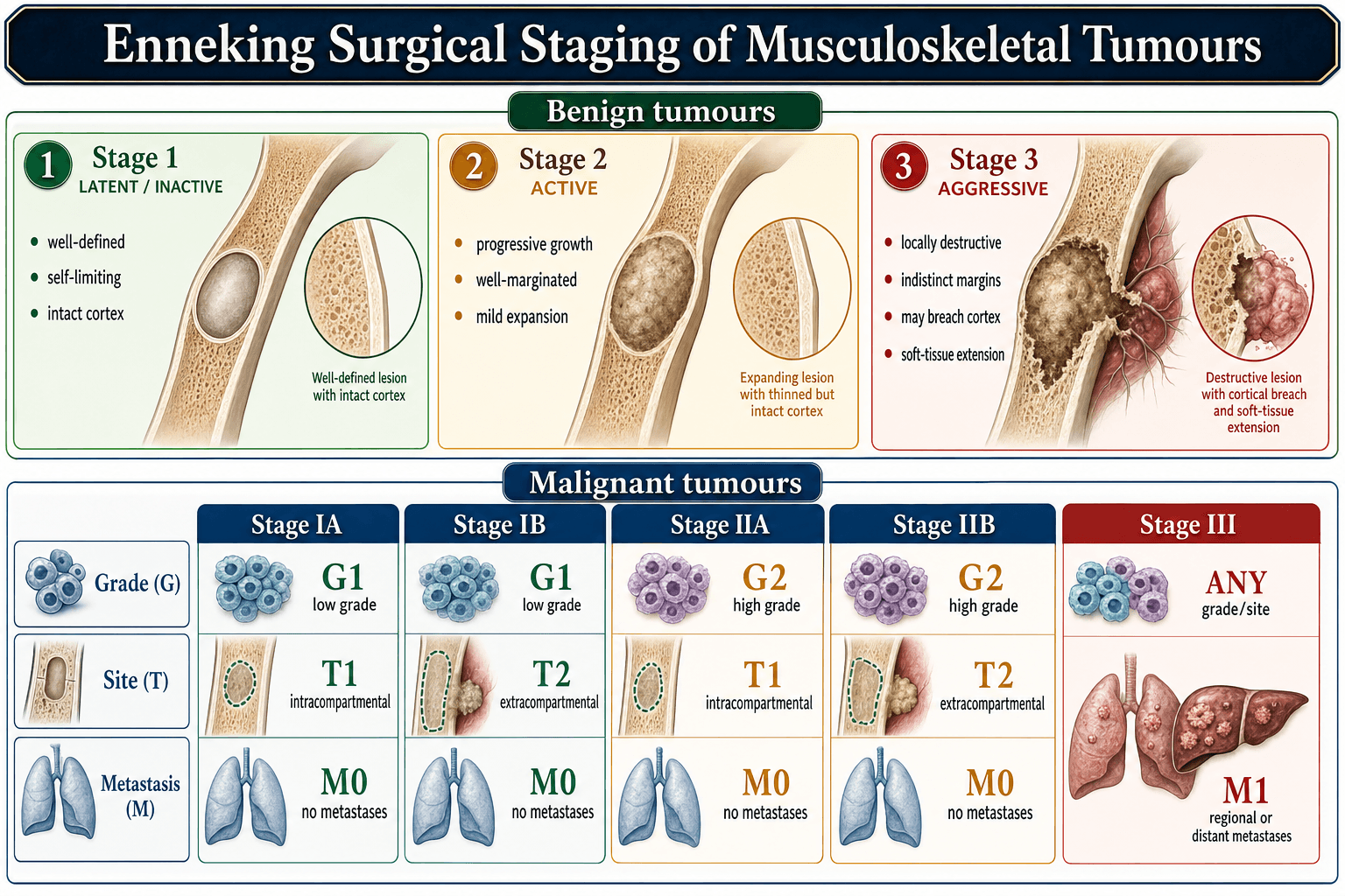
Pelvic Zones (Enneking-Dunham Classification)
The Enneking-Dunham classification divides the pelvis into anatomical zones for surgical planning:
Type I - Iliac Wing Resection
- Iliac wing above the acetabulum
- From iliac crest to sciatic notch
- Does NOT include acetabulum
- Gluteal muscles (detached)
- Iliac vessels (preserved, retracted)
- Sciatic nerve (usually preserved unless tumour extends posteriorly)
- Often NO formal reconstruction required
- Soft tissue repair of abdominal wall to remaining pelvis
- Excellent functional outcome as hip joint preserved
- Near-normal gait
- Full weight-bearing on preserved hip
- Minimal long-term disability
PELVISPELVIS - Pelvic Resection Zones (Enneking-Dunham)
Hook:P1-P4: Posterior ilium, Periacetabular, Pubic, Para-sacral - following the pelvic ring from back to front
Investigations and Preoperative Staging
Imaging
MRI (Essential)
- T1-weighted: Bone marrow extent, fat planes
- T2/STIR: Tumour extent, oedema
- Post-gadolinium: Vascularity, viable tumour
- MRA: Relationship to major vessels
- Tumour extent in bone and soft tissue
- Relationship to neurovascular bundle
- Skip lesions (additional foci)
- Joint involvement
- Define resection margins
- Assess neurovascular involvement
- Plan reconstruction approach
- 3D reformats for custom prosthesis design
MRI is mandatory for surgical planning - defines tumour extent and resectability.
Biopsy
- CT or ultrasound-guided
- Multiple cores for adequate tissue
- Place biopsy tract to allow excision with specimen
- Longitudinal incision in line with definitive surgery
- Meticulous haemostasis
- Close in layers without drain if possible
- Discuss with operating surgeon BEFORE biopsy
- Biopsy tract must be excised en bloc with tumour
- Avoid contaminating neurovascular structures
- Send tissue for histology, cytogenetics, and microbiology
Staging Workup
- Chest CT (pulmonary metastases)
- PET-CT or bone scan
- Bloods: FBC, UEC, LFT, LDH, ALP
- Consider bone marrow biopsy (Ewing sarcoma)
- Orthopaedic oncologist
- Medical oncologist
- Radiation oncologist
- Radiologist
- Pathologist
- Reconstructive surgeon (plastic/vascular)


Differential Diagnosis of a Destructive Pelvic Lesion
A destructive periacetabular or iliac lesion is not automatically a primary sarcoma. Distinguishing the differentials below determines whether hemipelvectomy is even appropriate.
- Typical Age / Clues
- Adult 40-70, indolent pain
- Imaging Signature
- Ring-and-arc / popcorn matrix, endosteal scalloping
- Implication for Surgery
- Wide resection - chemo/radio-resistant, surgery is curative treatment
- Typical Age / Clues
- Adolescent / young adult, rapid pain
- Imaging Signature
- Aggressive osteoid matrix, soft-tissue mass, periosteal reaction
- Implication for Surgery
- Neoadjuvant chemo then wide resection; margins can follow good response
- Typical Age / Clues
- Child / teenager, may have fever, raised inflammatory markers
- Imaging Signature
- Permeative lytic destruction, large soft-tissue mass
- Implication for Surgery
- Chemo +/- radiotherapy; surgery when achievable with acceptable morbidity
- Typical Age / Clues
- Older adult, known primary or anaemia/renal impairment
- Imaging Signature
- Often multiple lytic lesions; PET/skeletal survey positive elsewhere
- Implication for Surgery
- Biopsy and stage FIRST - hemipelvectomy almost never indicated
- Typical Age / Clues
- Adult, midline sacrococcygeal, slow growing
- Imaging Signature
- Midline destructive sacral mass, T2-bright
- Implication for Surgery
- En bloc sacral resection; margins drive recurrence, radio-resistant
- Typical Age / Clues
- Young adult 20-40, locally aggressive but benign
- Imaging Signature
- Eccentric lytic lesion, no matrix mineralisation
- Implication for Surgery
- Curettage/denosumab or resection - rarely needs hemipelvectomy
- Typical Age / Clues
- Any age, systemic features, raised CRP
- Imaging Signature
- Destruction with abscess, can mimic Ewing
- Implication for Surgery
- Diagnostic trap - biopsy with microbiology before any oncological surgery
Never plan a hemipelvectomy on imaging alone. A tissue diagnosis from a correctly placed biopsy (tract excisable en bloc) is mandatory - metastasis, myeloma and infection all mimic primary sarcoma and are not treated by amputation.
Management - Internal Hemipelvectomy Technique
Preoperative Preparation
Preoperative Planning:
-
MDT Discussion:
- Confirm diagnosis and staging
- Neoadjuvant therapy completion
- Surgical plan and reconstruction
-
Imaging Review:
- Define resection margins on MRI
- CT angiography for vascular planning
- 3D reconstruction for custom prosthesis
-
Patient Preparation:
- Medical optimisation
- Blood products arranged (6-10 units RBC minimum)
- Cell saver available
- ICU bed confirmed
-
Equipment:
- Tumour resection instruments
- Reconstruction implants (backup options)
- Vascular instruments available
- Nerve stimulator
Surgical Steps by Resection Type
Type I Iliac Wing Resection
Step 1: Anterior Approach
- Ilioinguinal or modified iliofemoral incision
- Identify and protect external iliac vessels
- Identify and protect femoral nerve
- Expose iliac crest and anterior ilium
Step 2: Posterior Exposure
- Extend incision or separate posterior approach
- Detach gluteal muscles from iliac crest
- Identify sciatic notch and sciatic nerve
- Control superior gluteal vessels
Step 3: Osteotomies
- Superior: Along iliac crest (or through crest for narrow margins)
- Inferior: Above acetabular dome (confirm with image intensifier)
- Posterior: Through sciatic notch, protecting sciatic nerve
- Anterior: Through ASIS or anterior ilium
Step 4: Specimen Removal
- Remove specimen en bloc
- Check margins grossly and with frozen section
- Haemostasis of bony surfaces
Step 5: Closure
- Reattach abdominal wall to remaining pelvis
- Layered closure
- Drain to surgical bed
Reconstruction Options for Type II Defects
Saddle Prosthesis
- Saddle-shaped femoral component
- Articulates with cut surface of remaining ilium
- No formal acetabular reconstruction
- Type II resection with preserved iliac wing
- Adequate remaining ilium for support
- Patient with good bone quality
- Prepare remaining ilium surface
- Insert femoral component (cemented or uncemented)
- Saddle rests on iliac surface
- Capsular repair for stability
- Ambulatory with walking aids
- Significant leg length discrepancy
- High revision rate (mechanical failure)
- Simple salvage if failure
Surgical Technique - External Hemipelvectomy
Indications for Hindquarter Amputation
Primary Indications:
- Tumour involving sciatic nerve and/or external iliac vessels
- Extensive soft tissue sarcoma precluding limb salvage
- Failed internal hemipelvectomy with recurrence
- Uncontrolled infection with tumour
Surgical Technique
Posterior (Gluteal) Flap Technique
Preferred Technique - provides excellent coverage
Step 1: Anterior Dissection
- Extended inguinal/ilioinguinal incision
- Ligate femoral vessels at inguinal ligament
- Divide femoral nerve
- Divide adductor muscles at origin
- Transect pubic symphysis or pubic ramus
Step 2: Posterior Dissection
- Posterior incision from PSIS to ischial tuberosity
- Preserve gluteal muscles on posterior flap
- Divide sciatic nerve high in pelvis
- Control gluteal vessels (maintain flap perfusion via inferior gluteal)
Step 3: Pelvic Division
- Sacroiliac joint disarticulation OR
- Osteotomy through sacral ala
- Divide sacrospinous and sacrotuberous ligaments
- Remove specimen en bloc
Step 4: Flap Closure
- Rotate posterior flap anteriorly
- Trim excess tissue for contour
- Tension-free closure
- Suction drains
Advantages:
- Excellent soft tissue bulk for sitting
- Reliable blood supply (inferior gluteal)
- Good wound healing
FLAPSFLAPS - External Hemipelvectomy Coverage
Hook:FLAPS for coverage - posterior Gluteal flap is the workhorse for hindquarter amputation
Controlling Catastrophic Pelvic Venous Haemorrhage
The topic repeatedly names the presacral venous plexus as the major bleeding source and quotes 3-10 litres of blood loss, but never develops how to control it - which is the single most important intraoperative survival skill in pelvic tumour surgery and a classic viva question.
- Why it is so dangerous. The pelvic veins - the presacral (sacral) venous plexus and internal iliac (hypogastric) veins - are a valveless, high-flow, low-pressure network that communicates with the vertebral (Batson) and basivertebral plexuses. A torn presacral vein retracts into the sacral foramen and cannot be clamped or ligated; blind clamping tears it further and endangers the sacral nerve roots. This is why it bleeds catastrophically and why conventional vascular control fails.
- Prevent it - proximal control first. Gain early retroperitoneal control of the common and internal iliac vessels BEFORE mobilising the tumour, and ligate the internal iliac artery (and vein) on the resection side. Consider preoperative selective arterial embolization (internal iliac) to cut inflow; some centres use intraoperative aortic or internal-iliac balloon occlusion for temporary inflow control during the critical resection.
- When presacral venous bleeding starts - do NOT blindly clamp. Apply direct pressure/packing first to control while the anaesthetist catches up. Definitive local options include sterile thumbtacks or bone wax pressed into the bleeding sacral foramen/cortex, muscle-fragment "welding" (a rectus/muscle pledget pressed onto the bleeding point and cauterised), and topical haemostatic agents. If it remains uncontrolled, pack the pelvis and use damage control - temporary closure, resuscitate on ICU, and a planned re-look at 24-48 hours.
- Resuscitate in parallel. Activate the massive transfusion protocol, give tranexamic acid, run the cell saver, and actively prevent the lethal triad of hypothermia, acidosis and coagulopathy.
Q: How do you control torn presacral venous plexus bleeding during a hemipelvectomy? A: Not by clamping or ligating - the valveless veins retract into the sacral foramina and blind clamping worsens the tear and risks the sacral roots. Prevent it with early proximal internal iliac control ± embolization/balloon occlusion; when it bleeds, pressure and packing first, then thumbtacks/bone wax into the foramen, muscle-fragment welding and haemostatic agents, and if still uncontrolled pack and damage-control with a planned re-look - all alongside a massive transfusion protocol, tranexamic acid and cell saver.
Sacral Nerve-Root Sacrifice: the Functional Map
The Type IV tab states that "S1 root sacrifice affects ankle dorsiflexion" and "S2-S4 sacrifice affects bladder/bowel function", but the functional consequences of sacral resection - central to consenting a Type IV or extended hemipelvectomy - are never developed (there is no dedicated sacrectomy topic to carry them).
- The single most important rule: unilateral is far better tolerated than bilateral. One intact side can maintain continence and useful function, so unilateral sacral-root sacrifice is generally acceptable, whereas bilateral loss of S2-S4 produces a neurogenic bladder and bowel and sexual dysfunction.
- Continence (bladder / bowel / sexual): S2-S4. These roots (parasympathetic plus the somatic pudendal supply) govern continence. As a working guide, preserving both S2 roots and at least one S3 usually maintains bladder and bowel control; bilateral sacrifice at or above S2-S3 does not. Counsel explicitly and plan for possible intermittent catheterisation or a stoma.
- Ambulation / motor. Useful gait needs the L5 and S1 roots; S1 contributes to plantarflexion (gastrocnemius-soleus) and hip/pelvic stability, so its preservation matters for walking. Resections below S1 typically preserve ambulation.
- Spinopelvic stability. A high sacral resection (through or proximal to S1, or crossing both sacroiliac joints) disconnects the spine from the pelvis - spinopelvic (lumbopelvic) dissociation - and requires lumbopelvic reconstruction/fixation (lumbar pedicle plus iliac screws, with or without fibular/allograft struts). Lower resections that keep S1 and at least one SI joint usually do not need spinopelvic fixation.
Q: What are the functional consequences of sacral-root sacrifice, and which must be preserved? A: Unilateral sacrifice is far better tolerated than bilateral. Continence depends on S2-S4 - keeping both S2 and at least one S3 usually preserves bladder/bowel/sexual function, whereas bilateral S2-S4 loss causes a neurogenic bladder/bowel. Ambulation needs L5-S1 (S1 for plantarflexion and pelvic stability). A resection through/above S1 or across both SI joints causes spinopelvic dissociation needing lumbopelvic fixation; below-S1 resections usually do not.
Complications
Intraoperative Complications
- Expected blood loss: 3-10 litres
- Cell saver essential
- Vascular surgery backup
- Risk of presacral venous plexus injury
- Management: Direct pressure, packing, vascular control
- Sciatic nerve (may be intentional sacrifice)
- Femoral nerve (must preserve for internal)
- Lumbosacral plexus injury
- Obturator nerve
- Ureter injury (preoperative stents may help)
- Bladder injury
- Rectal injury (especially posterior tumours)
- Requires intraoperative repair
Early Postoperative Complications
- Infection: 15-30%
- Dehiscence: 10-20%
- Flap necrosis: 5-15%
- Seroma/haematoma
- High risk (10-30% DVT)
- Pulmonary embolism risk
- Extended prophylaxis required (4-6 weeks)
- IVC filter consideration for high-risk
- Respiratory complications
- Renal failure (blood loss, contrast, myoglobin)
- Sepsis
- Mortality: 1-5%
Late Complications
- 15-25% with wide margins
- Higher with positive/close margins
- Salvage options limited
- Prosthetic dislocation: 10-30%
- Prosthetic loosening
- Prosthetic infection: 10-20%
- Allograft nonunion/fracture
- Chronic pain
- Phantom limb pain (external hemipelvectomy)
- Limb length discrepancy
- Gait abnormality
Postoperative Care and Rehabilitation
- ICU admission (24-48 hours minimum) after major resection
- Ongoing fluid resuscitation and transfusion; correct coagulopathy
- Multimodal analgesia (epidural or PCA), including neuropathic agents for phantom/sciatic pain
- Hourly wound and flap perfusion monitoring
- Drains removed when output minimal
- Monitor flap viability; early identification of wound complications
- Negative pressure wound therapy for dehiscence or high-risk wounds
- Mechanical plus extended pharmacological prophylaxis (4-6 weeks)
- Consider IVC filter in very high-risk or contraindicated-anticoagulation patients
- Early mobilisation when haemodynamically stable
- Wheelchair initially after external hemipelvectomy; protected/graduated weight-bearing dictated by reconstruction after internal hemipelvectomy
- Prosthetic assessment (only ~20-30% use a prosthesis long-term after hindquarter amputation)
- Psychological support and oncological surveillance imaging are essential
Outcomes and Prognosis
5-year survival for localised pelvic sarcoma is approximately 60-70%, dominated by tumour grade, margin quality and histology. High-grade and dedifferentiated tumours fare far worse; curatively resected external hemipelvectomy series report 5-year survival closer to 20%.
Local recurrence is 15-25% with wide margins and rises sharply with close/positive margins. Margin adequacy - not resection zone - is the principal modifiable driver of recurrence.
After internal hemipelvectomy, mean MSTS/TESS scores cluster around 60-77%; most patients ambulate with aids. After external hemipelvectomy only ~5-30% use a prosthesis, with most relying on crutches or a wheelchair.
Periacetabular endoprosthetic reconstruction carries high infection (often 15-30%) and dislocation rates, with 5-year implant survival around 50% in some series - a key counselling point.
Prognostic factors (most to least powerful): histological grade, then surgical margin status, then tumour size and stage, then anatomical resectability. Achieving a wide margin while preserving the limb is the central goal; amputation is justified when salvage would compromise the margin in high-grade disease.
Guidelines, Registries & Global Practice
Global Epidemiology
- Pelvic and sacral sites account for roughly 10-15% of all primary bone sarcomas.
- Chondrosarcoma is the most common primary pelvic bone sarcoma in adults; osteosarcoma and Ewing sarcoma predominate in adolescents and young adults.
- Bimodal age distribution: peak in the 2nd-3rd decade (Ewing/osteosarcoma) and 5th-7th decade (chondrosarcoma), with a slight male predominance.
- Pelvic sarcomas carry worse local control and survival than appendicular sarcomas because deep location delays diagnosis and complex anatomy limits achievable margins.
Guideline Comparison (Side by Side)
- NICE / BSG / BOA (UK)
- Mandatory referral to a designated bone sarcoma centre before biopsy
- NCCN / MSTS (US)
- Care at a specialist sarcoma/multidisciplinary centre strongly recommended
- ESMO-EURACAN (Europe)
- Reference-centre management within recognised networks (EURACAN)
- NICE / BSG / BOA (UK)
- Performed at, or directed by, the treating sarcoma unit
- NCCN / MSTS (US)
- Image-guided core biopsy planned so tract is excisable en bloc
- ESMO-EURACAN (Europe)
- Biopsy by the team that will perform definitive surgery
- NICE / BSG / BOA (UK)
- Wide margin limb-sparing resection where oncologically safe
- NCCN / MSTS (US)
- Wide margins; amputation only when salvage compromises margins
- ESMO-EURACAN (Europe)
- En bloc wide excision; reconstruction individualised
- NICE / BSG / BOA (UK)
- Neoadjuvant chemo for osteosarcoma/Ewing; none for conventional chondrosarcoma
- NCCN / MSTS (US)
- MAP for osteosarcoma; VDC/IE for Ewing; surgery alone for chondrosarcoma
- ESMO-EURACAN (Europe)
- Same chemo-sensitivity framework; consider trials for high-grade disease
Registry and Outcome Signals
- Volume-outcome relationship: national audits and sarcoma networks consistently show better local control and survival when pelvic sarcoma surgery is concentrated in high-volume reference centres.
- Reconstruction durability: implant and reconstruction outcomes are tracked in institutional and national bone-tumour databases rather than standard arthroplasty registries (NJR/AJRR/AOANJRR exclude most tumour endoprostheses); reported pelvic endoprosthesis infection rates are far higher than primary THA.
- Functional benchmark: MSTS and TESS scores after internal hemipelvectomy typically cluster around 60-77%, versus markedly lower self-reported function and prosthetic use after external hemipelvectomy.
High- vs Limited-Resource Practice Variation
Custom 3D-printed endoprostheses, navigation/patient-specific cutting guides, on-table vascular and plastic reconstruction teams, neoadjuvant chemotherapy and routine MDT planning enable higher limb-salvage rates.
Later presentation, limited access to custom implants and intraoperative frozen section shift practice toward biological reconstruction (irradiated autograft, fibular graft, arthrodesis), flail-hip resection arthroplasty, or external hemipelvectomy when salvage is unsafe.
Controversies and Areas of Uncertainty
Internal hemipelvectomy preserves the limb but high reconstruction-failure rates mean some salvaged limbs function worse than a well-rehabilitated amputation. The trade-off between limb preservation and durable function remains unresolved.
No reconstruction method (saddle, custom endoprosthesis, APC, irradiated autograft, arthrodesis, flail hip) is clearly superior. Endoprostheses give the best early function but the highest infection and dislocation burden.
Computer navigation and patient-specific guides improve margin accuracy in studies, but evidence that they improve survival or long-term function (rather than radiological margin) is still maturing.
Wide margin correlates with local control, but the exact safe distance and how to treat planned close margins around neurovascular structures (versus converting to amputation) is debated.
Resection arthroplasty has the lowest complication rate yet significant shortening and instability; whether to reconstruct at all in low-demand or high-infection-risk patients is individualised.
For close/positive margins or unresectable disease, the benefit of adjuvant radiotherapy (including particle therapy for chondrosarcoma/chordoma) over re-resection is not firmly established.
MCQ Practice Points
Q: What does Type II resection in the Enneking-Dunham pelvic classification involve? A: Periacetabular resection - removal of the acetabulum and surrounding bone. This is the most complex resection type and requires formal hip reconstruction (saddle prosthesis, custom prosthesis, APC) or acceptance of flail hip.
Q: What is the key difference between internal and external hemipelvectomy? A: Internal hemipelvectomy is limb-sparing - pelvic bone is resected but the lower extremity is preserved. External hemipelvectomy (hindquarter amputation) removes the entire lower limb through the pelvis. The key determinant is neurovascular bundle involvement.
Q: Which vessel is typically ligated during internal hemipelvectomy? A: The internal iliac artery is typically ligated as its branches supply the resected specimen. The external iliac artery must be preserved to maintain limb perfusion. Early vascular control is essential before tumour mobilisation.
Q: What is the preferred flap for wound coverage in external hemipelvectomy? A: The posterior (gluteal) flap is preferred as it provides excellent soft tissue bulk for the sitting surface and has reliable blood supply from the inferior gluteal artery. The anterior flap is used when posterior tissues are involved by tumour.
Q: Why is surgical resection the primary treatment for pelvic chondrosarcoma? A: Chondrosarcoma is resistant to chemotherapy and radiation therapy. Wide surgical margins are the only treatment proven to achieve local control and cure. This distinguishes it from osteosarcoma and Ewing sarcoma, which respond to neoadjuvant chemotherapy.
Clinical Decision Scenarios
Practise clinical reasoning and management decisions out loud
“A 45-year-old presents with a 6-month history of left hip pain. Imaging shows a destructive lesion of the left iliac wing extending to the acetabular dome. Biopsy confirms Grade II chondrosarcoma. How would you manage this patient?”
“Describe the surgical approach for a Type II internal hemipelvectomy. What are the key steps for vascular control?”
“A patient is undergoing internal hemipelvectomy. Intraoperatively, you find the tumour is encasing the external iliac vessels. What are your options?”
Types and Definitions
- Internal hemipelvectomy: Pelvic bone resection with limb preservation
- External hemipelvectomy: Hindquarter amputation - complete limb removal
- Key determinant: Neurovascular bundle involvement
Enneking-Dunham Classification
- Type I: Iliac wing (above acetabulum) - often no reconstruction needed
- Type II: Periacetabular - most complex, requires reconstruction
- Type III: Pubic rami/ischium - minimal reconstruction needed
- Type IV: Sacral ala - may need lumbopelvic fixation
Vascular Control
- Control common iliac vessels retroperitoneally FIRST
- Internal iliac artery typically LIGATED
- External iliac vessels MUST BE PRESERVED for limb salvage
- Presacral venous plexus - major bleeding risk
Type II Reconstruction Options
- Saddle prosthesis: Femur articulates with remaining ilium
- Custom endoprosthesis: Best function, highest complication rate
- Allograft-prosthetic composite (APC): Biological bone stock
- Flail hip: No reconstruction, lowest complications, worst function
External Hemipelvectomy Flaps
- Posterior (gluteal) flap: PREFERRED - good bulk, reliable supply
- Anterior (quadriceps) flap: When posterior tissues involved
- Goal: Adequate sitting surface with tension-free closure
Indications for Amputation
- Tumour involving sciatic nerve
- External iliac vessel encasement
- Failed limb salvage with recurrence
- Uncontrolled infection
Tumour Considerations
- Chondrosarcoma: Chemo-resistant - surgery alone
- Osteosarcoma: Neoadjuvant chemo (MAP), assess response
- Ewing sarcoma: Chemo/radio-sensitive, surgery preferred when feasible
Complications
- Blood loss: 3-10L expected - cell saver essential
- Wound complications: 30-50%
- Infection: 15-30%
- Local recurrence: 15-25%
- Mortality: 1-5%
Key Numbers
- 5-year survival (localised pelvic sarcoma): 60-70%
- Limb salvage rate: 60-70%
- Prosthesis use post-hindquarter amputation: 20-30%
- Major complication rate: 30-50%
Evidence Base
Enneking-Dunham Classification (Landmark Paper)
- Of over 200 patients evaluated, 57 were candidates for curative surgery - 25 underwent hemipelvectomy and 32 a non-amputative (limb-sparing) procedure
- Established the principle of zonal resection: iliac wing, periacetabular, and pubic regions
- Local recurrence 100% after oncologically inadequate resections versus 4% after adequate wide/radical resections
- Near-normal function when the hip joint was preserved; arthrodesis was achieved in only 50% of attempts
- Sciatic nerve resection was NOT a contraindication to a non-amputative procedure
Pelvic Chondrosarcoma - Landmark Outcomes Series
- 64 patients with localised pelvic chondrosarcoma; 51 limb-salvage, 13 hemipelvectomy
- Local recurrence 19% and distant metastases 17%
- Less than a wide surgical margin correlated with local recurrence (p=0.014)
- High tumour grade correlated with poor overall survival (p less than 0.001)
- All limb-salvage patients ambulatory; mean MSTS functional score 77%