Rare benign fibro-osseous lesion of anterior tibial cortex in children
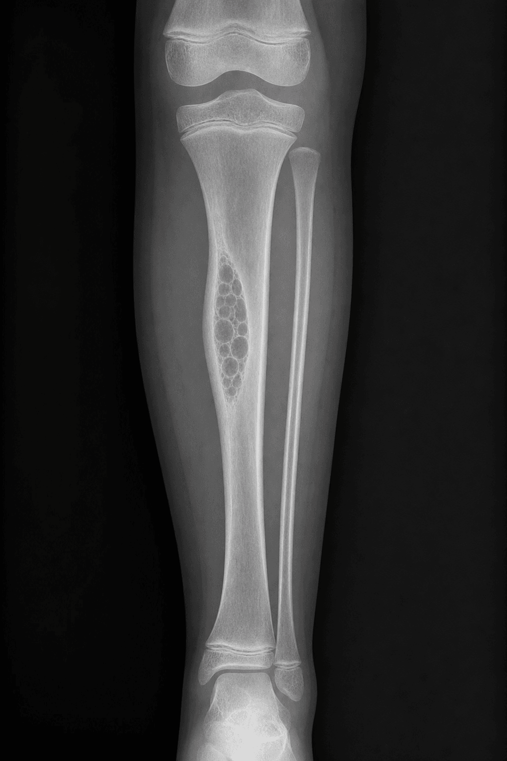
- Intracortical lucent lesion with sclerotic borders on X-ray
- Critical differential: Adamantinoma (epithelial islands, low-grade malignant)
- OFD shows only scattered keratin-positive cells; adamantinoma has true epithelial islands
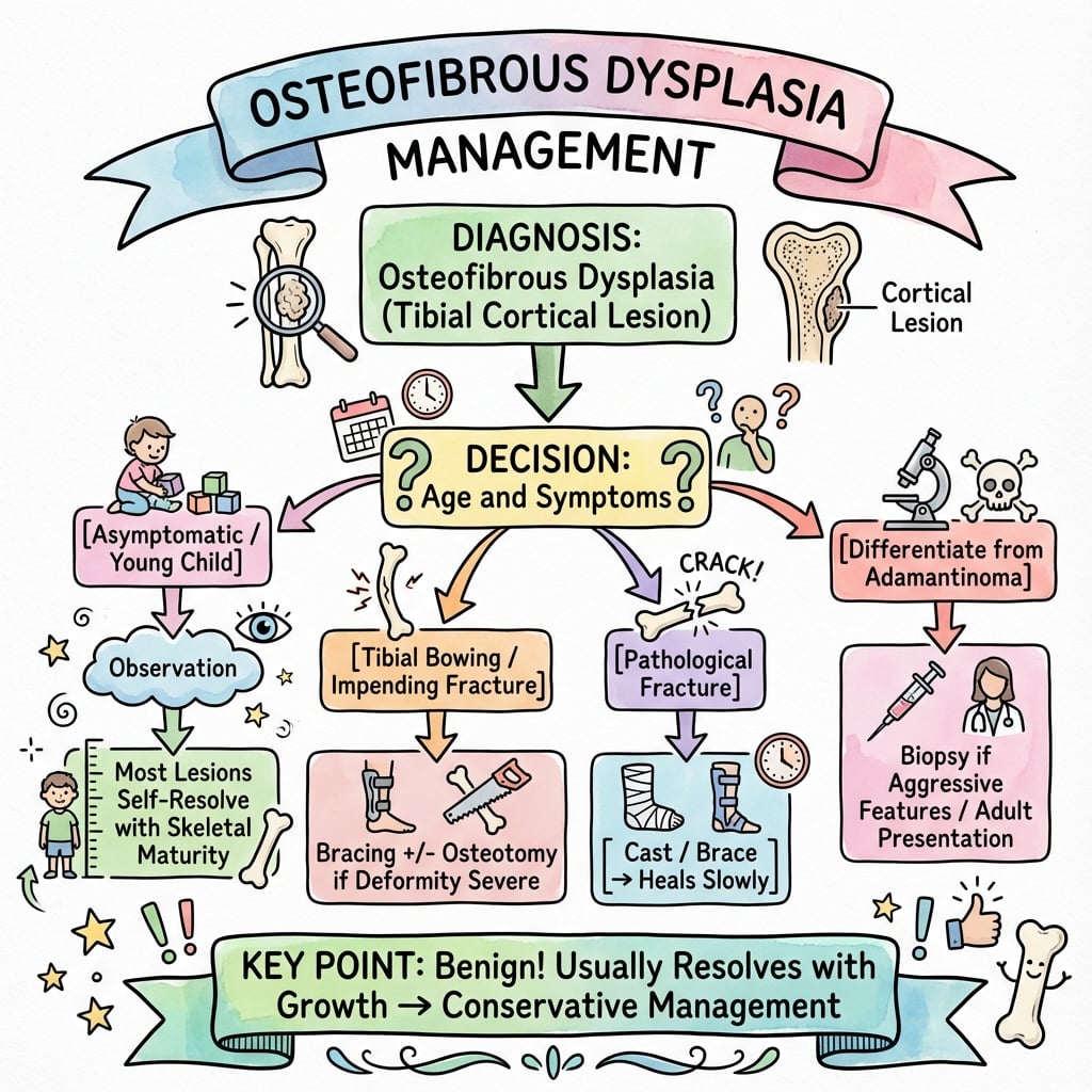
- Conservative observation - most resolve after skeletal maturity
- Surgery targets symptomatic deformity, not the lesion
- “Biopsy if atypical features OR age greater than 10 years
- “Fibula involvement in 8-10%
- “Anterior cortex preference with bowing
- “No epiphyseal involvement
Epidemiology & Pathogenesis
Demographics
- Peak incidence: 4-8 years (mean age 6 years)
- 90% diagnosed before age 10 years
- Rare in adults (usually progression from childhood)
- No gender predilection (equal male to female ratio)
- Tibia: 85-90% of cases (anterior cortex diaphysis/metaphysis)
- Fibula: 8-10% of cases
- Radius/ulna: Less than 2% (extremely rare)
- Other bones: Case reports only
- Extremely rare: Less than 200 cases in literature
- Represents less than 0.2% of benign bone tumors
- Geographic distribution: No known predilection
Pathogenesis
- Developmental Theory: Defect in cortical bone maturation
- Reactive Process: Response to microtrauma in growing bone
- Mesenchymal Origin: Abnormal differentiation of osteoblastic cells
- Genetic Factors: No consistent chromosomal abnormalities identified
- Active phase (childhood): High cellularity, prominent osteoid production
- Quiescent phase (adolescence): Reduced cellularity, increased bone formation
- Resolution phase (post-skeletal maturity): Fibrous tissue replacement with mature bone
- Three lesions sit on a debated spectrum: OFD (benign) — OFD-like (differentiated) adamantinoma (scattered keratin-positive epithelial cells, indolent, paediatric) — classic adamantinoma (frank epithelial islands, low-grade malignant, metastatic potential).
- Key nuance: OFD is NOT simply "cytokeratin negative." Sweet's AFIP series found isolated cytokeratin-positive stromal cells in 93% of OFD lesions; the discriminator from adamantinoma is the absence of hyperchromatic epithelial islands, not absence of all keratin staining.
- Molecular distinction from fibrous dysplasia is clear: OFD lacks the GNAS (Gsα) Arg201 mutation present in fibrous dysplasia, confirming they are separate entities.
- Direct malignant transformation of pure OFD to adamantinoma has not been demonstrated in the large pooled paediatric series — most reported "transformations" reflect under-sampling of an OFD-like adamantinoma at first biopsy.
Clinical Presentation
Symptoms & Signs
- Asymptomatic (40-50%): Incidental radiographic finding
- Painless Swelling (30-40%): Anterior tibial prominence
- Activity-Related Discomfort (20-30%): Mild aching with exercise
- Pathological Fracture (10-15%): Acute pain and deformity
- Anterior tibial cortex: Firm, non-tender mass
- Overlying skin: Normal, no warmth or erythema
- Anterior tibial bow: May be present with long-standing lesion
- Neurovascular status: Always normal
- Range of motion: Full and painless at adjacent joints
- Anterior tibial bowing (15-20% cases)
- Progressive with growth in active lesions
- Typically mild (less than 15 degrees angulation)
- Rarely requires corrective osteotomy
Clinical Course
- Childhood (Active Phase): Slow expansion with growth
- Adolescence: Stabilization in most cases
- Skeletal Maturity: Spontaneous resolution common
- Adult Follow-up: Residual cortical thickening, no active lesion
- Pathological fracture: 10-15% incidence
- Progressive deformity: 5-10% requiring intervention
- Recurrence after curettage: 50-60% (high rate)
- Malignant transformation: Extremely rare (less than 1%)
Campanacci - Defining the Entity and Its Natural History
- 35 patients (plus 22 prior literature cases) reframed under the unifying term osteofibrous dysplasia, separating it from fibrous dysplasia and adamantinoma
- Lesions seldom progress through childhood and any progression ceases after puberty; several regressed spontaneously
- Frequent recurrence after curettage or subperiosteal resection, but recurrences are usually non-progressive
- Conclusion: delay surgery as long as possible; radical surgery is not required even after recurrence, fracture or pseudarthrosis
Investigations
Plain Radiography
Characteristic Features:
- Location: Anterior tibial cortex, eccentric intracortical lesion
- Appearance: Well-defined lucent area with sclerotic margins
- Pattern: Geographic bone destruction (Lodwick grade IA)
- Cortical Changes: Thinning, expansion, scalloping of inner cortex
- Periosteal Reaction: Minimal to absent in uncomplicated cases
Differential Radiographic Features:
- ofd
- Anterior tibial cortex
- adamantinoma
- Any cortex, may be multicentric
- fibrous_dysplasia
- Medullary, not cortical
- ofd
- Well-defined, sclerotic rim
- adamantinoma
- Less defined, may breach cortex
- fibrous_dysplasia
- Ground glass, no sclerotic rim
- ofd
- Geographic, single lesion
- adamantinoma
- May be moth-eaten or permeative
- fibrous_dysplasia
- Ground glass, expansile
- ofd
- Less than 10 years typically
- adamantinoma
- 20-40 years peak
- fibrous_dysplasia
- Any age, often teenagers
- ofd
- Never present
- adamantinoma
- May be present in advanced cases
- fibrous_dysplasia
- Never present
Advanced Imaging
- Cortical Assessment: Precise delineation of cortical involvement
- Matrix: No calcification or ossification pattern
- 3D Reconstruction: Useful for surgical planning if needed
- Soft Tissue: Confirms absence of extraosseous component
- T1-Weighted: Low to intermediate signal intensity
- T2-Weighted: Variable signal (depends on fibrous versus osseous ratio)
- STIR Sequences: High signal in active lesions
- Contrast Enhancement: Moderate enhancement in active lesions
- Purpose: Exclude soft tissue mass (adamantinoma concern)
- Bone Scan: Increased uptake in active lesions
- Clinical Utility: Limited, not routinely required
- PET Scan: Not indicated for benign lesion
The main imaging goal is to differentiate osteofibrous dysplasia from adamantinoma. MRI showing NO soft tissue component strongly supports OFD diagnosis. If there is ANY soft tissue mass, you must suspect adamantinoma and proceed to biopsy.
Histopathology
- Well-circumscribed fibrous tissue within cortex
- Gritty texture due to osteoid formation
- No cystic changes or hemorrhage
- Sharp demarcation from surrounding bone
- Fibrous Stroma: Spindle cell proliferation, moderate cellularity
- Bone Formation: Trabeculae rimmed by osteoblasts (zoning phenomenon)
- Osteoid Deposition: Irregular seams surrounding fibrous tissue
- Cellular Atypia: None (bland spindle cells)
- Mitotic Activity: Rare to absent
- Cytokeratin (AE1/AE3): NEGATIVE (key distinguishing feature)
- Epithelial Membrane Antigen (EMA): NEGATIVE
- S100: Variable positivity in fibrous component
- CD34: May be positive in stromal cells
- Ki-67 Index: Low (less than 5%)
NOCYSTSOFD vs Adamantinoma - Histology
Hook:NO CYSTS for histologic differentiation
Differential Diagnosis
Key Differentials
- Age: 20-40 years (vs less than 10 for OFD)
- Location: Any tibial cortex, may be multicentric
- Radiology: Less well-defined, may have soft tissue mass
- Histology: Cytokeratin POSITIVE (epithelial component)
- Prognosis: Malignant potential, requires wide resection
- Location: Medullary, not intracortical
- Radiology: Ground-glass matrix, expansile
- Age: Adolescents/young adults typically
- Histology: "Chinese characters" pattern, no osteoblast rimming
- Behavior: Does not resolve spontaneously
- Location: Mandible/maxilla primarily, rare in long bones
- Radiology: Well-defined radiolucent to radiopaque
- Histology: Spherical ossicles (psammomatoid bodies)
- Age: Young adults
- Clinical: Activity-related pain, acute onset
- Radiology: Linear lucency, periosteal reaction
- MRI: Marrow edema, cortical fracture line
- Evolution: Healing within 6-8 weeks
- Location: Metaphysis, eccentric, medullary
- Radiology: Sclerotic scalloped margins, cortical thinning
- Age: Adolescents (10-15 years)
- Histology: Storiform pattern, foam cells, hemosiderin
(For the full behaviour and molecular basis of the two closest mimics, see the dedicated Fibrous Dysplasia, Adamantinoma and Non-Ossifying Fibroma topics; only their OFD-discriminating features are summarised here.)
The Zoning Phenomenon (Histological Hallmark)
The histopathology section names the "zoning phenomenon" as the defining microscopic feature, but the term is left undefined - and it is exactly what an examiner means when they slide a low-power photomicrograph across the desk and ask "what is the architectural clue that this is osteofibrous dysplasia and not fibrous dysplasia or adamantinoma?"
What "zoning" (Maturation Gradient) means. In osteofibrous dysplasia the lesion is organised as a centre-to-periphery maturation gradient rather than the haphazard pattern of fibrous dysplasia:
- Centre of the lesion: the most immature and cellular zone - a loose, mildly myxoid fibrous stroma containing irregular woven-bone trabeculae.
- Periphery of the lesion: the bone becomes progressively more mature and lamellar, with the outer trabeculae remodelling and blending into the surrounding host cortex.
- The unifying feature at every level: the woven-bone trabeculae are rimmed by a prominent layer of plump, active osteoblasts (osteoblastic rimming). This osteoblastic rimming is the single most useful discriminator.
- Fibrous dysplasia shows curvilinear "alphabet-soup"/"Chinese-character" woven bone that arises directly from the stroma without osteoblastic rimming and without a maturation gradient - the metaplastic bone is uniform and haphazard throughout. (It also carries the GNAS Arg201 mutation that OFD lacks - see the Sakamoto evidence.)
- Adamantinoma / OFD-like adamantinoma shares the fibro-osseous, osteoblast-rimmed background of OFD, so zonation alone does not exclude it; the malignant discriminator is the presence of hyperchromatic epithelial islands, not just the scattered keratin-positive stromal cells that OFD also contains (Sweet: 93% of OFD lesions). Zoning tells you "fibro-osseous, OFD-family"; the epithelial islands tell you "adamantinoma."
osteoblastic rimming + a peripheral maturation gradient = OFD. Lose the rimming and the gradient → think fibrous dysplasia. Add hyperchromatic epithelial islands → think adamantinoma. Small biopsies can miss both the gradient and rare epithelial islands, which is why radiology-pathology correlation and generous sampling are stressed elsewhere in this topic.
The "zoning phenomenon" is a maturation gradient: immature cellular woven bone centrally grading to mature lamellar bone peripherally, with woven trabeculae rimmed by prominent osteoblasts throughout. Osteoblastic rimming is the feature fibrous dysplasia lacks; hyperchromatic epithelial islands are the feature that upgrades the lesion to adamantinoma.
Dedifferentiated Adamantinoma - the Aggressive End of the Spectrum
- Three adamantinomas showed sarcomatoid (dedifferentiated) transformation of the epithelial component; one patient died of metastatic disease
- Dedifferentiated areas retained pankeratin positivity despite loss of overt epithelial morphology
- Illustrates the mesenchymal-epithelial plasticity that links OFD, OFD-like adamantinoma and classic adamantinoma
- A keratin-positive sarcomatoid tibial cortical tumour must include adamantinoma in the differential
Management
Conservative Treatment
- Indications: Asymptomatic, stable radiographic appearance, age less than 15 years
- Follow-up Schedule:
- First year: Every 3-6 months with X-rays
- Years 2-5: Every 6-12 months
- Until skeletal maturity: Annual radiographs
- Post-maturity: Discharge if stable
- No specific restrictions for asymptomatic lesions
- Avoid high-impact sports if cortical thinning severe (greater than 50%)
- Return to activities after pathological fracture healing
- Spontaneous resolution: 40-60% after skeletal maturity
- Stabilization: 30-40% with no further progression
- Progression: 10-20% may require intervention
Surgical Management
- Pathological fracture (not healing with conservative treatment)
- Progressive deformity (greater than 20 degrees angulation)
- Diagnostic uncertainty (cannot exclude adamantinoma)
- Persistent pain affecting quality of life
- Patient/family preference after counseling
- Technique: Cortical window, thorough curettage, autograft or allograft
- Recurrence Rate: 50-60% (very high)
- Indications: Small lesions, pathological fracture
- Outcomes: Often requires repeat procedures
- Technique: Wide resection with intercalary reconstruction
- Indications: Concern for adamantinoma, failed multiple curettages
- Recurrence: Less than 5% (definitive treatment)
- Morbidity: Significant, reconstruction challenges
- Indications: Established deformity (greater than 20 degrees) after lesion inactive
- Technique: Closing wedge or dome osteotomy
- Timing: After skeletal maturity preferred
- Outcomes: Good correction, does not address underlying lesion
Surgery Pearls:
- Curettage has HIGH recurrence (50-60%) especially if performed before age 10 years
- Consider observation until skeletal maturity in most cases
- En bloc resection reserved for adamantinoma concern or multiple recurrences
- Pathological fractures usually heal with conservative treatment
- Always send tissue for cytokeratin staining to exclude adamantinoma
Treatment Algorithm
Decision-Making Framework:
-
Initial Diagnosis (Age less than 10, typical radiographs):
- Observation with serial imaging
- No biopsy if classic presentation
-
Atypical Features (Age greater than 10, soft tissue mass, aggressive appearance):
- MRI to assess soft tissue component
- Biopsy mandatory (exclude adamantinoma)
- Consider en bloc resection if adamantinoma-like features
-
Pathological Fracture:
- Conservative fracture management first
- Consider curettage if non-union after 6 months
- Bone grafting to augment healing
-
Progressive Deformity:
- Observation until skeletal maturity if mild (less than 15 degrees)
- Corrective osteotomy after maturity if significant (greater than 20 degrees)
- Address lesion separately if still active
Largest Modern Series - Surgery Should Target Deformity, Not the Lesion
- International multicentre series of 101 tibial OFD lesions in 99 patients across three paediatric tertiary centres
- 41 lesions (40.6%) treated conservatively; anterior bowing under 10 degrees at presentation predicted successful non-operative management
- Progression or recurrence in only 9 lesions (8.9%); wide excision carried a high complication and surgical burden regardless of technique
- NO lesion transformed to adamantinoma, confirming OFD as a benign condition
- Surveillance should track angular deformity on full-length tibial radiographs; surgery is for symptomatic deformity, not the lesion itself
Protective Bracing & Fracture Prophylaxis in the Non-Operative Pathway
The MCQ points and natural-history discussion repeatedly invoke "observation, bracing if needed," but the role of orthotic protection is easily left undefined - a genuine gap for the candidate asked "the family accepts observation; is there anything else you offer?"
Rationale. The lesion weakens the anterior tibial cortex, so the two things that make a purely observed lesion "misbehave" are a pathological fracture (10-15%) and progressive apex-anterior bowing. An external orthosis cannot heal or shrink the lesion, but it can share load across the diaphysis, limit the bending moment on the weakened cortex, and buy time until the lesion becomes quiescent at skeletal maturity.
- When to consider it: a symptomatic or radiographically extensive lesion with significant cortical thinning, activity-related pain, or a healing/healed pathological fracture where the child is remobilising. It is an adjunct to observation, not a substitute for surveillance.
- What is used: a well-moulded clamshell (thermoplastic) tibial orthosis or a patellar-tendon-bearing (PTB)-style ankle-foot orthosis to offload the diaphysis; a simple below-knee brace during high-risk activity is a lower-burden option. Choice is pragmatic - there is no lesion-specific device and no high-level evidence favouring one design.
- What it does NOT do: bracing does not prevent the underlying deformity from progressing if the lesion is biologically active, and it does not replace the definitive answer to established bowing, which is corrective osteotomy near maturity (see Management). Prolonged rigid immobilisation risks stiffness and disuse osteopenia, so protection is graded to activity rather than continuous.
- Fracture that occurs anyway: the great majority (~90%) unite with a standard cast; bracing is the step-down as the child returns to weight-bearing, and non-union (rare) - not the brace - is what triggers surgery.
Bracing in osteofibrous dysplasia is protective, not curative: it offloads a thinned anterior cortex to reduce pathological-fracture risk and cushion the return to activity, but it neither eradicates the lesion nor corrects fixed bowing. If the examiner presses on "how does the brace change the natural history?", the honest answer is that surveillance and, ultimately, deformity-directed osteotomy - not the orthosis - determine the outcome.
Management Algorithm
Outcomes/Prognosis
Complications
- Usually after minor trauma
- Healing: 90% with conservative management (cast immobilization)
- Non-union: Rare (less than 5%), may require surgery
- Refracture: Possible if lesion remains active
- Anterior tibial bowing most common
- Progressive with skeletal growth
- Usually mild (less than 15 degrees)
- Functional impact: Minimal in most cases
- Highest in children under 10 years
- Multiple recurrences common
- May require en bloc resection ultimately
- Extremely rare, case reports only
- Usually represents misdiagnosis of adamantinoma
- Requires careful histological review
Prognosis
- Benign disease with spontaneous resolution potential
- No metastatic potential
- Normal life expectancy
- Minimal functional impairment in vast majority
- 70-80% stable or resolved by age 20 years
- 10-20% residual asymptomatic cortical thickening
- 5-10% require surgical intervention for complications
- Less than 5% develop adamantinoma (likely pre-existing)
RESOLVEFavorable Prognosis Indicators
Hook:RESOLVE for prognosis factors
Guidelines, Registries & Global Practice
Global Epidemiology
- OFD is rare worldwide - well under 0.2% of primary bone tumours - with consistent demographics across published series from Europe, North America and Asia: tibial predominance, anterior cortex of the middle third, and presentation in childhood/adolescence (mean age 7-13 years depending on series).
- No geographic or ethnic predilection and no sex predilection are reported. The largest single dataset is the 101-lesion international series pooling UK and Australian tertiary centres (Dala-Ali, 2022).
Side-by-Side Guidance
There is no single dedicated society guideline for this rare lesion; practice derives from bone-tumour society principles and the major case series. The recommendations below are broadly concordant rather than conflicting.
- principle
- Diagnose on radiology-pathology correlation; histology to exclude adamantinoma
- source
- WHO Classification of Bone Tumours; AAOS review (Most 2010)
- principle
- Avoid biopsy in classic young presentation; biopsy if atypical age, soft-tissue mass or progression
- source
- ESMO/EURACAN sarcoma principles; bone-tumour unit referral pathways
- principle
- Observation with serial full-length tibial radiographs tracking angular deformity
- source
- Dala-Ali 2022; Campanacci 1981
- principle
- Target symptomatic deformity (osteotomy near maturity); avoid lesion-directed curettage (high recurrence)
- source
- Dala-Ali 2022; Park 1993
- principle
- Refer to specialist sarcoma centre; wide en bloc excision with staging (chest CT)
- source
- Hazelbag 1994; Houdek 2018; BSG/ESMO principles
Registry Notes
- OFD is too rare for dedicated registry survival data. National bone-tumour and sarcoma registries (e.g. the UK NSSG/regional bone-tumour services, European EURACAN reference network, Scandinavian sarcoma group databases) capture it mainly to flag and track the OFD-like adamantinoma it can mimic, where long-term recurrence (beyond 15 years, Houdek 2018) justifies registry surveillance.
High- vs Limited-Resource Practice Variation
- Well-resourced settings: ready access to MRI, immunohistochemistry (cytokeratin/EMA) and specialist sarcoma MDTs allows confident observation of classic lesions and early detection of atypical ones; deformity is managed with planned osteotomy and growth modulation.
- Limited-resource settings: plain radiography remains the mainstay; the key practice point is to AVOID intralesional curettage of a presumed benign lesion that could be an under-sampled adamantinoma, and to refer atypical, painful or progressive cortical tibial lesions to a centre with histopathology and limb-salvage capability rather than treating locally.
Why This Topic Matters in Exams
Globally (FRCS, FRACS, EBOT, ABOS, DNB/MS), examiners test the OFD-adamantinoma-fibrous dysplasia triad: location (cortical vs medullary), cytokeratin nuance, GNAS molecular distinction, the observation-first natural history, and the danger of mislabelling an atypical lesion.
Controversies & Areas of Uncertainty
- Is OFD a precursor to adamantinoma? Unresolved. Large paediatric series (Park, Dala-Ali) report NO transformation, arguing OFD is a self-limiting benign lesion. Pathology series (Hazelbag) document OFD-like adamantinomas recurring as classic adamantinoma, supporting a precursor or sampling-bias model. The pragmatic consensus: pure OFD does not need to be treated as pre-malignant, but an atypical or relapsing lesion warrants thorough sampling to exclude OFD-like adamantinoma.
- OFD vs OFD-like adamantinoma on a small biopsy. Scattered keratin-positive cells occur in both; the malignant label requires hyperchromatic epithelial islands. Limited biopsies under-sample, so radiology-pathology correlation and, where feasible, generous sampling are essential.
- Role and timing of biopsy. Classic radiology in a young child can be observed without biopsy. Biopsy is reserved for atypical age (over the second decade), a soft-tissue mass, aggressive radiology, or progression - precisely because needling a benign lesion risks recurrence and offers little when the picture is typical.
- What to operate on. Modern thinking (Dala-Ali) shifts the surgical target from the lesion to the deformity: intralesional curettage has high recurrence and rarely changes natural history, while wide excision carries substantial morbidity. Corrective osteotomy for established angular deformity, ideally near maturity, is increasingly favoured over lesion-directed surgery.
- Surveillance metric. Anterior angular deformity on full-length tibial radiographs (not lesion size alone) is the most useful parameter to follow, with bowing under 10 degrees predicting successful conservative management.
MCQ Practice Points
Q: What is the classic location and appearance of osteofibrous dysplasia?
A: Anterior tibial cortex in children, typically first decade. Creates eccentric, cortical-based, intracortical lytic lesion with anterior bowing of tibia. Ground-glass or multiloculated appearance. Distinguished from fibrous dysplasia by cortical location (FD is medullary). May involve fibula. Self-limiting in most cases.
Q: What is the relationship between osteofibrous dysplasia and adamantinoma?
A: They lie on a spectrum: benign OFD, OFD-like (differentiated) adamantinoma, and classic adamantinoma. OFD contains only scattered keratin-positive stromal cells (seen in ~93% of cases), whereas adamantinoma shows true hyperchromatic epithelial islands and is a low-grade malignancy with metastatic potential. An atypical lesion in an older patient (over age 20) or with rapid growth or a soft-tissue mass warrants biopsy to exclude adamantinoma.
Q: What is the typical natural history of osteofibrous dysplasia?
A: Self-limiting in most children - lesions stabilize or regress with skeletal maturity. Progressive anterior tibial bowing may occur. Conservative management preferred: Observation, bracing if needed. Surgery reserved for pathological fracture, significant deformity, or suspicion of adamantinoma. High recurrence rate if curettage performed before maturity.
Q: How do you differentiate osteofibrous dysplasia from fibrous dysplasia radiographically?
A: Osteofibrous dysplasia: Cortical-based (eccentric), anterior tibia specific, multiloculated, causes anterior bowing, children only. Fibrous dysplasia: Medullary-based (central), any bone, ground-glass matrix, "shepherd's crook" proximal femur, any age. Both show fibrous tissue replacing bone but location is key differentiator.
Q: What histological feature distinguishes osteofibrous dysplasia from fibrous dysplasia?
A: Osteofibrous dysplasia: Woven bone trabeculae rimmed by prominent osteoblasts (osteoblastic rimming), may have cytokeratin-positive epithelial cells. Fibrous dysplasia: Chinese letter/alphabet soup woven bone pattern WITHOUT osteoblastic rimming. Presence of epithelial cells raises concern for adamantinoma spectrum.
At a Glance
Osteofibrous dysplasia is a rare benign fibro-osseous lesion affecting the anterior tibial cortex in 85-90% of cases, with peak incidence in the first decade of life (mean age 4-8 years). Radiographs show an intracortical lucent lesion with sclerotic borders causing anterior tibial bowing. The critical differential is adamantinoma - osteofibrous dysplasia is cytokeratin negative while adamantinoma is positive. Most cases are managed conservatively with observation as spontaneous resolution commonly occurs after skeletal maturity. Surgery is reserved for pathological fracture (10-15%) or progressive deformity.
TIBIAOsteofibrous Dysplasia Location - TIBIA
Hook:Think TIBIA for this tibial lesion
CYTOOFD vs Adamantinoma - CYTO
Hook:CYTOkeratin is the key differentiator
OBSERVEOFD Management - OBSERVE
Hook:Most cases managed with observation
Osteofibrous Dysplasia
Exam Essentials:
- Benign fibro-osseous lesion of anterior tibial cortex (85-90% cases)
- Peak incidence first decade of life (mean age 4-8 years)
- Intracortical lucent lesion with sclerotic borders on X-ray
- Differentiate from adamantinoma (cytokeratin positive)
- Conservative management with observation in most cases
- Spontaneous resolution common after skeletal maturity
- Pathological fracture in 10-15% of cases
Visual One-Pager
- 6-year-old child with painless anterior tibial swelling
- Incidental radiographic finding during trauma workup
- Anterior tibial cortex involvement with intracortical lucency
- Well-defined sclerotic margins on plain films
- X-ray: Intracortical lucent lesion with sclerotic rim
- CT: Cortical thinning and expansion without soft tissue mass
- MRI: Low T1, variable T2 signal, no soft tissue component
- Biopsy: Fibrous tissue with osteoid rimming, cytokeratin negative
- Initial Assessment: Clinical examination, plain radiographs
- Advanced Imaging: CT/MRI to rule out adamantinoma
- Biopsy Consideration: If atypical features or age greater than 10 years
- Observation: Serial radiographs every 6-12 months
- Surgical Intervention: Only for pathological fracture or progressive deformity
Clinical Decision Scenarios
Practise clinical reasoning and management decisions out loud
“A 6-year-old boy presents after a soccer injury. Ankle radiographs show an incidental 3 cm well-defined intracortical lucent lesion in the anterior tibial diaphysis with sclerotic margins. The child is asymptomatic regarding the lesion.”
“A 9-year-old girl had curettage and bone grafting for osteofibrous dysplasia 18 months ago. Follow-up radiographs show recurrence with similar intracortical lucency. Parents are frustrated and want definitive treatment.”
“A 17-year-old presents with several months of anterior shin pain and swelling. Radiographs show a multilocular, eccentric cortical lesion in the mid-tibial diaphysis with some ill-defined margins. MRI shows a small soft-tissue component. The referring clinician has labelled it osteofibrous dysplasia.”
Must-Know Facts
- LOCATION: Anterior tibial cortex (85-90%), children less than 10 years
- RADIOLOGY: Intracortical lucency, well-defined, sclerotic rim, geographic pattern
- HISTOLOGY: Osteoblast rimming + scattered keratin+ cells; adamantinoma has true epithelial islands
- NATURAL HISTORY: Spontaneous resolution 40-60% after skeletal maturity
- TREATMENT: Observation preferred, curettage has 50-60% recurrence
- COMPLICATION: Pathological fracture 10-15%, heals with conservative Rx
- KEY DIFFERENTIAL: Adamantinoma (older, cytokeratin +, soft tissue mass possible)
Diagnostic Workup
- PLAIN X-RAY: First-line, usually diagnostic in classic presentation
- MRI: If atypical (age greater than 10, soft tissue suspected, aggressive features)
- BIOPSY: Not needed if classic (age less than 10, typical location/radiology)
- BIOPSY INDICATED: Age greater than 10, soft tissue mass, multicentric, aggressive
- IMMUNOSTAIN: Cytokeratin (AE1/AE3) must be negative to confirm OFD
- CT: Helpful for surgical planning if intervention considered
Management Algorithm
- OBSERVATION: First-line for asymptomatic, classic presentation
- FOLLOW-UP: X-ray every 6-12 months until skeletal maturity, then discharge if stable
- SURGERY INDICATIONS: Pathological fracture (non-healing), progressive deformity (greater than 20°), diagnostic uncertainty, persistent pain
- CURETTAGE: High recurrence (50-60%), especially age less than 10 years
- EN BLOC RESECTION: Definitive (less than 5% recurrence), reserved for adamantinoma concern or multiple recurrences
- FRACTURE MANAGEMENT: Conservative first (cast), 90% heal without surgery
Viva Traps to Avoid
- DON'T: Rush to biopsy classic presentation in young child
- DON'T: Offer curettage as 'cure' - high recurrence rate, inform consent
- DON'T: Miss adamantinoma - check age, soft tissue, cytokeratin on histology
- DON'T: Recommend prophylactic surgery - observation superior natural history
- DO: Explain spontaneous resolution potential to family
- DO: Document shared decision-making if family requests surgery
- DO: Review original histology if recurrence (confirm diagnosis)
Quick Differentials
- ADAMANTINOMA: Age 20-40 years, cytokeratin +, may have soft tissue, malignant potential
- FIBROUS DYSPLASIA: Medullary location, ground-glass matrix, no resolution
- STRESS FRACTURE: Acute pain, linear lucency, heals 6-8 weeks
- NON-OSSIFYING FIBROMA: Metaphysis, medullary, scalloped margins, age 10-15 years
- OSSIFYING FIBROMA: Jaw bones, rare in long bones, psammomatoid bodies
Evidence Base
Park / Mayo Clinic - 80-Case Clinicopathologic Reference
- 80 long-bone OFD cases; recurrence common after incomplete excision/biopsy regardless of regimen (9 of 18 consultation cases)
- Two paediatric lesions histologically matured into fibrous dysplasia on later sampling
- Adamantinoma did NOT develop in any of the 41 cases with follow-up; OFD does not progress to adamantinoma
- Surgery reserved for extensive lesions, pseudarthrosis or accentuated tibial bowing; overall prognosis good even with recurrence
Sweet (AFIP) - Cytokeratin-Positive Cells Are the Norm in OFD
- 30 cortical OFD cases (mean age 13.4 years); tibia involved in all, ipsilateral fibula in 17%
- Isolated cytokeratin-positive stromal cells found in 28 of 30 lesions (93%) - hyperchromatic epithelial islands were absent in all
- 50 control fibro-osseous lesions (fibrous dysplasia, fibroxanthoma, ossifying fibroma) had NO keratin-positive cells
- Dispels the exam oversimplification that OFD is uniformly cytokeratin negative; the discriminator from adamantinoma is absent epithelial islands
Sakamoto - GNAS (Gsα) Mutation Separates OFD from Fibrous Dysplasia
- 7 of 7 fibrous dysplasia lesions carried the Gsα Arg201 missense mutation (Arg-to-His or Arg-to-Cys)
- 0 of 7 osteofibrous dysplasia lesions and normal bone controls carried the mutation
- Confirms distinct molecular pathogenesis for OFD versus fibrous dysplasia
- Arg201 testing is a useful molecular discriminator between the two fibro-osseous lesions
Most / Mayo - Standard Review of the OFD-Adamantinoma Spectrum
- Defines OFD, OFD-like (differentiated) adamantinoma and classic adamantinoma along a single disease spectrum
- Adamantinoma is a low-grade malignancy of epithelial lineage requiring wide resection and reconstruction
- OFD management ranges from observation to surgery depending on patient age and lesion extent
- Authoritative AAOS review that frames the modern diagnostic and management algorithm
Hazelbag - Adamantinoma Behaviour and the Precursor Hypothesis
- 32 long-bone adamantinomas; intralesional or marginal excision was the strongest risk factor for local recurrence and metastasis (p under 0.001)
- Overall metastasis rate 29%; local recurrence in 9 of 28 followed patients, all after inadequate margins
- Two OFD-like adamantinomas with only scattered keratin-positive cells recurred as full adamantinoma, supporting a precursor relationship
- Establishes that adamantinoma demands WIDE excision, unlike benign OFD
Houdek / Mayo - Long-Term Adamantinoma Outcomes (the Danger if OFD Is Misdiagnosed)
- 46 extremity adamantinomas; tibia most common (n=31); mean age 24 years, mean follow-up 16 years
- 10-year disease-specific survival 92% and recurrence-free survival 72%; 39% required reoperation
- Older (over 20 years) and male patients had higher local recurrence; recurrences occurred beyond 15 years post-op
- Justifies prolonged surveillance and reinforces why correctly separating adamantinoma from benign OFD matters