Superior / deltotrapezial approach · Rockwood III–VI · advanced
- Rockwood III is the key controversy — most centres now offer non-operative management first for low-demand patients, with surgery reserved for those failing 3 months of physiotherapy or for high-demand workers and athletes. Rockwood IV, V, and VI are operative indications.
- Two coracoclavicular ligaments: the conoid (posteromedial, cone-shaped, resists superior translation) and the trapezoid (anterolateral, horizontal fibres, resists axial compression). Both must be reconstructed for full multi-directional stability.
- Modern suspensory fixation (Dog-Bone button, TightRope, MINAR) has replaced the Weaver-Dunn transfer as the gold standard in most high-volume centres — superior biomechanical strength and a lower loss-of-reduction rate.
- Hardware migration after coracoclavicular screw fixation (Bosworth screw) is a recognised complication requiring early screw removal at 6–8 weeks — still used in acute settings in some centres.
When & Why
The indication for ACJ reconstruction is a symptomatic acromioclavicular dislocation graded by the Rockwood classification. The decision to operate is driven entirely by the grade, by the demands of the patient, and by whether the injury is acute or chronic. Decision by grade - Grade I (AC ligament sprain, CC intact, no displacement): sling 1–2 weeks, early mobilisation, analgesia. Return to sport at 2–3 weeks. No surgery.
- Grade II (AC ligament torn, CC sprained, less than 25% superior displacement on stress views): sling 2–3 weeks, physiotherapy from week 2. Return to sport at 4–6 weeks. Symptomatic AC joint arthritis develops in up to 40% at 5 years and may require later distal clavicle excision.
- Grade III (both AC and CC torn, 25–100% superior displacement): genuinely controversial — see the table below.
- Grade IV (posterior displacement of the clavicle into the trapezius, with posterior skin tenting): requires reduction and stabilisation regardless of demand level.
- Grade V (100–300% superior displacement, with muscle tenting, severe deformity and possible brachial plexus traction symptoms): always operative.
- Grade VI (inferior, subcoracoid or subclavicular dislocation from a high-energy mechanism, often with rib fractures, pneumothorax or brachial plexus injury): rare, and always operative.
- Non-operative
- No significant difference vs surgery on DASH (Tamaoki Cochrane, PMID 31604007)
- Operative
- No additional functional benefit at 1 year (low-quality evidence, Tamaoki PMID 31604007)
- Non-operative
- Faster recovery at 6 weeks in conservatively treated patients (Tamaoki PMID 31604007)
- Operative
- Delayed early recovery; higher-demand sport return more reliable in selected patients
- Non-operative
- Residual cosmetic deformity common but usually acceptable
- Operative
- Deformity corrected, but a higher rate of adverse events (RR 2.82, Tamaoki PMID 31604007)
- Non-operative
- Most patients — trial non-operative management first
- Operative
- High-demand manual worker, overhead athlete, or heavy labourer failing 3 months of conservative care
- Non-operative
- First-line treatment for type III
- Operative
- After failure of a 3-month conservative trial, or up-front for selected high-demand athletes
- Non-operative
- Ceccarelli 2008 (PMID 19384625)
- Operative
- Beitzel/Mazzocca 2013 systematic review (PMID 23369483)
Imaging that drives the decision. The Zanca view is the gold-standard radiograph for ACJ grading: a 10–15° cephalic-tilt AP taken bilaterally on a single film at 50% reduced penetration, so the coracoclavicular (CC) distance can be compared side to side (normal 11–13mm bilaterally). Stress views holding 5–10kg weights may unmask displacement in equivocal Grade II/III cases. Significant CC injury is present when there is greater than 5mm of absolute asymmetry or a greater than 25% relative increase; Grade V is defined as a CC distance greater than double the contralateral side (100–300% displacement). Timing changes everything. The later the surgery, the more complex the reconstruction required:
Direct repair is possible, the anatomy is clear and the CC ligament ends are accessible. Suspensory fixation alone may suffice without a graft.
The inflammatory phase makes the dissection technically harder. The repair is usually augmented with a graft or synthetic tape.
The CC ligament is retracted and fibrosed and cannot be repaired directly. A formal reconstruction with a hamstring autograft or synthetic ligament is mandatory, and the AC capsule is attenuated.
Choosing the reconstruction. Once the decision to operate is made, the choice of construct is the next decision:
- Mechanism
- Transfers the coracoacromial ligament to the resected distal clavicle
- Pros
- Familiar, no foreign body, biological
- Cons
- Weak (200–300 N); 30–40% loss of reduction; high revision rate
- Evidence
- Historical gold standard — largely superseded
- Mechanism
- CA ligament transfer plus suture reinforcement around the coracoid and through the clavicle
- Pros
- Stronger than classic Weaver-Dunn; no hardware migration
- Cons
- Still weaker than native CC; suture cut-through risk
- Evidence
- Intermediate option; used when no button or graft is available
- Mechanism
- Suture tape through coracoid and clavicle tunnels; buttons flip to hold the construct
- Pros
- Rapid; excellent immediate strength; reproducible
- Cons
- Coracoid fracture 4–10%; loss of reduction 10–15%; no biological healing
- Evidence
- Salzmann 2010 (PMID 20442326); Beitzel 2013 (PMID 23369483)
- Mechanism
- Free graft looped through the coracoid base and clavicle tunnels, tied or docked
- Pros
- Biological healing; multi-directional strength; low long-term failure
- Cons
- Donor site; longer operative time; requires tunnels; not for acute repairs
- Evidence
- Best biomechanics vs Weaver-Dunn; Mazzocca 2005 (PMID 16282577); Millett 2015 (PMID 25998014)
- Mechanism
- Cancellous screw across the CC space providing temporary reduction
- Pros
- Cheap; fast; good acute reduction
- Cons
- Must be removed at 6–8 weeks; migration risk; no biological healing; re-displacement on removal
- Evidence
- Largely historical; still used in some acute high-grade injuries
The Operation
The goal is to restore the coracoclavicular relationship anatomically — reconstructing both the conoid and trapezoid ligament functions — while protecting the brachial plexus and subclavian vessels that lie immediately posteromedial to the coracoid, and finishing with a meticulous repair of the deltotrapezial fascia for horizontal-plane stability. The exposure is a superior saber-cut approach with a deltotrapezial split, laid out in full in the first steps below.
Operative sequence — suspensory / anatomic reconstruction
- Beach-chair (semi-reclined 45–70°) or modified supine; head in a padded ring, neck neutral; ipsilateral arm draped free for intra-operative mobility.
- Drape fluoroscopy (mini or full C-arm) into the field for AP and Zanca views of the AC joint.
- Prepare from neck to hand; include the ipsilateral leg to thigh if a hamstring graft is planned.
- Palpate and mark the AC joint, the distal clavicle, the coracoid tip and the planned saber-cut incision along Langer lines.
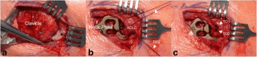
- Make a saber-cut incision 6–8cm along Langer lines from the AC joint, curving gently superior and parallel to the clavicle (an alternative is a transverse superior incision at the AC joint level). Deepen through skin and subcutaneous fat to the deltotrapezial fascia.
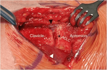
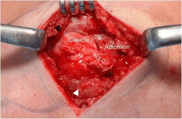
- Identify the AC joint by palpation. Incise the deltotrapezial fascia longitudinally along the ANTERIOR border of the clavicle (rather than directly over the joint) to preserve tissue for later repair.
- Split the deltoid fibres antero-inferiorly no more than 2–3cm and retract with blunt retractors. Expose the superior surface of the distal clavicle and the coracoid process.
- This deltotrapezial plane is the structural layer overlying the joint; it is the key exposure layer and must be repaired at closure for horizontal-plane stability.
- Dissect carefully along the undersurface of the clavicle, staying ON BONE. Identify the coracoacromial ligament anterolaterally (its origin at the coracoid tip is a landmark structure).
- Palpate the coracoid base with a curved elevator or a finger, then pass a right-angle clamp gently around the base of the coracoid.
- The brachial plexus and subclavian vessels lie immediately posteromedial — never pass medial to the coracoid without tactile confirmation of coracoid bone.
- Place the coracoid drill guide (or drill free-hand with fluoroscopy) at the BASE of the coracoid process, about 1cm from the tip.
- Drill from superior to inferior with a 4mm cannulated drill, angled antero-inferiorly (never posteromedially toward the subclavian artery), to a depth of about 15–20mm, using a depth stop.
- Confirm position with fluoroscopy before completing the tunnel. Pass a shuttle suture (PDS 0 or FiberWire) through the tunnel for later graft retrieval.
- Make two drill holes in the clavicle at the native ligament footprints: the CONOID position (a posteromedial tunnel, 45–55mm from the AC joint) and the TRAPEZOID position (an anterolateral tunnel, 25–35mm from the AC joint, anterior to the conoid).
- Use a 4.5mm drill with a depth stop; direct the drill anteriorly and inferiorly to protect the subclavian vessels posterior to the clavicle.
- Confirm both positions with fluoroscopy and pass shuttle sutures through each tunnel.
- Suture tape / Dog-Bone: load a double-loaded FiberTape or TightRope through the coracoid tunnel using the flip technique, then pass the superior button through the clavicle tunnel (one tape through each tunnel for a double-button technique, or a single clavicle tunnel for a Dog-Bone).
- Hamstring autograft: harvest semitendinosus (30–35cm) from the ipsilateral knee using a standard technique, tube the graft to 5–6mm diameter, loop it through the coracoid tunnel via the shuttle, and pass both limbs through the two clavicle tunnels (one limb per tunnel).
- Under fluoroscopic Zanca guidance, reduce the clavicle to the anatomic position by pushing the elbow proximally (lifting the arm weight) and pressing the clavicle inferiorly. The CC distance should equal the contralateral side.
- With reduction maintained, flip the inferior button (if suspensory) or tie the graft under sufficient tension.
- Cycle the shoulder 10–15 times and recheck reduction — the AC joint must remain reduced through the functional arc.
- If the distal clavicle is arthritic or the injury is chronic, excise 5–7mm of distal clavicle (Mumford).
- Repair the AC ligaments and joint capsule with number 1 absorbable sutures to reconstruct the horizontal plane.
- Repair the deltotrapezial fascia meticulously with mattress sutures of number 1 PDS or Vicryl — this horizontal-plane reconstruction is critical and must not be neglected. Close the skin in layers.
The brachial plexus and subclavian vessels lie immediately posteromedial to the coracoid base and posterior to the clavicle in the costoclavicular space. Never pass an instrument medial to the coracoid without tactile bone contact, and drill the coracoid and clavicle tunnels with a depth stop angled antero-inferiorly — never posteromedially. The musculocutaneous nerve enters the conjoint tendon 3–8cm distal to the coracoid tip, so never retract aggressively medial to the conjoint. A Bosworth coracoclavicular screw must be removed at 6–8 weeks — mediastinal migration with great-vessel injury has been fatal.
Place two clavicle tunnels at the anatomic conoid (posteromedial, 45–55mm from the AC joint) and trapezoid (anterolateral, 25–35mm from the AC joint) footprints. A single-tunnel reconstruction fails to control anteroposterior translation and axial rotation and has a higher loss-of-reduction rate. The anatomic two-tunnel construct restores translation closer to the intact joint than a modified Weaver-Dunn (Mazzocca 2005, PMID 16282577).
Vertical stability comes from the CC ligaments (conoid and trapezoid); horizontal (anteroposterior) stability comes from the AC capsule — especially the superior and posterior bands — and the deltotrapezial fascia. Isolated vertical CC fixation leaves residual horizontal instability, a recognised cause of failure. Repair the AC capsule and deltotrapezial fascia at closure (Mazzocca 2007, PMID 17251175).
Reference technique — Weaver-Dunn (historic). Still examined as the original transfer: (1) saber incision and deltotrapezial split as above; (2) excise 1–1.5cm of distal clavicle; (3) identify the coracoacromial ligament from the acromion; (4) detach it from the acromion with a small bone block using an osteotome, preserving the coracoid origin; (5) gouge a canal 1–1.5cm deep in the distal medullary clavicle; (6) pass the CA ligament into the canal and secure it with multiple number 2 non-absorbable sutures through drill holes in the clavicle; (7) augment with a CC suture around the coracoid and through the clavicle (the modified Weaver-Dunn); (8) repair the deltotrapezial fascia. It is biomechanically weak — the CA ligament transfer restores only about a quarter of the native CC load-to-failure — which is its high loss-of-reduction rate. Arthroscopic-assisted technique (contemporary evolution). Several centres now perform the reconstruction arthroscopically-assisted or fully arthroscopic: the coracoid tunnel is drilled under arthroscopic visualisation from a posterior glenohumeral portal, giving direct confirmation of tunnel position and lowering coracoid fracture risk, while the clavicle tunnels are still made through a small superior open incision with fluoroscopy. Advantages are direct visualisation of the coracoid base, concurrent treatment of intra-articular pathology (SLAP tears, rotator cuff disease), and smaller incisions with equivalent reduction and functional outcomes (Salzmann 2010, PMID 20442326). The trade-offs are a steep learning curve, longer operative time during learning, and the continued need for a superior clavicle mini-incision. After securing any construct, perform dynamic fluoroscopy — a Zanca view through abduction and cross-body adduction — to confirm reduction is maintained through the functional arc.
Aftercare & Complications
Rehabilitation principle — progressive loading without traction. For the first 6 weeks the construct is protected from the deforming force of arm weight: gravity pulls the arm (and clavicle) inferiorly relative to the scapula, so the sling supports the forearm and elbow to offload the reconstruction. | Phase | Timing | Immobilisation | Therapy | |-------|--------|----------------|---------| | 1 — Protection | 0–6 weeks | Broad arm sling worn for 4–6 weeks | Elbow/wrist/hand exercises from day 1; pendulums from week 2. No active abduction, no overhead motion, no lifting greater than 1kg, no cross-body adduction | | 2 — Mobilisation | 6–12 weeks | Sling off at 6 weeks | Active-assisted forward flexion and abduction in a pain-free range; periscapular (serratus, lower trapezius) activation; rotator cuff resistance band from week 8 | | 3 — Strengthening | 12–24 weeks | — | Full active ROM expected by week 12; progressive resistance; overhead press and bench press deferred to week 16; sport-specific training from week 16 | Hamstring donor site (if applicable): knee ROM from day 1, quadriceps strengthening from week 2, no running until week 8. | Activity | Timeframe | |----------|-----------| | Driving (manual) | 6–8 weeks | | Office work (one-handed) | 2–4 weeks | | Light manual work | 12 weeks | | Heavy manual work | 4–6 months | | Contact sport | 4–6 months | | Overhead sport | 5–6 months | | Full unrestricted return | 6 months | Physiotherapy principles. In the protection phase, therapy focuses on cervical and periscapular re-education (posture correction and lower trapezius activation to avoid shoulder protraction, which increases CC distraction forces), rotator cuff activation at zero load (external rotation isometrics in the sling from week 2), and proprioception (joint position sense is disrupted after high-grade injuries; gentle joint mobilisation from week 4 aids neuromuscular re-education). Expected outcomes. Rockwood III (operative group): 85–90% good-to-excellent results at 2 years, mean ASES improvement of 25–30 points, CC distance restored to within 3–5mm of the contralateral side in 75–80%, and return to pre-injury activity in 80–85%. Rockwood V: 80–85% satisfactory outcomes, with any pre-operative neurovascular symptoms resolving in 85–90% after reduction and a higher risk of secondary AC arthritis than Grade III. Chronic reconstruction (greater than 3 months): anatomic CC reconstruction with a free tendon graft gives good-to-excellent outcomes in those who avoid a complication (mean ASES around 94 at 2 years; Millett 2015, PMID 25998014), but the procedure is technically more demanding and carries a clinically meaningful revision-requiring complication rate (around 22%); roughly 75–80% return to pre-injury function. Complications
- Incidence
- 10–25% (suspensory); 30–40% (Weaver-Dunn)
- Prevention
- Anatomic tunnel positioning; adequate graft tension; protect the repair 6 weeks; deltotrapezial fascia repair; two-tunnel technique for rotational control
- Management
- If asymptomatic: observe — many do not need revision. If symptomatic with pain or recurrent deformity: revision with an augmented biological graft; assess for coracoid fracture first on CT
- Incidence
- 4–10% (suspensory fixation)
- Prevention
- 4mm tunnel maximum; place the tunnel at the coracoid base 1cm from the tip (not the apex); do not overtighten; warn the patient pre-op; check coracoid size on pre-op CT or X-ray
- Management
- Undisplaced: conservative (6 weeks sling), abandon suspensory fixation and switch to a graft through the intact coracoid or a clavicle-coracoid screw. Displaced: ORIF with a mini-screw if bone allows, or revision reconstruction
- Incidence
- Up to 5% if not removed
- Prevention
- Schedule screw removal at 6–8 weeks; document in the operative note; patient education; mark removal on the theatre list before discharge
- Management
- Urgent removal if symptomatic or migrating medially. If mediastinal migration: emergency cardiothoracic referral — reported deaths from great-vessel injury
- Incidence
- 25–30% at 5 years
- Prevention
- Preserve the AC joint if possible; excise the distal clavicle proactively in a chronic or arthritic joint; anatomic reduction reduces cartilage shear stress
- Management
- Isolated AC arthritis: image-guided steroid injection first line; if that fails, arthroscopic or open distal clavicle excision (Mumford) — 85% satisfaction at 2 years
- Incidence
- 1–3%
- Prevention
- Prophylactic cefazolin 2g IV at induction; careful tissue handling; avoid dead space; closed suction drain in complex reconstructions; synthetic graft increases biofilm risk
- Management
- Superficial: oral antibiotics and wound care. Deep: washout and debridement, retain the graft if fixation is stable, organism-specific IV antibiotics for 6 weeks; remove the implant if infected and the reconstruction has failed
- Incidence
- Less than 0.5% (catastrophic if it occurs)
- Prevention
- Never pass instruments medial to the coracoid without tactile bone contact; drill with a depth stop and anterior angulation; limit dissection in the costoclavicular space
- Management
- Intra-operative vascular injury: direct pressure and a vascular surgery call. Brachial plexus traction (commoner): most resolve with conservative management; EMG at 4–6 weeks if persisting
- Incidence
- 3–8% (biological graft)
- Prevention
- Protected rehabilitation; no overhead loading for 12 weeks; adequate graft diameter (5mm minimum); secure fixation with whipstitch preparation
- Management
- Symptomatic: revision reconstruction. Asymptomatic: observe if the Grade III outcome is acceptable; operate if there is Grade IV/V re-displacement
Viva & Exam Focus
ROCKWOODROCKWOOD — acromioclavicular injury classification
WEAVERWEAVER — Weaver-Dunn key steps
The operative versus non-operative debate for Rockwood III remains unresolved. The systematic review by Ceccarelli et al. (PMID 19384625), the Cochrane meta-analysis by Tamaoki et al. (PMID 31604007), and the current-concepts systematic review by Beitzel and Mazzocca et al. (PMID 23369483) show no consistent superiority of surgery at 1–2 year follow-up. International expert consensus (ISAKOS upper-extremity committee) recommends non-operative treatment as first line for type III, with surgery reserved for failure of conservative management or for selected high-demand patients. Examiners expect you to articulate both sides and individualise the decision.
Clinical Decision Scenarios
Practise clinical reasoning and management decisions out loud
“A 28-year-old recreational cyclist presents after a fall onto the shoulder with pain and a visible step deformity at the AC joint. Zanca views show 80% superior displacement of the clavicle relative to the acromion, with the CC distance increased to 14mm compared with 11mm on the other side. How do you counsel him?”
“A 35-year-old construction worker presents with a 6-month history of right shoulder pain, a visible superior deformity, and inability to work at height or lift over 5kg. He had a Rockwood V ACJ dislocation treated non-operatively by another surgeon. Examination confirms complete superior dislocation with 200% displacement on Zanca view and a positive piano-key sign. Describe your management and reconstruction technique.”
“A 42-year-old patient returns 14 days after ACJ suspensory reconstruction with increasing neck and right arm pain, paraesthesia in the medial forearm (C8/T1 distribution) and grip weakness. Her wound is clean and she is afebrile, with no documented intra-operative complication. What do you suspect, and how do you investigate and manage this?”
Rockwood classification & operative indications
- Grade I: AC sprain, CC intact — non-operative, sling 1–2 weeks
- Grade II: AC torn, CC sprained, less than 25% displacement — non-operative, sling 2–3 weeks
- Grade III: both AC and CC torn, 25–100% displacement — CONTROVERSIAL: non-operative first for most; operate if failing 3 months or for a high-demand worker/athlete
- Grade IV: posterior displacement into the trapezius — always operative
- Grade V: 100–300% superior displacement — always operative
- Grade VI: inferior (subcoracoid) dislocation, high energy — always operative (rare)
Coracoclavicular ligament anatomy
- Conoid: posteromedial, cone-shaped, inserts on the conoid tubercle 45–55mm from the AC joint — resists superior translation
- Trapezoid: anterolateral, horizontal fibres, inserts on the trapezoid line 25–35mm from the AC joint — resists axial load
- Two-tunnel reconstruction (one at each footprint) is required for full rotational and vertical stability
- Normal CC distance 11–13mm; greater than 5mm asymmetry is abnormal
Reconstruction options
- Weaver-Dunn (CA ligament transfer): historic gold standard, weak (200–300 N), 30–40% loss of reduction — largely superseded
- Suspensory fixation (Dog-Bone / TightRope / MINAR): suture tape through coracoid and clavicle tunnels; excellent initial strength; coracoid fracture risk 4–10%
- Biological graft (semitendinosus autograft): the best long-term biological reconstruction; looped through the coracoid and two clavicle tunnels
- Bosworth screw: acute temporary fixation, must be removed at 6–8 weeks (migration risk, including mediastinal migration)
Key steps — suspensory reconstruction
- Saber-cut superior incision 6–8cm; deltotrapezial split along the anterior clavicle border
- Coracoid tunnel at the base (1cm from the tip), 4mm drill, antero-inferior angulation, depth stop mandatory
- Two clavicle tunnels at the conoid (45–50mm) and trapezoid (25–30mm) positions from the AC joint
- Reduce under fluoroscopy (Zanca view) before securing the construct; cycle the shoulder 10–15 times and recheck
- Repair the deltotrapezial fascia meticulously — critical for horizontal-plane stability
Danger zones
- Subclavian artery: posterior to the clavicle during tunnel drilling — always drill anteriorly and inferiorly with a depth stop
- Musculocutaneous nerve: enters the conjoint tendon 3–8cm from the coracoid tip — never retract medial to the conjoint without bone contact
- Brachial plexus (lower trunk): posteromedial to the coracoid base — maintain tactile bone contact at all times during coracoid dissection
- Bosworth screw migration: mediastinal migration reported — mandatory removal at 6–8 weeks
Complications (with incidence)
- Loss of reduction: 10–25% suspensory; 30–40% Weaver-Dunn — the most common failure mode
- Coracoid fracture: 4–10% — related to tunnel size, position and over-tensioning
- Hardware migration (Bosworth screw): up to 5% if not removed — potentially fatal if mediastinal
- AC arthritis: 25–30% at 5 years — manage with an intra-articular injection or distal clavicle excision
- Thoracic outlet syndrome: from re-displacement — the most important early complication to exclude post-op
Post-op milestones
- Weeks 0–6: broad arm sling, elbow/wrist/hand exercises, pendulums from week 2
- Week 6: sling off, active-assisted ROM commenced
- Week 12: full active ROM, progressive resistance training
- Week 16: overhead press and bench press permitted
- Months 4–6: contact sport and full manual work return
- Month 6: full unrestricted activity
Exam evidence anchors
- Mazzocca AJSM 2006 (PMID 16282577): anatomic free-graft CC reconstruction restores AP translation closer to the intact state than a modified Weaver-Dunn — the biomechanical basis for modern anatomic reconstruction
- Ceccarelli 2008 (PMID 19384625): systematic review — clinical results comparable for Grade III operative vs non-op, with more complications after surgery; supports conservative first-line
- Beitzel/Mazzocca systematic review 2013 (PMID 23369483): consensus on non-op for I–II, non-op first for III, operative for IV–VI; insufficient evidence for anatomic vs non-anatomic
- Millett 2015 (PMID 25998014): anatomic CC reconstruction outcomes — ASES 58.9 to 93.8 at 2 years, 22.6% revision-requiring complications
- Tamaoki Cochrane 2019 (PMID 31604007): no 1-year DASH difference, faster 6-week recovery with conservative care, higher adverse events with surgery (RR 2.82)
- Weaver-Dunn original 1972 (PMID 4652050): the historical reference for the classic coracoacromial ligament transfer
Background & Evidence
The Rockwood classification in full. Rockwood graded ACJ injuries by the structures torn and the direction and degree of displacement, and the grade drives management:
- Ligament injury
- AC sprain, CC intact
- Displacement
- No displacement
- Management
- Sling 1–2 weeks; non-operative
- Ligament injury
- AC torn, CC sprained
- Displacement
- Less than 25% superior on stress views
- Management
- Sling 2–3 weeks; non-operative
- Ligament injury
- AC and CC both torn
- Displacement
- 25–100% superior
- Management
- Controversial — non-op first; operate if failing 3 months or high-demand
- Ligament injury
- AC and CC torn
- Displacement
- Posterior (into the trapezius)
- Management
- Always operative
- Ligament injury
- AC and CC torn
- Displacement
- 100–300% superior
- Management
- Always operative
- Ligament injury
- AC and CC torn
- Displacement
- Inferior (subcoracoid)
- Management
- Always operative (rare, high-energy)
Coracoclavicular ligament anatomy (the critical stabiliser). The CC ligament is the primary restraint to vertical displacement and comprises two distinct parts. The conoid ligament is posteromedial and cone-shaped, with a broad base on the posteromedial coracoid and its apex on the conoid tubercle of the clavicle about 45–55mm from the AC joint; its vertical collagen fibres make it the primary restraint to superior translation. The trapezoid ligament is anterolateral and trapezoidal, arising from the superior anterior coracoid and inserting on the trapezoid line of the clavicle about 25–35mm from the AC joint, anterior to the conoid insertion; its horizontal fibres make it the primary restraint to axial compression and lateral displacement. The 3–5mm interligamentous space between them contains a fat pad and small communicating vessels and is the anatomic plane for graft passage. The coracoacromial ligament runs from the anterolateral coracoid tip to the undersurface of the acromion, forming the coracoacromial arch; it has no role in AC joint stability (it is a roof, not a tether) and is harvested for the Weaver-Dunn transfer. Bony anatomy. The AC joint is a diarthrodial joint between the lateral clavicle (convex) and the medial acromial facet (flat to concave), with an average joint gap of 3–5mm and an intra-articular disc present in about 70% that degenerates with age and is gone by the fourth decade. Acromial morphology is described by Bigliani as flat (Type I), curved (Type II) or hooked (Type III, associated with impingement). The distal clavicle is elliptical in cross-section and normally overrides the acromion by 0–3mm in the coronal plane. Deltotrapezial fascia. The deltotrapezial fascia (deltotrapezial aponeurosis) is the conjoined fascia of the deltoid origin (anteroinferior) and the trapezius insertion (posterosuperior) at the clavicle and acromion. It is the primary incision layer in a superior exposure, a structural contributor to horizontal-plane stability, and the layer that must be repaired at closure to restore the dynamic muscular stabilisers. Relevant neurovascular anatomy. The suprascapular nerve (upper trunk, C5–C6) passes through the suprascapular notch to supply supraspinatus and infraspinatus; it is not directly at risk unless dissection extends far medially. The lateral pectoral nerve (lateral cord, C5–C7) runs with the pectoral branch of the thoracoacromial artery and is at risk with excessive medial dissection around the coracoid base. The musculocutaneous nerve enters the conjoint tendon 3–8cm distal to the coracoid tip. The subclavian artery and vein lie posterior to the clavicle in the costoclavicular space and are relevant during clavicle tunnel drilling. Chronic superior clavicle displacement in Rockwood V can cause thoracic outlet syndrome from brachial plexus traction, which resolves after reduction. Biomechanics of ACJ stability. Vertical (superior-inferior) stability is provided primarily by the conoid ligament (force to failure about 400–600 N), with the trapezoid a secondary vertical restraint and the deltotrapezial fascia a minor supplementary check. Horizontal (anteroposterior) stability is provided primarily by the AC ligaments, particularly the superior and posterior bands, which resist AP translation and axial rotation; the CC ligaments provide minimal horizontal restraint. This distinction is critical — reconstruction must address BOTH planes. Rotational stability is resisted by the posterior AC capsule and posterior deltotrapezial fascia, which is why the double-tunnel (conoid + trapezoid position) technique is biomechanically superior to a single-tunnel construct. | Structure | Approximate load to failure | |-----------|-----------------| | Conoid ligament (isolated) | ~500 N | | Trapezoid ligament (isolated) | ~300 N | | Intact CC ligament complex | ~500–725 N | | Coracoacromial ligament transfer (Weaver-Dunn) | ~100–150 N (about 25% of native CC) | | Single suspensory cortical button construct | ~500–1000 N (initial, construct-dependent) | | Doubled semitendinosus / looped tendon graft | Exceeds the native CC complex on cyclic testing | Figures are approximate; cadaveric values vary by specimen age, loading direction and construct (see Mazzocca 2005, PMID 16282577). Clinical implication. The Weaver-Dunn coracoacromial ligament transfer restores only a fraction of native CC strength, which explains its high loss-of-reduction rate. Modern suspensory and biological graft techniques more closely approximate or exceed native biomechanical behaviour, and on cyclic loading the anatomic free-graft reconstruction restores anteroposterior translation closer to the intact state than the Weaver-Dunn (Mazzocca 2005, PMID 16282577).
References
- Weaver JK, Dunn HK. "Treatment of acromioclavicular injuries, especially complete acromioclavicular separation." J Bone Joint Surg Am. 1972;54(6):1187-1194. PMID: 4652050. [Original description of the coracoacromial ligament transfer to the resected distal clavicle] 2. Mazzocca AD, Arciero RA, Bicos J. "Evaluation and treatment of acromioclavicular joint injuries." Am J Sports Med. 2007;35(2):316-329. PMID: 17251175. doi:10.1177/0363546506298022. [Comprehensive review of ACJ anatomy, biomechanics, classification and treatment; emphasises horizontal as well as vertical instability and the limitations of the Weaver-Dunn] 3. Mazzocca AD, Santangelo SA, Johnson ST, Rios CG, Dumonski ML, Arciero RA. "A biomechanical evaluation of an anatomical coracoclavicular ligament reconstruction." Am J Sports Med. 2006;34(2):236-246. PMID: 16282577. doi:10.1177/0363546505281795. [Cadaveric study: anatomic free-graft CC reconstruction has less anterior/posterior translation and more closely approximates the intact joint than the modified Weaver-Dunn] 4. Rios CG, Arciero RA, Mazzocca AD. "Anatomy of the clavicle and coracoid process for reconstruction of the coracoclavicular ligaments." Am J Sports Med. 2007;35(5):811-817. PMID: 17293463. doi:10.1177/0363546506297536. [Defines the conoid and trapezoid footprints: conoid about 47mm (M) / 43mm (F) and trapezoid about 25mm (M) / 23mm (F) from the lateral clavicle; conoid/trapezoid-to-clavicle-length ratios constant at about 0.31 and 0.17] 5. Beitzel K, Cote MP, Apostolakos J, et al. "Current concepts in the treatment of acromioclavicular joint dislocations." Arthroscopy. 2013;29(2):387-397. PMID: 23369483. doi:10.1016/j.arthro.2012.11.023. [Systematic review: consensus for non-operative type I-II and initial non-operative type III, operative type IV-VI; insufficient evidence for early vs delayed and anatomic vs non-anatomic] 6. Millett PJ, Horan MP, Warth RJ. "Two-year outcomes after primary anatomic coracoclavicular ligament reconstruction." Arthroscopy. 2015;31(10):1962-1973. PMID: 25998014. doi:10.1016/j.arthro.2015.03.034. [31 shoulders (Rockwood III and V) with free tendon allograft; ASES improved 58.9 to 93.8; 22.6% required revision surgery for graft attenuation, clavicle fracture, distal clavicle hypertrophy or adhesive capsulitis] 7. Martetschläger F, Horan MP, Warth RJ, Millett PJ. "Complications after anatomic fixation and reconstruction of the coracoclavicular ligaments." Am J Sports Med. 2013;41(12):2896-2903. PMID: 24007761. doi:10.1177/0363546513502459. [59 procedures; overall complication rate 27.1%; coracoid fracture, clavicle fracture and graft rupture described; construct survivorship 86.2% at 12 months and 83.2% at 24 months] 8. Ceccarelli E, Bondi R, Alviti F, Garofalo R, Miulli F, Padua R. "Treatment of acute grade III acromioclavicular dislocation: a lack of evidence." J Orthop Traumatol. 2008;9(2):105-108. PMID: 19384625. doi:10.1007/s10195-008-0013-7. [Systematic review: clinical results comparable between operative and non-operative grade III, with more complications in the surgical group; non-operative treatment remains valid first-line] 9. Tamaoki MJS, Lenza M, Matsunaga FT, Belloti JC, Matsumoto MH, Faloppa F. "Surgical versus conservative interventions for treating acromioclavicular dislocation of the shoulder in adults." Cochrane Database Syst Rev. 2019;10:CD007429. PMID: 31604007. doi:10.1002/14651858.CD007429.pub3. [Cochrane review, 6 trials, 357 patients: no difference in 1-year DASH function; conservatively treated patients recovered faster at 6 weeks; higher adverse-event rate with surgery (RR 2.82)] 10. Salzmann GM, Walz L, Buchmann S, Glabgly P, Venjakob A, Imhoff AB. "Arthroscopically assisted 2-bundle anatomical reduction of acute acromioclavicular joint separations." Am J Sports Med. 2010;38(6):1179-1187. PMID: 20442326. doi:10.1177/0363546509355645. [23 patients (mostly Rockwood V); Constant score improved 34.3 to 94.3 at 24 months; supports the anatomic 2-tunnel/double-bundle concept and stresses precise tunnel and button placement]
Evaluation and Treatment of Acromioclavicular Joint Injuries
- Comprehensive review of ACJ anatomy, biomechanics, classification and management
- Emphasises that ACJ injury disrupts both vertical (CC ligament) and horizontal (AC capsule) stability
- Documents the unpredictable long-term results of the Weaver-Dunn procedure and its modifications
- Provides the conceptual basis for anatomic conoid and trapezoid reconstruction
A Biomechanical Evaluation of an Anatomical Coracoclavicular Ligament Reconstruction
- Cadaveric study of 42 fresh-frozen shoulders comparing anatomic CC reconstruction, arthroscopic reconstruction and a modified Weaver-Dunn
- The modified Weaver-Dunn had significantly greater laxity than the anatomic free-graft reconstruction
- The anatomic free-graft reconstruction had significantly less anterior and posterior translation than the Weaver-Dunn
- The anatomic reconstruction more closely approximates the intact joint
Anatomy of the Clavicle and Coracoid Process for Reconstruction of the Coracoclavicular Ligaments
- 120 dry and 19 fresh cadaveric clavicles/scapulae analysed to define CC ligament footprints
- Conoid tuberosity about 47.2mm (male) / 42.8mm (female) from the lateral clavicle edge; trapezoid about 25.4mm (male) / 22.9mm (female)
- Conoid-to-clavicle-length ratio constant at about 0.31 and trapezoid ratio about 0.17 across sexes
- Mean coracoid length 45.2mm, width 24.9mm, height 11.9mm
Surgical Versus Conservative Interventions for Treating Acromioclavicular Dislocation in Adults (Cochrane Review)
- Six trials, 357 mainly young adult patients with acute ACJ dislocation
- No significant difference in 1-year shoulder function (DASH) between surgery and conservative treatment (low-quality evidence)
- Conservatively treated patients had better function at 6 weeks, indicating earlier recovery
- Greater risk of adverse events with surgery (RR 2.82, 95% CI 1.65–4.82)
Two-Year Outcomes After Primary Anatomic Coracoclavicular Ligament Reconstruction
- 31 shoulders (Rockwood grade III and V) reconstructed with free tendon allograft
- Mean ASES improved from 58.9 to 93.8 and SF-12 PCS from 45.3 to 54.4 at minimum 2 years
- 22.6% required a subsequent procedure (graft rupture/attenuation, clavicle fracture, distal clavicle hypertrophy, adhesive capsulitis)
- Patients without a complication achieved excellent outcome scores (median satisfaction 9/10)