Subcutaneous Border | FCU-ECU Internervous Plane | Workhorse for Nightstick & Both-Bone Forearm Plating
Surgical Imaging
Indications & Rationale
Plate fixation of displaced/angulated isolated (nightstick) ulnar shaft fractures; the ulnar side of both-bone forearm fractures; Monteggia fracture-dislocations (ulnar fixation restores the radiocapitellar relationship); ulnar shortening osteotomy (ulnocarpal impaction); ulnar non-union/malunion and diaphyseal lesion surgery.
The ulna's subcutaneous border means bone is reached directly with no muscle to traverse, along a true internervous plane (FCU/ECU). It is extensile over the whole shaft and is technically the most straightforward forearm exposure.
Most isolated, minimally displaced nightstick fractures heal with functional bracing (99% union; over 96% good/excellent — Sarmiento 1998). Reserve surgery for displacement over 50% shaft width, angulation over 10 degrees, both-bone injuries, Monteggia patterns and open fractures.
For both-bone and Monteggia injuries, the forearm is a ring: ulnar length, alignment and bow must be restored anatomically to preserve forearm rotation and the proximal/distal radioulnar joints.
Surgical Anatomy
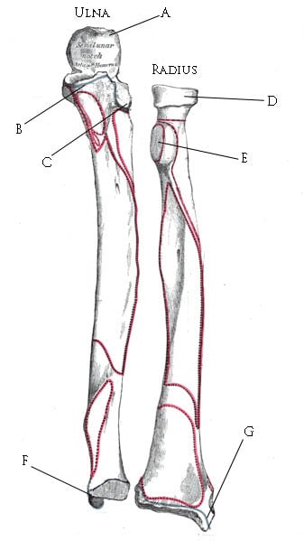
- Volar boundary: Flexor carpi ulnaris (FCU) — innervated by the ulnar nerve.
- Dorsal boundary: Extensor carpi ulnaris (ECU) — innervated by the posterior interosseous nerve (deep branch of the radial nerve).
- The plane runs directly over the palpable subcutaneous (dorsal) border of the ulna from the olecranon to the styloid — a true internervous interval at every level, so the approach is fully extensile without crossing a nerve territory.
- Ulnar nerve and ulnar artery: Run on the volar surface of FCU (the nerve deep to FCU in the forearm). Protected by keeping FCU intact and retracting it volarward; greatest risk with deep volar dissection and proximally near the cubital tunnel.
- Dorsal ulnar cutaneous nerve (DUCN): Arises from the ulnar nerve ~5 cm proximal to the wrist, passes dorsally deep to FCU then subcutaneously to the ulnar dorsum of the hand — at risk in the distal third; injury causes numbness/neuroma over the dorsoulnar hand.
- Posterior interosseous nerve / artery: In the depth of the dorsal forearm — respected by staying on the periosteum of the ulna and not straying radially into the interosseous space unnecessarily.
The Approach — Step by Step
- Supine with the arm across the chest or on a hand table; forearm pronated to bring the subcutaneous border uppermost. Tourniquet as required; image intensifier available.
- Landmark: palpate the subcutaneous border of the ulna from the olecranon to the ulnar styloid — the skin incision lies directly over it for the relevant segment.
Dangers & How to Avoid Them
Structures at risk
Stay on the subcutaneous border and keep flexor carpi ulnaris (with the ulnar nerve and artery beneath it) retracted volarward — the ulnar neurovascular bundle is volar to FCU and should never be in the dorsal working field. In the distal third, actively protect the dorsal ulnar cutaneous nerve.
Operate or Brace? Indication Evidence
Isolated (nightstick) ulnar shaft fracture
Outcomes & Evidence
Isolated ulnar shaft fractures heal predictably — most non-operatively
Based on an article retrieved from PubMed: Sarmiento et al. (DOI) for the non-operative outcome benchmark that frames the operative indications. The internervous plane (FCU/ECU), the subcutaneous-border anatomy, the volar position of the ulnar neurovascular bundle and the course of the dorsal ulnar cutaneous nerve reflect standard, well-established surgical-anatomy teaching (Hoppenfeld/AO descriptions).
Viva Scenarios
Practise clinical reasoning and management decisions out loud
“You are about to plate an isolated displaced ulnar shaft fracture. The examiner asks: 'Describe your approach and its internervous plane.'”
“A fit adult has an isolated, minimally displaced nightstick fracture. The examiner asks whether you would operate.”
Viva & Exam Focus
FUNDUlnar shaft plane & dangers
Hook:Plate the subcutaneous border between FCU and ECU — keep the FUNDamentals: FCU/ulnar bundle volar, ECU/PIN dorsal, brace the simple nightstick.
- True internervous plane: FCU (ulnar nerve) vs ECU (posterior interosseous nerve).
- Ulnar nerve and artery are VOLAR to FCU.
- Most isolated nightstick fractures heal non-operatively (99% with bracing).
- Restore the ulnar bow/length for both-bone and Monteggia injuries (the forearm 'ring').