Distal, diaphyseal and basal osteotomies of the fifth metatarsal for symptomatic lateral prominence | intermediate
Surgical Imaging
The trap: The fifth metatarsal receives its endosteal supply from a single nutrient artery entering the middle third of the diaphysis. A transverse osteotomy through this zone disrupts both endosteal and periosteal supply at the watershed, with nonunion rates of 25-30 percent reported in some series.
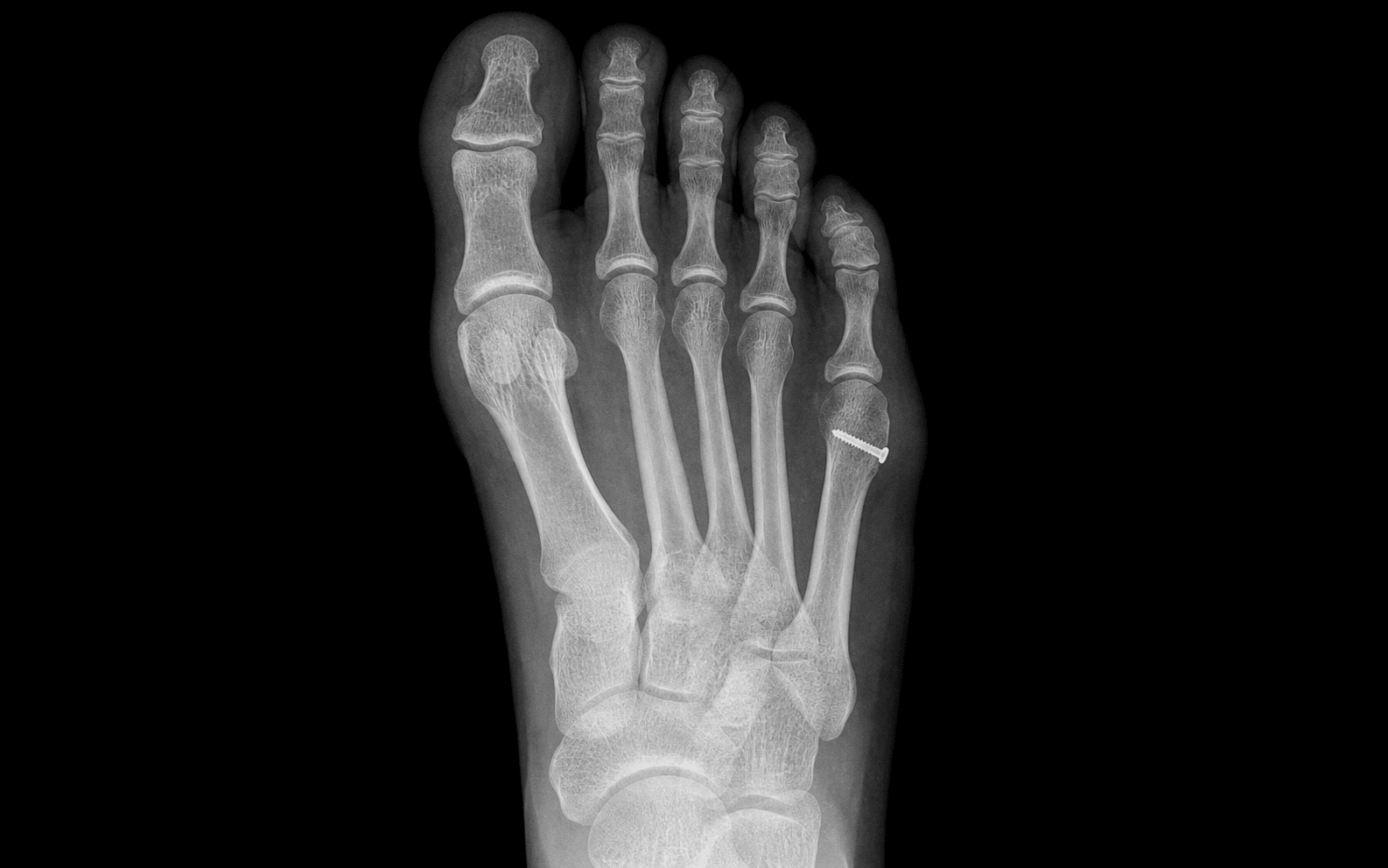
The fix: Use a long oblique (Coughlin) osteotomy, a distal chevron, or a basal osteotomy — never a transverse mid-shaft cut. Preserve the metaphyseal blood supply by minimising lateral soft-tissue stripping. If a screw is placed, consider a solid core titanium 2.5-3.0 mm screw with a partially threaded design for compression.
Location: The dorsolateral cutaneous nerve (a branch of the sural nerve) crosses the fifth metatarsal shaft obliquely from proximal-medial to distal-lateral, running in the subcutaneous tissue 5-10 mm lateral to the extensor digitorum longus tendon to the fifth toe.
Risk: A dorsal or dorsolateral incision placed too laterally, or aggressive subcutaneous dissection, can transect this nerve and produce a painful neuroma over the lateral border of the foot. Identify and protect the nerve throughout the approach.
Why it matters: Transfer metatarsalgia under the fourth metatarsal head is a recognised complication of bunionette correction, particularly with distal osteotomies that are over-shortened or dorsiflexed. The fifth metatarsal normally bears about 1/5 of forefoot load; reducing its length shifts load to the fourth.
Prevention: Avoid excessive shortening — a long oblique osteotomy is intrinsically stable and minimally shortening. If a chevron is used, plan the cut so that translation does not exceed about 3-4 mm and never dorsiflex the capital fragment. Smooth plantar condylectomy only when the condyle is clearly the source of pain.
Cause: The fifth metatarsal head is supplied by metaphyseal vessels entering the distal flare, supplemented by endosteal supply from the diaphyseal nutrient artery. A distal chevron osteotomy with extensive lateral soft-tissue stripping, or a transverse mid-shaft osteotomy, can devascularise the head.
Prevention: Limit lateral periosteal stripping; preserve the metaphyseal blood supply. Use a medial-to-lateral translation rather than a lateral-based wedge. If a screw is inserted, place it from medial to lateral in the distal fragment to avoid the lateral blood supply.
Why it happens: Recurrence rates of 5-15 percent are reported. The most common cause is under-correction of the 4-5 intermetatarsal angle, especially when a distal chevron is used for a Type 3 deformity with an IMA greater than 8-9 degrees. Adjacent hallux valgus deformity (a frequent association) also drives recurrence if not addressed.
Fix: Pre-operative weight-bearing AP radiographs to measure 4-5 IMA, 4-5 DMAA, and the fifth metatarsophalangeal angle. Match the osteotomy to the deformity: a Type 3 with IMA greater than 8-9 degrees needs a diaphyseal or basal osteotomy. Assess and correct first-ray pathology in the same sitting.
The trap: A painful prominence under the fifth metatarsal head is not always a bunionette. Exclude (1) a tailor's bunion (true bunionette) with a prominent lateral condyle, (2) a hammertoe or adductovarus fifth toe causing a dorsal keratosis at the PIP, (3) an interdigital (Morton) neuroma between the 4th and 5th web space with secondary pressure, (4) a Freiberg infraction of the second metatarsal head producing referred pain.
The exam answer: Always document the location of the keratosis (plantar-lateral vs dorsal vs interdigital) and the underlying bony anatomy on weight-bearing films. A true bunionette correction addresses the lateral condyle and the intermetatarsal angle; treating a hammertoe as a bunionette leaves the patient in pain.
B.U.N.I.O.N.E.T.T.EBUNIONETTE — Coughlin Types and Osteotomy Choice
F.O.R.E.F.O.O.TFOREFOOT — Surgical Approach to the Fifth Metatarsal
K.I.S.SKISS — Keep It Simple, Stable, Short
Surgical Indications
Absolute Indications
- Symptomatic bunionette with persistent pain at the lateral fifth metatarsal head that has failed non-operative management (footwear modification, padding, orthoses, corticosteroid injection)
- Lateral keratosis that fails to off-load with shoe modification, padding, or custom orthoses
- Recurrent callus formation at the lateral fifth metatarsal head with associated bursitis
- Progressive deformity with worsening 4-5 intermetatarsal angle on serial radiographs
Relative Indications
- Patient preference for definitive correction after non-operative measures have failed
- Concurrent first-ray pathology (hallux valgus, metatarsus primus varus) that can be addressed in the same surgical sitting
- Inflammatory arthropathy (rheumatoid, psoriatic) with symptomatic lateral fifth metatarsal head prominence
- Bunionette in the setting of a cavus foot or forefoot adductus, where correction of the fifth metatarsal is part of a global forefoot reconstruction
Contraindications
Absolute:
- Active soft-tissue or bone infection in the operative field
- Non-reconstructable peripheral vascular disease
- Non-ambulatory or medically unfit patient
Relative:
- Painless prominence with cosmetic concerns only — counsel that the goal of surgery is pain relief, not cosmesis
- Severe peripheral neuropathy (e.g. Charcot-Marie-Tooth) where recurrence is high and rehabilitation is challenging
- Smoker with poor healing capacity — counsel on smoking cessation; the fifth metatarsal is notoriously prone to delayed union
- Active inflammatory arthropathy flare — optimise medical management before surgery
Non-Operative Management — Brief Overview
- Footwear: Wide, low-heeled shoes with a soft upper; avoid narrow toe-box dress shoes and high heels
- Padding: Doughnut pad around the lateral prominence to off-load the keratosis
- Orthoses: Custom-made insoles with a metatarsal pad and a fifth-metatarsal relief area
- Corticosteroid injection: For associated adventitial bursitis, not as a primary treatment of the bunionette itself
- Outcomes: Non-operative management is the first-line treatment in most patients; surgery is reserved for those with persistent pain that limits activity or footwear
Evidence for Operative Management
Distal Chevron Osteotomy
- Indicated for Coughlin Type 2 bunionette (lateral diaphyseal bow) and for mild-to-moderate Type 3 deformity (4-5 IMA less than 8-9 degrees)
- A 60-degree V-shaped osteotomy at the distal metaphysis; capital fragment translated 3-4 mm medially
- Reported satisfaction: 80-95 percent in case series
- AVN of the head is rare (less than 1 percent) when the metaphyseal blood supply is preserved
- Transfer metatarsalgia: 5-10 percent if the osteotomy is over-shortened or dorsiflexed
Diaphyseal Oblique Rotational (Coughlin) Osteotomy
- Indicated for Type 3 bunionette with 4-5 IMA greater than 8-9 degrees
- Long oblique osteotomy from distal-lateral to proximal-medial, allowing rotation of the distal fragment
- Can correct IMA up to about 10-12 degrees
- Intrinsically stable; fixation with 2.5-3.0 mm cortical or headless screw
- Recurrence: 5-10 percent; transfer metatarsalgia: 5-15 percent if over-shortened
Basal Osteotomy
- Indicated for severe Type 3 deformity (4-5 IMA greater than 10-12 degrees) or revision surgery
- Closing or opening wedge at the proximal fifth metatarsal base
- Powerful correction but slower healing (8-12 weeks) and risk of nonunion (5-10 percent)
- Fixation with a mini-fragment plate or a single oblique screw
Lateral Condylectomy
- Indicated for Type 1 bunionette (enlarged lateral condyle with normal IMA)
- A medial-to-lateral excision of the prominent lateral condyle; preserves the fifth MTP joint
- Limited correction — does not address an increased IMA
- Adjunctive procedure when a Type 1 component is present in a higher-grade deformity
Minimally Invasive / Percutaneous Techniques
- Growing interest in percutaneous fifth metatarsal osteotomies (Bosch-type, distal metatarsal osteotomy) for bunionette
- Smaller scars, less soft-tissue stripping
- Limited long-term data; relies heavily on intra-operative fluoroscopy
- Consider as an alternative in selected patients with appropriate surgeon experience
Bunionette Osteotomies — Indications and Outcomes
Key Evidence
Treatment of bunionette deformity with longitudinal diaphyseal osteotomy with distal soft tissue repair
Subcapital oblique osteotomy for correction of bunionette deformity: medium-term results
Distal Chevron metatarsal osteotomy for bunionette
The intraosseous blood supply of the fifth metatarsal: implications for proximal fracture healing
The tailor's bunionette deformity: a field guide to surgical correction
Clinical Decision Scenarios
Practise clinical reasoning and management decisions out loud
“A 52-year-old woman presents with a 2-year history of pain and a callus over the lateral border of her right fifth metatarsal head. She has tried wide shoes, padding, and a custom orthosis without relief. On weight-bearing AP radiograph, the 4-5 intermetatarsal angle measures 11 degrees and the fifth metatarsal head shows a normal lateral condyle with a mild lateral bow of the diaphysis. How do you classify and manage this bunionette?”
“A 38-year-old competitive runner has a Type 1 bunionette with a prominent lateral condyle, a normal 4-5 intermetatarsal angle of 7 degrees, and a callus on the lateral fifth metatarsal head. He has failed non-operative management. What is the appropriate surgical option, and what complications are you most concerned about in this patient?”
“A 28-year-old smoker presents 4 months after a transverse mid-shaft osteotomy of the fifth metatarsal for a bunionette. The osteotomy has not united, the hardware is intact but loose, and the patient has pain at the osteotomy site with weight-bearing. How do you manage this nonunion?”
References
-
Coughlin MJ (1991). Treatment of bunionette deformity with longitudinal diaphyseal osteotomy with distal soft tissue repair. Foot Ankle. 1991 Feb;11(4):195-203. PMID 1855704. DOI 10.1177/107110079101100402. — Longitudinal diaphyseal osteotomy of the fifth metatarsal with distal soft tissue repair for Type 3 bunionette.
-
Cooper MT, Coughlin MJ (2013). Subcapital oblique osteotomy for correction of bunionette deformity: medium-term results. Foot Ankle Int. 2013 Oct;34(10):1376-80. PMID 23650648. DOI 10.1177/1071100713489121. — Medium-term follow-up of the subcapital oblique osteotomy for bunionette deformity.
-
Kitaoka HB, Holiday AD Jr, Campbell DC 2nd (1991). Distal Chevron metatarsal osteotomy for bunionette. Foot Ankle. 1991 Oct;12(2):80-5. PMID 1773999. DOI 10.1177/107110079101200204. — Distal chevron osteotomy reliably corrects mild-to-moderate bunionette; AVN rare with limited lateral soft-tissue stripping.
-
Smith JW, Arnoczky SP, Hersh A (1992). The intraosseous blood supply of the fifth metatarsal: implications for proximal fracture healing. Foot Ankle. 1992 Mar-Apr;13(3):143-52. PMID 1601342. DOI 10.1177/107110079201300306. — Anatomical study of the diaphyseal blood supply; single nutrient artery enters the middle third; transverse mid-shaft osteotomies disrupt the endosteal supply.
-
Roukis TS (2005). The tailor's bunionette deformity: a field guide to surgical correction. Clin Podiatr Med Surg. 2005 Apr;22(2):223-45, vi. PMID 15833418. DOI 10.1016/j.cpm.2004.10.004. — Comprehensive review of bunionette correction techniques; matched osteotomy choice to anatomical deformity is the most important determinant of outcome.