Adding an acetabular component to a hemiarthroplasty, with or without stem revision · Advanced
- The classic presentation is progressive groin pain from acetabular cartilage erosion — thigh pain points to stem loosening, buttock pain to referred spine pathology.
- The central operative decision is whether to retain a well-fixed, modular, taper-compatible stem or revise it. Retention works only if fixation, modularity, taper compatibility and taper condition are ALL satisfactory.
- Ream to restore the anatomic centre of rotation — do not follow the eroded, medialized centre, or you will medialize the cup and risk medial wall perforation.
- Dislocation risk (5-10 percent) is far higher than primary THA — strongly consider a dual mobility cup.
- For a cemented stem that must come out (loose, monoblock, or corroded taper), an extended trochanteric osteotomy is the safest route to the cement mantle.
When & Why
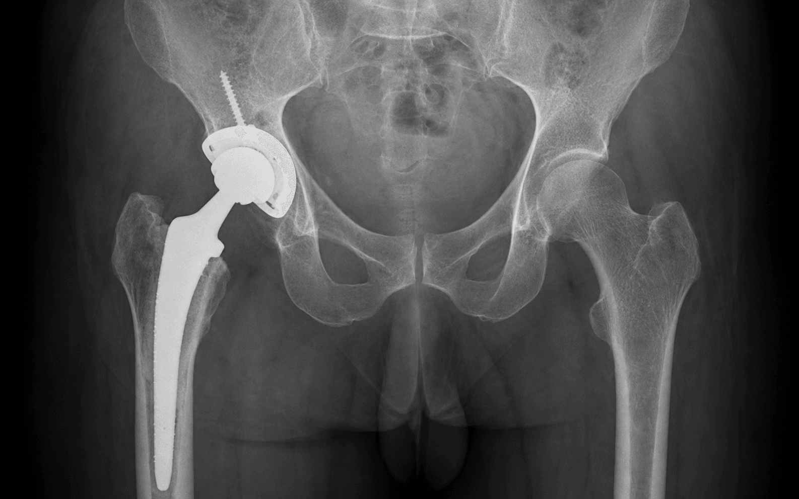
The indication. Conversion of a hemiarthroplasty to a total hip arthroplasty (THA) is performed for a painful hemiarthroplasty where the femoral stem is sound but the native acetabulum has failed, OR where both components have failed. The single most common indication is acetabular cartilage erosion — the metal head progressively wears the native cartilage, producing groin pain and medial migration of the head on the AP radiograph.
- Typical presentation
- Groin pain, medial head migration on radiograph
- Urgency
- Semi-elective
- Typical presentation
- Multiple dislocations, frank instability
- Urgency
- Semi-elective
- Typical presentation
- Thigh pain, stem subsidence
- Urgency
- Elective
- Typical presentation
- Acute pain, unable to weight-bear
- Urgency
- Urgent
- Typical presentation
- Pain, systemic symptoms, raised CRP
- Urgency
- Staged / urgent
Timing. Average time to conversion is commonly reported at around 6-8 years; cohort series (for example Diwanji, mean 7.2 years) report similar intervals. A substantial proportion of conversions occur within the first 5 years. Late presentation may carry severe erosion or protrusio. Patient factors favouring conversion over simple observation. Age less than 70 years with preserved cognition, high functional demand, pre-existing hip arthritis, and long life expectancy tilt the balance toward conversion THA rather than accepting a painful hemiarthroplasty. Clinical assessment — let the pain point to the diagnosis. - Groin pain suggests acetabular erosion (the commonest cause).
- Thigh pain suggests stem loosening.
- Buttock pain may be referred from the spine.
- Document any dislocation history (number, direction, mechanism) and functional status (walking distance, aids, ADLs). Imaging and laboratory protocol. Exclude infection before any conversion.
- Purpose
- Compare both hips; assess erosion
- Key findings
- Medial migration, broken Shenton's line
- Purpose
- Stem version, head position
- Key findings
- Anterior or posterior migration
- Purpose
- Acetabular columns
- Key findings
- Defect location and extent
- Purpose
- Bone stock, stem loosening
- Key findings
- Paprosky classification, bone loss
The Operation
The goal is to add a stable acetabular component to the existing hip, retaining the femoral stem wherever it is safe to do so, reconstructing the eroded acetabulum at the anatomic centre of rotation, and protecting the sciatic nerve throughout. The exposure is laid out in full below.
Operative sequence — stem retention (and the stem-revision branch)
- Lateral decubitus, using the previous incision — typically the posterior approach used for the hemiarthroplasty. Extend proximally or distally as needed for exposure.
- Have an extensile approach ready in case the stem must be revised.
- Identify and protect the sciatic nerve early. In revision surgery it may be adherent to scar tissue posteriorly — careful blunt dissection only; palpate the nerve before any sharp dissection posterior to the hip. Keep the knee flexed during retraction to reduce tension.
- Release the short external rotators and tag them for later repair (posterior approach).
- Perform a capsulectomy around the femoral neck and acetabulum, then dislocate the hip with flexion and internal rotation.
- Remove the modular hemiarthroplasty head and note the taper condition.
- Test stem stability with rotational torque and axial stress. Any toggle or rotation means the stem must be revised.
- Clean and dry the taper and inspect it under magnification. Any visible corrosion, fretting or pitting means the stem must be revised — corrosion products drive adverse local tissue reactions (ALTR).
- If the stem is well-fixed, modular, taper-compatible with an undamaged taper and in acceptable position, retain it and continue to Step 4. If not, proceed to the stem-revision branch (Steps 9-11), then return to the acetabular work.
- Elevate the labrum and capsular remnants and identify the transverse acetabular ligament.
- Place retractors on the anterior wall (protected — over the bone, not soft tissue) and the posterior wall.
- Assess the acetabular cartilage wear and the bone stock.
- Remove the remaining articular cartilage with curettes.
- Ream starting from the peripheral rim — do NOT follow the eroded, medialized centre of rotation, or you will medialize the cup and risk breaching a paper-thin medial wall.
- Ream sequentially to healthy bleeding bone and assess the rim for defects.
- Standard bone stock (Paprosky I-IIA): cementless press-fit cup, line-to-line or 1-2 mm under-ream, with screws for supplemental fixation. Target 40-45 degrees inclination and 15-20 degrees anteversion.
- Medial wall deficiency (Paprosky IIC): consider a protrusio ring or medialised cup design, bone-graft a large medial defect, and use a larger cup to span it.
- Significant bone loss (Paprosky III): trabecular metal augments, jumbo cups, or cup-cage / reconstruction cage constructs.
- Select a THA head compatible with the retained stem taper and trial neck lengths for stability (no impingement, adequate tension), equal leg length, and adequate offset.
- Given the elevated dislocation risk, favour a larger head (36 mm) and/or a dual mobility construct. If there is any instability at trial, convert to dual mobility rather than accept marginal stability.
- Seat the final head with a single firm impaction and reduce the hip.
- Test stability in flexion and internal rotation (posterior) and extension and external rotation (anterior); document the range of motion achieved.
- Repair the posterior capsule and short external rotators (posterior approach) and close in layers over a drain.
- For a monoblock stem (Thompson, Austin Moore), apply axial traction and rotation to remove the entire stem with the head.
- For a modular stem with an incompatible or corroded taper, remove the head with a head extractor; if the taper is corroded, the stem must come out.
- Mark the osteotomy 10-12 cm from the tip of the trochanter distally; make longitudinal saw cuts on the anterolateral and posterolateral cortex and complete them with thin osteotomes, hinging the fragment on its lateral soft-tissue sleeve. This gives direct access to the cement mantle and protects from femoral perforation.
- Remove bulk cement with osteotomes, adherent cement with an ultrasonic device, and residual cement with a high-speed burr — clear all cement to its distal extent.
- For a cementless stem, disrupt the interface with flexible osteotomes (an ETO if it is extensively ingrown) and extract with axial pull, using a trephine for bony ingrowth if needed.
- Choose the stem on remaining bone: metaphyseal fixation if the diaphysis is intact, diaphyseal (fully porous) fixation if the metaphysis is deficient; modular stems allow independent offset and length adjustment.
- Secure the ETO with cerclage wires (typically two to three), then proceed to the acetabular work (Steps 4-8).
The sciatic nerve lies about 2-3 cm posterior to the hip capsule and is at increased risk in revision surgery because it may be adherent to scar. Identify and palpate it early in a posterior approach, keep the knee flexed during retraction to reduce tension, and limit lengthening to 4 cm to avoid a traction injury. A common peroneal division injury presents as foot drop.
Use the previous approach. If a lateral approach was used for the original hemiarthroplasty, a posterior approach can still be used for the conversion if preferred — just be aware of the previous scar and the gluteal insertion.
The medial wall may be eroded to paper-thinness, and protrusio may be present. Do NOT breach the medial wall. If protrusio is severe, use bone graft or a medialised cup design rather than aggressive reaming.
Clean the taper with saline, dry it thoroughly, and inspect under loupe magnification. Any visible corrosion or pitting means revise the stem — corrosion products cause an adverse local tissue reaction.
Aftercare & Complications
Rehabilitation. Protected weight-bearing until an ETO has healed (typically 6-8 weeks) if the stem was revised; otherwise weight-bearing as tolerated with the usual hip precautions for 6-12 weeks. Standard thromboprophylaxis extended to 35 days, and serial radiographs to monitor the ETO and stem position. Instability prevention. Dislocation is the dominant complication of conversion THA. The risk factors and their mitigation:
- Risk level
- High
- Management
- Dual mobility or constrained liner
- Risk level
- High
- Management
- Dual mobility
- Risk level
- High
- Management
- Constrained liner
- Risk level
- Moderate
- Management
- Consider constrained liner
- Risk level
- Moderate
- Management
- Repair posterior structures meticulously
- Risk level
- Moderate
- Management
- Consider dual mobility
- Recognition
- Acute pain, shortening, deformity, patient unable to move the hip
- Prevention
- Dual mobility cup, larger heads, repair posterior structures, restore offset
- Management
- Closed reduction, hip precautions, revise if recurrent (constrained liner, correct component position)
- Recognition
- Crack during cement removal or stem extraction, sudden loss of fixation
- Prevention
- Extended trochanteric osteotomy for cemented stems, prophylactic cerclage, avoid excessive torque
- Management
- Cerclage wire fixation, possible plating, longer stem to bypass the fracture
- Recognition
- Patient aware of a limb-length difference, gait asymmetry, back pain
- Prevention
- Preoperative templating, intraoperative measurement against fixed landmarks
- Management
- Shoe raise if less than 2 cm and symptomatic, revision if greater than 2 cm and symptomatic
- Recognition
- Foot drop (common peroneal), hamstring weakness (tibial), posterior thigh numbness
- Prevention
- Identify the nerve, limit lengthening to 4 cm, flex the knee during retraction, consider monitoring
- Management
- Observe 3-6 months (most recover), AFO for foot drop, explore if no recovery
- Recognition
- Sudden give during reaming, blood from the medial wall, pelvic pain
- Prevention
- Careful reaming with protrusio, know medial wall thickness from CT, avoid over-reaming
- Management
- Bone-graft the defect, larger cup to span, cage construct if major, vascular surgery if arterial injury
- Recognition
- Persistent pain, wound problems, raised inflammatory markers, fever
- Prevention
- Prophylactic antibiotics, meticulous technique, avoid haematoma, optimise nutrition
- Management
- Aspiration, debridement if early (less than 4 weeks), two-stage revision if chronic
- Recognition
- Groin pain, raised metal ions, pseudotumour on MRI
- Prevention
- Manufacturer-matched heads only, clean and dry the taper, single impaction
- Management
- Revise the head to ceramic if possible, revise the stem if corrosion is severe, debride the pseudotumour
- Recognition
- Thigh pain, progressive shortening, stem migration on serial radiographs
- Prevention
- Adequate press-fit, correct stem size, ETO healed before full weight-bearing
- Management
- Protected weight-bearing, longer stem if progressive, ensure the ETO has healed
- Recognition
- Stiffness, reduced range of motion at 6-12 weeks, visible on radiograph
- Prevention
- Indomethacin 75 mg daily for 6 weeks, or single-dose radiation 7 Gy
- Management
- Observe if asymptomatic, resect if Brooker III-IV and limiting function (wait 12-18 months for maturity)
- Recognition
- Progressive pain, radiolucency around components on serial radiographs
- Prevention
- Adequate primary fixation, appropriate constraint, avoid excessive lengthening
- Management
- Revision when symptomatic, addressing bone defects with augments or allograft
Viva & Exam Focus
GROINGROIN — causes of pain after a hemiarthroplasty
TAPERTAPER — stem retention checklist (all must be met)
Lies about 2-3 cm posterior to the hip capsule, at increased risk in revision surgery because it may be adherent to scar. A traction injury during leg lengthening greater than 4 cm gives a foot drop. Key: identify the nerve early in a posterior approach, limit lengthening to 4 cm, flex the knee during retraction, consider nerve monitoring.
Lies anterior to the hip capsule, about 4-5 cm from the acetabulum, at risk with aggressive anterior retraction — especially with a medialized, eroded acetabulum. Key: place anterior retractors on bone only, and remember protrusio brings the medial wall closer to the vessels.
Exits the greater sciatic notch and runs with the superior gluteal vessels about 3-5 cm above the acetabular rim. Injury causes abductor weakness and a Trendelenburg gait. Key: do not extend a split in gluteus medius more than 5 cm proximal to the tip of the greater trochanter.
Often eroded or paper-thin in conversion cases, and may show protrusio. Aggressive reaming risks medial wall perforation and injury to intrapelvic vessels (obturator, iliac). Key: assess medial wall thickness on CT preoperatively, ream carefully, consider a medialised cup design.
A cement mantle and osteoporotic bone raise the fracture risk during stem removal, especially for cemented stems. Key: if a cemented stem must come out, use an extended trochanteric osteotomy, and have plating and cerclage available.
Clinical Decision Scenarios
Practise clinical reasoning and management decisions out loud
“A 75-year-old woman presents with progressive groin pain 7 years after a cemented hemiarthroplasty for a displaced intracapsular femoral neck fracture. Her inflammatory markers are normal. How do you assess and manage this patient?”
“During conversion THA you retain the well-fixed stem and insert the acetabular component. At trial reduction the hip feels unstable even with the shortest head option. How do you troubleshoot this?”
“A 68-year-old man had a cemented Thompson hemiarthroplasty 5 years ago. He now presents with groin pain and thigh pain. Radiographs show acetabular erosion AND stem subsidence of 8 mm compared with the immediate post-operative films. How do you manage this case?”
Indications
- Groin pain equals acetabular erosion (most common)
- Thigh pain equals stem loosening
- Recurrent dislocation, periprosthetic fracture, infection
- Average time to conversion: 6-8 years
TAPER stem retention criteria
- Taper compatible — matching THA head available
- Anchored well — no loosening or subsidence
- Position acceptable — version, offset, leg length
- Examined taper — no corrosion or damage
- Removable head — modular design, not monoblock
Stem revision indications
- Monoblock design (Thompson, Austin Moore)
- No compatible THA head available
- Taper corrosion or damage
- Stem loosening or subsidence
- Malposition affecting stability
Acetabular bone stock (Paprosky)
- I-IIA: standard cementless cup
- IIB: cup plus screws, possible augment
- IIC: medial augment or protrusio design
- III: jumbo cup, augments, cage constructs
Key technical points
- Use the previous approach (usually posterior)
- Identify and protect the sciatic nerve (may be in scar)
- ETO for cemented stem removal
- Ream to restore the anatomic centre of rotation, not the erosion
- Consider dual mobility for instability risk
Dislocation prevention
- Dislocation risk 5-10 percent versus 1-2 percent for primary THA
- Dual mobility reduces risk to less than 1 percent
- Repair the posterior structures
- Consider a larger head (36 mm)
- Elevated-rim liner if a standard head is used
Cemented stem removal
- Extended trochanteric osteotomy is essential
- Mark 10-12 cm from the trochanter tip
- Anterolateral and posterolateral saw cuts
- Complete with osteotomes
- Cerclage wire fixation of the ETO (2-3 wires)
Danger structures
- Sciatic nerve — may be adherent to scar
- Medial wall — often thin from erosion
- Femoral vessels — closer if protrusio
- Superior gluteal nerve — 5 cm proximal limit
Background & Evidence
Epidemiology and registry context. Conversion and revision THA after a hemiarthroplasty carries a higher cumulative revision risk than primary THA for osteoarthritis. Major joint registries (NJR England and Wales, AJRR USA, AOANJRR Australia, SHAR Sweden, NZJR New Zealand) consistently show this elevated risk, driven largely by dislocation and the technical demands of the eroded acetabulum. The fracture-related diagnosis itself is an independent dislocation risk factor. The Paprosky acetabular bone-loss classification guides acetabular reconstruction and is central to pre-operative planning:
- Description
- Supportive rim, minimal bone loss
- Treatment
- Standard cementless cup
- Description
- Superior dome loss, less than 30 percent
- Treatment
- Cementless cup plus screws
- Description
- Less than 50 percent superolateral loss
- Treatment
- Cementless cup, possible augment
- Description
- Medial wall deficient
- Treatment
- Medial augment or protrusio cup
- Description
- Greater than 40 percent host bone contact
- Treatment
- Jumbo cup, augments
- Description
- Less than 40 percent host bone contact
- Treatment
- Cage, cup-cage, or custom triflange
Key evidence. Diwanji's series of 25 bipolar hemiarthroplasty conversions (mean follow-up 7.2 years) showed reliable pain relief and functional improvement (mean Harris Hip Score 41 to 85), with a complication profile approximating revision rather than primary THA. Archibeck's series of 102 conversion or salvage THAs after failed fracture fixation reported an early surgical complication rate of 11.8 percent, dominated by dislocation and periprosthetic fracture. Registry work from the Swedish Hip Arthroplasty Register (Haller) established that a femoral neck fracture as the primary diagnosis carries a markedly higher revision-for-dislocation risk (relative risk 3.9 versus osteoarthritis), that small (22 mm) heads double that risk, and that dual mobility cups markedly reduce recurrent instability when used for revision.
References
- National joint replacement registries (NJR England and Wales, AJRR USA, AOANJRR Australia, SHAR Sweden, NZJR New Zealand). Annual reports. Used as global registry evidence for revision and dislocation rates after conversion and revision THA. 2. Bhandari M, Devereaux PJ, Tornetta P 3rd, et al. Operative management of displaced femoral neck fractures in elderly patients. An international survey. J Bone Joint Surg Am. 2005;87(9):2122-2130. 3. Archibeck MJ, Carothers JT, Tripuraneni KR, White RE Jr. Total hip arthroplasty after failed internal fixation of proximal femoral fractures. J Arthroplasty. 2013;28(1):168-171. PMID 22682040. 4. Tidermark J, Ponzer S, Svensson O, et al. Internal fixation compared with total hip replacement for displaced femoral neck fractures in the elderly. A randomised, controlled trial. J Bone Joint Surg Br. 2003;85(3):380-388. 5. Diwanji SR, Kim SK, Seon JK, et al. Clinical results of conversion total hip arthroplasty after failed bipolar hemiarthroplasty. J Arthroplasty. 2008;23(7):1009-1015. PMID 18534504. 6. Parvizi J, Picinic E, Sharkey PF. Revision total hip arthroplasty for instability: surgical techniques and principles. J Bone Joint Surg Am. 2008;90(5):1134-1142. 7. Hailer NP, Weiss RJ, Stark A, Kärrholm J. The risk of revision due to dislocation after total hip arthroplasty depends on surgical approach, femoral head size, sex, and primary diagnosis. An analysis of 78,098 operations in the Swedish Hip Arthroplasty Register. Acta Orthop. 2012;83(5):442-448. PMID 23039167. 8. Civinini R, Carulli C, Matassi F, et al. A dual-mobility cup reduces risk of dislocation in isolated acetabular revisions. Clin Orthop Relat Res. 2012;470(12):3542-3548. PMID 22700131. 9. Hailer NP, Weiss RJ, Stark A, Kärrholm J. Dual-mobility cups for revision due to instability are associated with a low rate of re-revisions due to dislocation: 228 patients from the Swedish Hip Arthroplasty Register. Acta Orthop. 2012;83(6):566-571. PMID 23116439. 10. Younger TI, Bradford MS, Magnus RE, Paprosky WG. Extended proximal femoral osteotomy. A new technique for femoral revision arthroplasty. J Arthroplasty. 1995;10(3):329-338. PMID 7673912.
Total hip arthroplasty after failed internal fixation of proximal femoral fractures
- Retrospective series of 102 conversion/salvage THAs after failed internal fixation of hip fracture (39 intertrochanteric, 63 femoral neck)
- Early surgical complication rate 11.8% (12 of 102): dislocation 4.9%, periprosthetic fracture 3.9%, haematoma 2.0%, infection 1%
- Failure mechanisms included osteonecrosis, post-traumatic arthritis, early fixation failure and nonunion
Clinical results of conversion total hip arthroplasty after failed bipolar hemiarthroplasty
- 25 conversions of bipolar hemiarthroplasty to THA, mean follow-up 7.2 years
- Indications: acetabular erosion with well-fixed stem (13), erosion with femoral loosening (8), periprosthetic fracture (4)
- Mean Harris Hip Score improved from 41 to 85; complications included 4 dislocations and 1 acetabular loosening
The risk of revision due to dislocation after total hip arthroplasty depends on surgical approach, femoral head size, sex, and primary diagnosis (78,098 operations, Swedish Hip Arthroplasty Register)
- Registry analysis of 78,098 primary THAs; femoral neck fracture as the primary diagnosis carried a markedly higher revision-for-dislocation risk (RR 3.9 vs osteoarthritis)
- 22mm heads doubled dislocation revision risk versus 28mm (RR 2.0); only 1 of 287 dual-mobility cups was revised for dislocation
- Posterior and minimally invasive approaches increased dislocation revision risk versus the direct lateral approach
Dual-mobility cups for revision due to instability are associated with a low rate of re-revisions due to dislocation: 228 patients from the Swedish Hip Arthroplasty Register
- 228 cup revisions for recurrent dislocation using a dual-mobility cup
- 2-year survival with re-revision for dislocation as endpoint was 99%; only 2% re-revised for dislocation
- Younger age (50-59) and prior hip revision were risk factors for re-revision for any reason
A dual-mobility cup reduces risk of dislocation in isolated acetabular revisions
- 33 isolated acetabular revisions reconstructed with a dual-mobility cup, mean follow-up 3 years
- No dislocations occurred; component survivorship 97% at 5 years with a 3% re-revision rate
- Mean Harris Hip Score improved from 48 to 86 with no progressive osteolysis, migration or loosening