Posterior approach with olecranon chevron osteotomy for articular access; bicolumnar plating with two 3.5 mm reconstruction plates; ulnar nerve identification and anterior transposition mandatory throughout | Advanced trauma procedure
- AO/OTA classification C1-C3: C1 = simple articular + simple metaphyseal, C2 = simple articular + complex metaphyseal, C3 = complex articular with multi-fragment comminution. C3 is the most technically demanding.
- Bicolumnar plating principle: both medial and lateral columns must be independently supported by two plates placed at 90 degrees to one another (perpendicular plating) or in a parallel configuration. Each column carries half the load.
- Olecranon chevron osteotomy provides the best articular access for C2 and C3 fractures — chevron cut at 70 degrees, apex pointing distally, pre-drill and K-wire before saw use to ensure accurate reattachment.
- Ulnar nerve transposition is mandatory: identify the nerve at the start of every case, protect throughout all dissection steps, and transpose anteriorly subcutaneously or submuscularly at closure to prevent post-operative cubital tunnel syndrome.
- Functional arc for elbow: 100 degrees of flexion (30 to 130 degrees) plus 100 degrees of forearm rotation (50 pronation, 50 supination) accomplishes most activities of daily living (Morrey 1981). Goal of reconstruction is a stable, congruent joint moving within this arc.
- “Two plates capture both columns — historically two 3.5 mm reconstruction plates at 90 degrees (perpendicular or orthogonal), each engaging the articular block with at least two screws. The classic biomechanical principle (Schemitsch 1994) is that two plates must sit on separate columns and different surfaces; strict 90-degree orientation is NOT required for adequate rigidity.
- “Parallel plating (medial and lateral plates in the sagittal plane, O'Driscoll/Sanchez-Sotelo principle-based technique) maximises distal articular fixation and supracondylar stability — biomechanically stiffer than orthogonal in varus and axial loading (Zalavras 2011); both constructs are clinically acceptable.
- “Olecranon osteotomy gives unmatched articular exposure for C3 fractures — the trick to mention in the exam: pre-drill the osteotomy site and insert a K-wire guide before the saw cut to guarantee reattachment accuracy.
- “In elderly patients with C3 fractures and poor bone quality, primary total elbow replacement (TER) is a validated alternative with superior 2-year MEPS outcomes versus ORIF (McKee 2009 RCT); Cobb and Morrey 1997 first established primary TER as a valid option.
When & Why
Indication. Operative fixation of a displaced intra-articular (AO/OTA C-type) distal humerus fracture in a physiologically active patient. The goal is a stable, congruent joint moving within the functional arc of 30 to 130 degrees of flexion. Most displaced intra-articular fractures in active patients are treated operatively; non-operative management is reserved for the elderly, low-demand patient in whom reconstruction is not feasible and for whom a "bag of bones" (so-called "benign neglect") approach with early movement is acceptable. Absolute indications - All AO C-type intra-articular fractures in physiologically active patients
- Open fractures (urgent debridement and stabilisation)
- Associated neurovascular injury (brachial artery, ulnar nerve)
- Fracture-dislocation of the elbow Relative indications - B-type fractures with displacement greater than 2 mm of articular step-off
- Elderly patients — an individualised decision (ORIF versus TER, see below) The decision in the elderly patient — ORIF or total elbow replacement (TER)? In patients older than 65 years with C3 fractures and osteoporotic bone, primary TER is a validated alternative to ORIF, supported by level-1 evidence. The landmark randomised trial (McKee 2009) compared ORIF with primary semiconstrained TER in patients older than 65 with displaced intra-articular (OTA 13C) fractures: TER produced significantly better Mayo Elbow Performance Scores at 3, 6, 12 and 24 months (86 versus 73 at 2 years), and a quarter of fractures randomised to ORIF proved unreconstructable and were converted to TER on the table. Cobb and Morrey (1997) had first established primary TER as a valid option in 20 elderly patients (15 excellent, 5 good results). Pajarinen and Bjorkenheim (2002) confirmed that bicolumnar ORIF gives satisfactory outcomes but is technically demanding. The choice is individualised to age, bone quality, fracture reconstructability on CT, and functional demand:
The standard for younger, active patients with reconstructable fractures. Aims for anatomic articular reduction and rigid fixation to allow early movement. Fails when bone quality cannot hold fixation.
Preferred in patients older than 65 with an unreconstructable C3 fracture and osteoporotic bone. Predictable pain relief and function, but carries a permanent 2 kg lifting restriction and revision is difficult.
Age greater than 65, osteoporosis, articular comminution not amenable to stable fixation on CT, and low functional demand favour TER. Age less than 65 and high demand favour ORIF. Counsel and consent for both options pre-operatively.
Consent specifically for ulnar nerve symptoms (most common nerve injury), stiffness and the possible need for later capsular release, heterotopic ossification, wound problems, non-union, and — if TER is chosen — the lifelong 2 kg lifting restriction, component wear or loosening, and the difficulty of revision surgery. Setup. Lateral decubitus (most common) with the operative elbow flexed 90 degrees over a padded bolster, giving full posterior access and room for intraoperative fluoroscopy on the opposite side (prone with the arm on an arm board is an equivalent alternative, preferred by some for bilateral injuries). Proximal brachium tourniquet at 250 to 300 mmHg after exsanguination. Pad all bony prominences and place an axillary roll in the lateral position.
The Operation
The goal: expose the fracture through a posterior approach, gain articular access via an olecranon chevron osteotomy, reassemble the articular surface anatomically, and stabilise both columns independently with two plates — all while protecting the ulnar nerve from the first incision to closure. The exposure and the operative sequence are laid out in full below (and on the posterior approach to the elbow page).
Anatomy that drives the fixation. The distal humerus is a triangle: two diverging columns bracing an articular arc. The medial column runs from the medial supracondylar ridge to the medial epicondyle and trochlea (thicker cortex; origin of the common flexor mass and ulnar collateral ligament, with the ulnar nerve immediately posterior in the cubital tunnel). The lateral column runs to the lateral epicondyle and capitellum (broader and more anterior; origin of the common extensors and lateral collateral ligament complex). The trochlea is a 300-degree spool articulating with the olecranon notch; the capitellum is almost entirely anterior cartilage. The olecranon fossa (posterior, 10 to 12 mm deep) and coronoid fossa (anterior) receive the olecranon and coronoid tips in full extension and flexion respectively — no screw may enter them, or motion is blocked. The normal valgus carrying angle is 11 to 15 degrees in males and 13 to 16 degrees in females; restore it on the post-operative AP film. The trochlea has a dual blood supply (medial branches of the superior and inferior ulnar collateral arteries; lateral branches of the radial recurrent and middle collateral arteries), but its central "bare area" covering the distal 1.5 cm is relatively avascular — preserve soft-tissue attachments where possible to avoid avascular necrosis.
- Location / why it is at risk
- Runs in the cubital tunnel posterior to the medial epicondyle; enters the forearm between the two heads of flexor carpi ulnaris 3 to 4 cm distal to the epicondyle. Lies immediately posterior to the medial column and is vulnerable during all medial dissection, retraction and drilling.
- How to protect it
- Identify the nerve FIRST before any retraction or drilling; mobilise 8 to 10 cm proximally and 5 cm distally (divide the arcade of Struthers proximally if present); protect with a vessel loop throughout; transpose anteriorly (subcutaneous or submuscular) at closure.
- Location / why it is at risk
- Pierces the lateral intermuscular septum approximately 10 cm proximal to the lateral epicondyle and lies anterior to brachialis and brachioradialis. At risk with excessive proximal dissection on the lateral column.
- How to protect it
- Limit lateral dissection to the lateral epicondyle and distal humerus. Do not extend the posterior incision proximal to 10 cm above the joint without identifying the nerve. Use blunt proximal retraction only.
- Location / why it is at risk
- Anterior to the distal humerus in the antecubital fossa; the median nerve lies medial to the brachial artery, both deep to the bicipital aponeurosis. At risk with severe anterior fracture displacement or anterior capsule dissection.
- How to protect it
- Assess and document neurovascular status pre-operatively. With high-energy anterior displacement, elevate brachialis subperiosteally from lateral to medial if the anterior capsule must be released. Check the radial pulse after retractor placement.
- Location / why it is at risk
- The bare area of the trochlear notch (the narrowest cartilage-free zone, about 2 cm from the olecranon tip). Incorrect technique risks non-union, prominent hardware and malunion step-off.
- How to protect it
- Use a chevron cut at 70 degrees (apex pointing distally). Pre-drill and insert K-wire guides before the oscillating saw. Reattach with a tension band wire or a 6.5 mm cancellous screw with tension band. Confirm anatomic reduction fluoroscopically before accepting fixation.
- Location / why it is at risk
- The posterior capsule, brachialis belly and fracture haematoma are the commonest sites. Incidence 10 to 20 percent after distal humerus ORIF; higher with concurrent head injury, burns or delayed surgery.
- How to protect it
- Prophylaxis with indomethacin 25 mg three times daily for 6 weeks (start within 24 to 48 hours); single-dose radiotherapy 7 Gy is an alternative when NSAIDs are contraindicated. Early active mobilisation from day 1 to 2 is critical — prolonged immobilisation promotes ectopic bone.
Operative sequence
- Lateral decubitus (most common): patient on their side with the operative elbow flexed 90 degrees over a padded bolster; allows full posterior access and intraoperative fluoroscopy.
- Prone with the arm on an arm board is equally effective and preferred by some for bilateral injuries.
- Tourniquet at the proximal brachium (250 to 300 mmHg), exsanguinate with elevation.
- Image intensifier on the opposite side for AP and lateral views.
- Pad all bony prominences; check the axillary roll in the lateral position.
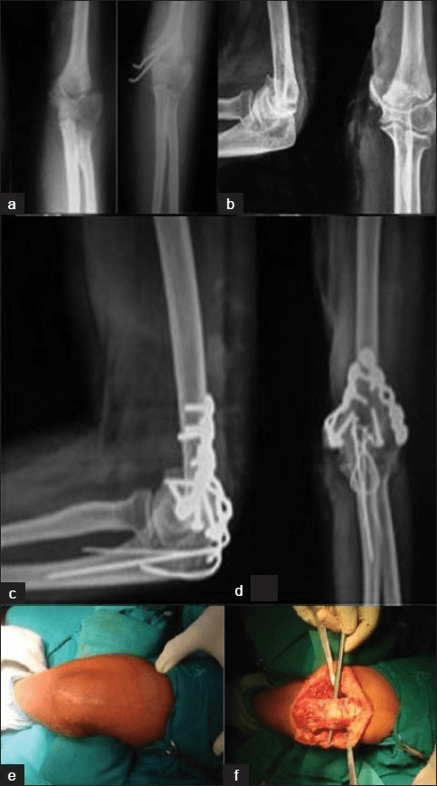
- Straight posterior midline incision from 8 to 10 cm proximal to the olecranon tip to 4 to 5 cm distal to the tip.
- Raise full-thickness skin flaps medially and laterally to the epicondyles.
- Identify the ulnar nerve immediately — it lies in the subcutaneous tissue posterior to the medial epicondyle — and vessel-loop it before any further dissection.
- Identify the nerve posterior to the medial epicondyle.
- Mobilise 8 to 10 cm proximally and 5 to 6 cm distally, dividing the arcade of Struthers proximally if present.
- Protect with a vessel loop throughout all subsequent steps.
- Do NOT transpose yet — mobilise only; transpose at closure after the implants are seated.
- Identify the bare area of the trochlear notch (cartilage-free zone, approximately 2 cm from the olecranon tip) — this is the osteotomy site.
- Pre-drill two 1.6 mm K-wires across the proposed osteotomy as guides for reattachment.
- Mark the chevron: apex pointing distally, limbs at 70 degrees — use electrocautery.
- Complete the osteotomy with an oscillating saw (two oblique cuts), then complete the apex with a narrow osteotome to leave a rough cancellous surface that enhances healing.
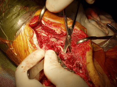
- Reflect the olecranon fragment proximally with the triceps attached — this exposes the entire distal humeral articular surface. Tag the fragment with heavy suture for later retrieval.
- Identify all articular fragments using fluoroscopy and direct vision.
- Reassemble the articular surface anatomically using pointed reduction forceps, provisional K-wires and lag screws (2.0 to 2.7 mm cancellous or Herbert screws).
- Restore the trochlea first (the medial spool), then the capitellum (lateral sphere).
- Confirm articular congruity with direct vision and fluoroscopy before column plating.
- Articular lag screws must not protrude into the olecranon or coronoid fossae (impingement in motion).
- Contour a 3.5 mm reconstruction plate (or a pre-contoured locking plate) to the medial column.
- Seat the plate along the posterior medial column, extending distally to the medial epicondyle.
- Place at least two locking or bicortical screws into the articular block distally, ideally with the most distal screw passing across the midline to engage the lateral column.
- Place at least three bicortical screws in the humeral shaft proximally.
- For perpendicular plating: the plate sits on the posterior surface of the lateral column; distal screws engage the lateral column and capitellum, passing medially to engage the medial column.
- For parallel plating: the plate sits on the lateral surface of the lateral column, in the same sagittal plane as the medial plate.
- Again use at least two articular block screws distally and three shaft screws proximally.
- Each plate must independently support its column — do NOT rely on cross-column inter-plate compression.
- AP and lateral views confirming: articular step-off less than 1 mm, restoration of the normal carrying angle, no screws in the olecranon or coronoid fossae, and adequate plate seating on both columns.
- Perform a full passive range-of-motion test on the table: aim for 0 to 130 degrees. Any block suggests screw impingement or hardware prominence and must be corrected before closure.
- Retrieve the olecranon fragment and reduce it anatomically under direct vision using the pre-drilled K-wire guides.
- Tension band wire technique: two parallel K-wires plus a figure-of-eight 18-gauge wire through a transosseous tunnel — simple, low-profile and reliable.
- Plate fixation (preferred for larger fragments or osteoporotic bone): a 3.5 mm or 2.7 mm plate along the dorsal surface of the olecranon.
- Confirm anatomic reduction of the articular surface (no step-off at the trochlear notch) and fluoroscopic confirmation in AP and lateral.
- Perform anterior subcutaneous transposition: create a subcutaneous pocket anterior to the medial epicondyle and route the nerve gently anteriorly without kinking.
- Secure with a fascial sling (not too tight — the nerve should move freely). Submuscular transposition under the flexor-pronator mass gives more protection in high-risk cases but adds dissection.
- Layered wound closure: deep fascia, subcutaneous tissue, skin.
- Place a drain if there is significant ooze (remove within 24 to 48 hours).
- Apply a posterior slab at 90 degrees of flexion for comfort (remove at 2 to 3 days and start physiotherapy).
Identify and vessel-loop the ulnar nerve at the very start, before any retraction, drilling or plating on the medial column. It lies in the cubital tunnel posterior to the medial epicondyle and is the structure most often injured in distal humerus surgery. Mobilise it 8 to 10 cm proximally and 5 to 6 cm distally, protect it throughout, and transpose it anteriorly at closure — failure to transpose risks post-operative cubital tunnel syndrome from scarring and implant proximity. Document ulnar nerve function (ring and small finger sensation, intrinsic power) before and after the operation.
Pre-drill the olecranon osteotomy site and insert K-wire guides BEFORE making the saw cut. When you reduce the fragment at the end, the pre-placed guides guarantee anatomic reattachment with no articular step-off at the trochlear notch — the single most reliable way to avoid an olecranon osteotomy non-union.
Two plates on separate columns and different surfaces give rigid fixation; strict 90-degree (perpendicular) orientation is NOT required (Schemitsch 1994). Parallel plating (both plates on the column surfaces in the sagittal plane, O'Driscoll/Sanchez-Sotelo) maximises distal articular fixation and is biomechanically stiffer in varus and axial loading, particularly with metaphyseal comminution (Zalavras 2011). Both constructs are clinically acceptable — the choice is guided by fracture pattern, soft-tissue access and surgeon preference.
Aftercare & Complications
Rehabilitation | Phase | Timing | Immobilisation | Therapy | |-------|--------|----------------|---------| | 1 | 0 to 7 days | Posterior slab at 90 degrees for comfort only (NOT for immobilisation); drain out at 24 to 48 hours | Elevation; start indomethacin; document ulnar nerve function | | 2 | 7 days to 6 weeks | Slab removed at 2 to 3 days | Active-assisted flexion-extension and forearm rotation; target 0 to 130 degrees by 6 to 8 weeks | | 3 | 6 to 12 weeks | Splint for heavy tasks only | Light resistance (theraband, putty); dynamic splinting if the arc plateaus at less than 100 degrees | | 4 | 3 to 6 months | None | Progressive return to activities of daily living; heavy loading after 12 to 16 weeks radiographic union | Avoid passive forced stretching in the early weeks — it promotes heterotopic ossification. Forearm rotation (pronation and supination) is exercised early because it is important for capitellar articular congruity. Return to function - Light activities (writing, eating): 2 to 4 weeks
- Driving: 8 to 12 weeks (when safe to grip the wheel and react)
- Office work: 4 to 6 weeks
- Manual labour: 4 to 6 months
- Implant removal: only for symptomatic prominence; minimum 18 to 24 months after surgery and after confirmed fracture union Radiographic follow-up - Week 2: check the wound and confirm implant position
- Week 6: confirm early fracture healing and adjust physiotherapy
- Month 3: confirm bridging callus and clear for progressive loading
- Month 6: final check — confirm full union and arc of motion Heterotopic ossification prophylaxis | Agent | Dose | Duration | Notes | |-------|------|----------|-------| | Indomethacin | 25 mg three times daily | 6 weeks | Most evidence; avoid in renal impairment and peptic ulcer; use a proton pump inhibitor concurrently | | Celecoxib | 200 mg once daily | 6 weeks | COX-2 selective with better gastrointestinal tolerability; less evidence for the elbow | | Radiotherapy | 7 Gy single dose | Within 72 hours | Reserved for high-risk patients (head injury) or when NSAIDs are contraindicated | Complications
- Incidence / risk factors
- 2 to 10 percent; higher with inadequate fixation, smoking and osteoporosis
- Prevention
- Rigid bicolumnar fixation (two plates, minimum three shaft screws each); bone graft if a defect is present; avoid over-distraction
- Management
- Revision ORIF with bone grafting (iliac crest or allograft); consider conversion to TER in the elderly with failed ORIF and poor bone stock
- Incidence / risk factors
- 10 to 20 percent; risk factors: head injury, burns, delayed surgery, extensive soft-tissue stripping
- Prevention
- Indomethacin 25 mg three times daily for 6 weeks (start within 24 to 48 hours); single-dose radiotherapy 7 Gy if NSAIDs contraindicated; early mobilisation from day 1 to 2
- Management
- Mild: physiotherapy, stretching and serial splinting. Severe (blocks motion): surgical excision after maturation (minimum 12 to 18 months, confirmed by bone-scan quiescence); prescribe indomethacin post-excision as recurrence is high without prophylaxis
- Incidence / risk factors
- 10 to 15 percent transient; 2 to 5 percent permanent; risk increases without transposition
- Prevention
- Identify and vessel-loop the nerve at the start; protect throughout; anterior transposition at closure; avoid excessive retraction
- Management
- Transient neuropraxia: observe, splint to protect the hand, neurophysiology at 6 weeks. No recovery by 3 months: surgical exploration, decompression or re-transposition. Permanent motor loss: hand therapy and tendon transfers
- Incidence / risk factors
- 20 to 30 percent have functionally significant stiffness (arc less than 100 degrees); the commonest complication
- Prevention
- Early active mobilisation from day 2 to 3; avoid prolonged immobilisation; HO prophylaxis; anatomic articular reconstruction
- Management
- Physiotherapy (dynamic splinting, serial casting) for 6 to 12 months first. Open or arthroscopic capsular release after 12 months if the arc remains less than 70 degrees and the patient is motivated; remove prominent hardware
- Incidence / risk factors
- 2 to 5 percent with tension band; higher with inadequate fixation or non-compliance
- Prevention
- Chevron cut (maximises cancellous contact); tension band or plate fixation; anatomic reduction using pre-drilled K-wire guides; protect from early varus or valgus stress
- Management
- Symptomatic non-union: revision fixation with bone graft. Prominent hardware: remove after union (minimum 12 months). Asymptomatic fibrous non-union with full function: observe
- Incidence / risk factors
- Superficial 3 to 5 percent; deep 1 to 2 percent; higher risk with diabetes, obesity, immunosuppression and prolonged tourniquet
- Prevention
- Full-thickness skin flaps; meticulous haemostasis; tourniquet off before closure; drain if ooze; prophylactic cephalosporin at induction; optimise diabetic control
- Management
- Superficial: oral antibiotics and wound care. Deep infection with implant: IV antibiotics, washout and debridement (retain the implant if the fracture is unhealed; remove after union if persistent). Exposed implant: negative-pressure wound therapy and plastic surgery
Viva & Exam Focus
BOLTBOLT — Bicolumnar Fixation Principles
Clinical Viva Scenarios
Practise clinical reasoning and management decisions out loud
“A 30-year-old right-hand-dominant carpenter falls from a ladder and sustains an AO C3 distal humerus fracture. Describe your positioning, approach and fixation strategy.”
“A 72-year-old woman with osteoporosis sustains an AO C3 distal humerus fracture. How do you decide between ORIF and primary total elbow replacement?”
“A patient returns to your clinic 6 months after distal humerus ORIF with a flexion arc of only 40 to 90 degrees (a 50-degree arc). How do you manage this?”
Indication
- All AO C-type intra-articular fractures in active patients; open fractures; neurovascular injury; fracture-dislocation
- B-type fractures with displacement greater than 2 mm of articular step-off
- Functional target: 30 to 130 degree arc (Morrey 1981)
ORIF versus TER decision
- TER in age greater than 65 with C3, osteoporotic bone and an unreconstructable articular surface
- McKee 2009 RCT: TER superior 2-year MEPS (86 versus 73); reoperation 27 percent ORIF versus 12 percent TER (not significant); 24 percent of ORIF cases unreconstructable and converted to TER intraoperatively
- Cobb and Morrey 1997: uniformly good to excellent results with primary TER in 20 elderly patients
- TER restriction: 2 kg permanent lifting limit — discuss pre-operatively
- Age less than 65: always attempt ORIF first; failed ORIF revision to TER through a scarred field is a major salvage
Positioning and approach
- Lateral decubitus: arm over a padded bolster, full posterior access, fluoroscopy on the opposite side
- Straight posterior midline incision: 8 to 10 cm proximal to the olecranon to 4 to 5 cm distal
- Raise full-thickness flaps medially and laterally to the epicondyles
- Olecranon chevron osteotomy for C2 and C3: best articular exposure
- Chevron at 70 degrees, apex pointing distally, at the bare area of the trochlear notch (about 2 cm from the tip)
- Pre-drill K-wire guides before the saw cut — ensures accurate reattachment
Ulnar nerve management
- Identify the ulnar nerve FIRST before any retraction, drilling or plating — vessel-loop immediately
- Mobilise 8 to 10 cm proximally (divide the arcade of Struthers if present) and 5 to 6 cm distally
- Protect with a vessel loop throughout all steps; never blindly retract the medial soft tissues
- Transpose anteriorly (subcutaneous) at closure — prevents post-operative cubital tunnel syndrome from implant proximity
- Submuscular transposition under the flexor-pronator mass gives more protection in high-risk cases
- Document ulnar nerve function (ring and small finger sensation, intrinsic power) pre- and post-operatively
Plating construct
- Two plates minimum — each independently supports its column; one plate alone is insufficient
- Perpendicular plating (90-90): medial plate on the posterior medial column, lateral plate on the posterior lateral column
- Parallel plating: both plates on the column surfaces in the sagittal plane — biomechanically stiffer in varus and axial loading (Zalavras 2011); principle-based technique maximises distal articular fixation (Sanchez-Sotelo 2007)
- Each plate: at least two screws in the articular block distally (crossing to engage the opposite column) and three shaft screws proximally
- 3.5 mm reconstruction plates or pre-contoured locking plates; locking preferred in osteoporotic bone
- Confirm no screws in the olecranon or coronoid fossae on fluoroscopy and a full passive ROM test on the table
Olecranon osteotomy repair
- Reduce anatomically using the pre-drilled K-wire guides — ensures no articular step-off at the trochlear notch
- Tension band wire: two parallel K-wires plus a figure-of-eight 18-gauge wire through a transosseous tunnel — simple and reliable
- Plate fixation preferred for large fragments or osteoporotic bone (3.5 mm or 2.7 mm dorsal plate)
- Confirm anatomic articular reduction and hardware position fluoroscopically before accepting
- Non-union rate 2 to 5 percent; the chevron cut maximises cancellous contact area to minimise this risk
- Prominent hardware (K-wire tips) is common — warn the patient; remove after union if symptomatic
Heterotopic ossification
- Incidence 10 to 20 percent after distal humerus ORIF — highest risk: head injury, burns, delayed surgery, extensive stripping
- Prophylaxis: indomethacin 25 mg three times daily for 6 weeks, start within 24 to 48 hours; use a PPI concurrently; avoid in renal impairment
- Alternative: single-dose radiotherapy 7 Gy within 72 hours (if NSAIDs are contraindicated or the patient is high-risk)
- Early active mobilisation from day 2 to 3 is critical — prolonged immobilisation promotes HO
- Established HO causing stiffness: excise only after maturation (12 to 18 months, quiescent bone scan)
- Prescribe indomethacin for 6 weeks after excision to prevent recurrence
Rehabilitation
- Posterior slab at 90 degrees for comfort only (2 to 3 days) — NOT for prolonged immobilisation
- Physiotherapy commences day 2 to 3: active-assisted flexion-extension and forearm rotation
- Goal: 0 to 130 degree arc by 6 to 8 weeks (functional threshold 30 to 130 degrees, Morrey 1981)
- Dynamic splinting if motion plateaus at less than a 100-degree arc at 3 months
- Stiffness (arc less than 70 degrees at 12 to 18 months, confirmed union, quiescent HO): open capsular release
- Return to light activity 4 to 6 weeks; manual labour 4 to 6 months; implant removal only if symptomatic, minimum 18 to 24 months
Background & Evidence
Classification (AO/OTA). Distal humerus fractures are classified by articular involvement. The C-type (complete articular) fractures — where the articular surface is completely separated from the diaphysis — are the ones that demand this operation, and they are graded by articular and metaphyseal complexity:
- Pattern
- Metaphyseal fracture sparing the articular surface
- Typical management
- Posterior plating; bicolumnar technique helpful but articular reconstruction not required
- Pattern
- Lateral column involvement
- Typical management
- Single-column or buttress plating
- Pattern
- Medial column involvement
- Typical management
- Single-column or buttress plating
- Pattern
- Capitellar or trochlear shear
- Typical management
- Headless lag screws or anterior plating
- Pattern
- Simple articular + simple metaphyseal
- Typical management
- Bicolumnar ORIF; the easiest C-type to fix
- Pattern
- Simple articular + complex (comminuted) metaphyseal
- Typical management
- Bicolumnar ORIF with bridging plate length for the metaphyseal comminution
- Pattern
- Complex (multi-fragment) articular
- Typical management
- The most demanding bicolumnar ORIF; consider primary TER in age greater than 65 with osteoporosis
Perpendicular (orthogonal) versus parallel plating. The biomechanical question that underpins the construct:
- Configuration
- Medial plate on the posterior medial column; lateral plate on the posterior or dorsolateral lateral column at 90 degrees
- Biomechanics and evidence
- With cortical contact, medial-and-lateral and 90-degree constructs give equivalent rigidity; strict 90-degree orientation is NOT required for rigidity (Schemitsch 1994). The classic AO construct — each plate on a separate column and a different surface.
- Configuration
- Both plates on the respective column surfaces in the sagittal plane (O'Driscoll / Sanchez-Sotelo principle-based technique)
- Biomechanics and evidence
- Significantly stiffer in varus cyclic loading and a higher ultimate torque and load to failure, with no posterior-plate screw loosening, particularly with a metaphyseal defect (Zalavras 2011). Maximises distal articular fixation and makes each distal screw contribute to supracondylar stability (Sanchez-Sotelo 2007).
References
TER versus ORIF for intra-articular distal humeral fractures in the elderly — multicentre RCT
- 42 patients older than 65 with OTA 13C fractures randomised to ORIF versus primary semiconstrained total elbow arthroplasty (TER)
- 5 of 21 (24 percent) randomised to ORIF were converted to TER intraoperatively because stable fixation could not be achieved
- TER gave significantly better Mayo Elbow Performance Scores at 3, 6, 12 and 24 months (86 versus 73 at 2 years); DASH better short-term but not significantly different at 2 years
- Reoperation 4 of 15 (27 percent) ORIF versus 3 of 25 (12 percent) TER — not statistically significant (P =.2)
Total elbow arthroplasty as primary treatment for distal humeral fractures in elderly patients
- 20 consecutive patients (21 elbows), mean age 72, primary TER for acute comminuted distal humeral fractures; 9 had rheumatoid arthritis
- 15 excellent and 5 good Mayo Elbow Performance Scores, no fair or poor results; mean arc 25 to 130 degrees at mean 3.3 years
- One revision (fractured ulnar component after a fall); no radiographic loosening
Complex distal humeral fractures: internal fixation with a principle-based parallel-plate technique
- 34 complex distal humeral fractures (26 AO C3, 14 open) fixed with two parallel plates in the sagittal plane
- Union in 31 of 32 fractures followed; no hardware failure or fracture displacement in any patient; mean MEPS 85, mean arc 99 degrees
- Technique principles: maximise screw fixation in the distal articular fragments and make each distal screw also contribute to supracondylar stability
Biomechanical evaluation of parallel versus orthogonal plate fixation of intra-articular distal humerus fractures
- 14 matched cadaver pairs with a simulated intra-articular fracture and metaphyseal defect, fixed parallel versus orthogonal
- Parallel constructs significantly stiffer in varus cyclic loading (P =.002) and a higher ultimate torque and axial load to failure
- Screw loosening occurred in all posterior plates of orthogonal constructs but in none of the parallel constructs
Biomechanical evaluation of methods of internal fixation of the distal humerus
- Cadaver study of five plate configurations using two plate designs
- With cortical contact, two plates placed medial-and-lateral OR at 90 degrees gave equivalent rigidity
- Two-plate constructs do not require 90-degree orientation but must sit on separate bony columns and on different surfaces
Operative treatment of type C intercondylar distal humerus fractures
Series of 18 AO type C intercondylar distal humerus fractures managed with bicolumnar ORIF at a mean 2-year follow-up. Satisfactory outcomes are achievable with appropriate technique but the procedure is technically demanding, and the olecranon osteotomy approach is emphasised for articular exposure.
ORIF of severe distal humerus fractures in elderly patients — a critical analysis
Series demonstrating high complication rates (stiffness, heterotopic ossification, non-union) with ORIF in elderly patients, supporting total elbow replacement as the preferred option in this population.
Outcomes of distal humerus fractures managed by plating in patients above 60 years old
Series in patients older than 60 demonstrating acceptable ORIF outcomes when adequate fixation is achieved, but identifying predictors of failure that support the TER selection criteria.
Operative treatment of distal humeral fractures — contemporary review
Contemporary review of ORIF technique, implant options and outcomes; confirms bicolumnar plating as the gold standard for C-type fractures.
A biomechanical study of normal functional elbow motion
The classic study establishing the functional arc of the elbow: 100 degrees of flexion (30 to 130 degrees) and 100 degrees of forearm rotation (50 degrees pronation, 50 degrees supination) accomplish most activities of daily living — the target of distal humeral reconstruction.