Dorsal midline or medial approach to first metatarsophalangeal joint | intermediate
- Grade IV hallux rigidus (Coughlin and Shurnas classification) is the primary indication — end-stage articular destruction with loss of motion, bone-on-bone, and marked osteophytosis. In the original 2003 series, Grade IV and Grade III with under 50% metatarsal head cartilage remaining were the threshold for arthrodesis rather than cheilectomy
- Position is critical and is the most examinable point: 15 to 20 degrees dorsiflexion relative to the first metatarsal shaft, 5 to 10 degrees valgus, and neutral rotation — the hallux tip must clear the ground during gait
- Dorsal plate plus an interfragmentary (lag) screw is a mechanically attractive construct that resists the dorsal bending moment at push-off; clinical union rates are high for both plate and crossed-screw fixation and no construct is decisively superior in comparative studies — favour the plate for revision, poor bone stock or graft interfaces
- Failed Keller's resection arthroplasty is a salvage indication requiring bone graft to restore length; neuromuscular hallux valgus with MTPJ OA and rheumatoid forefoot are other key indications
- “Describe the Coughlin and Shurnas (2003) classification fully — examiners expect you to articulate why Grade IV demands arthrodesis over cheilectomy or interpositional arthroplasty
- “Intraoperative position check: with the foot flat on the table the hallux pulp should just clear the floor by 1 to 2 mm — too flat produces IPJ arthritis, too dorsiflexed causes dorsal footwear pressure and lesser-ray transfer metatarsalgia
- “Know the evidence-based review (McNeil, Baumhauer, Glazebrook 2013) that grades arthrodesis as the only fair-evidence (Grade B) procedure for hallux rigidus, and the Gibson RCT showing fusion superior to total toe replacement in pain relief and revision rate
- “Cite specific non-union risk factors: smoking, osteoporosis, poor bone contact, and inadequate fixation — dorsal plate plus compression screw reduces non-union versus screws alone
When & Why
Primary indication. Symptomatic end-stage first metatarsophalangeal joint (MTPJ) disease — most commonly Coughlin and Shurnas Grade IV hallux rigidus (bone-on-bone, rigid, globally painful joint) — that has failed non-operative care (stiff-soled or rocker-bottom footwear, NSAIDs, orthoses, and where appropriate a cheilectomy or injection). Absolute indications
- Grade IV hallux rigidus: bone-on-bone with a rigid, globally painful first MTPJ
- Failed cheilectomy for advanced hallux rigidus with persistent or worsening symptoms
- Rheumatoid arthritis forefoot with first MTPJ destruction (often combined with lesser MTPJ procedures)
- Salvage of a failed Keller's (resection) arthroplasty — requires structural bone graft to restore length Relative indications
- Grade III hallux rigidus in heavy-demand patients where cheilectomy alone is unlikely to maintain function
- Hallux valgus with coexisting first MTPJ osteoarthritis where correction and arthrodesis provide a combined solution
- Neuromuscular hallux valgus with MTPJ degeneration (e.g. cerebral palsy, poliomyelitis) where soft-tissue correction alone is insufficient
- First ray hypermobility with symptomatic MTPJ osteoarthritis
- Septic arthritis with irreversible joint destruction after infection clearance The threshold rule (Coughlin and Shurnas, 2003). Arthrodesis is indicated for Grade IV, or for Grade III with under 50 percent of the metatarsal head cartilage remaining at surgery. Cheilectomy remains the procedure of choice for Grades I and II and for selected Grade III joints where more than half the head cartilage is preserved. The full five-grade classification is detailed under Background and Evidence. Contraindications
- Active infection at the operative site (relative — treat the infection first; delayed arthrodesis is acceptable)
- Severe peripheral vascular disease precluding healing (absolute)
- Avascular necrosis of the first metatarsal head without revascularisation potential (relative)
- Established interphalangeal joint (IPJ) arthritis — not an absolute contraindication, but the patient must be counselled that fusion accelerates IPJ loading
- Unrealistic expectations about post-fusion footwear limitations Consent specifically for: permanent stiffness of the joint (the mechanism of pain relief), a non-union rate of roughly 2 to 10 percent (higher in smokers), hardware prominence requiring later removal, transfer metatarsalgia or IPJ arthritis if the fusion is malpositioned, wound problems in thin rheumatoid skin, and the higher complexity of any Keller's salvage. Setup. Supine, foot at the end of the table so the surgeon can sit or stand at the foot. Thigh or calf tourniquet for a bloodless field. Image intensifier (mini C-arm) available for intraoperative position and fixation checks. Have local reamings available, and iliac crest graft if a Keller's salvage is planned. Anaesthesia: general, spinal or ankle block. Position the foot so it can be laid flat on the table or a sterile back surface for the ground-clearance test.
The Operation
The goal is to excise the arthritic joint surfaces to bleeding cancellous bone, hold the hallux in the functional fusion position, and achieve union. The exposure is straightforward but the position is the operation — every step serves the final alignment. The standard route is a dorsal midline approach (preferred when a dorsal plate is used); a medial approach is an alternative for crossing-screw fixation. Surgical anatomy that drives the technique
- Osseous. The first metatarsal head is large and rounded with medial and lateral condyles and a plantar sulcus cradling the sesamoids; its articular surface extends further plantarward than dorsally, so plantar cartilage must also be removed. The proximal phalanx base is concave — the rationale for cup-and-cone (dome) reaming, which reproduces this convex–concave geometry and maximises cancellous contact.
- Soft tissue. The medial capsule is thick (contains the medial collateral ligament); the dorsal capsule is thinner and elevated as a full-thickness flap. The plantar plate resists dorsiflexion. Extensor hallucis longus (EHL) crosses dorsally and is retracted; flexor hallucis longus (FHL) runs plantarly in its fibro-osseous tunnel and is not directly at risk.
- At risk. The medial plantar digital nerve runs along the medial plantar surface of the phalanx and metatarsal head — vulnerable during medial periosteal dissection and phalanx reaming. The first dorsal metatarsal artery is safe with standard approaches.
Operative sequence
- Supine, foot at the end of the table so you can sit or stand at the foot; thigh or calf tourniquet for a bloodless field.
- Image intensifier (mini C-arm) available for intraoperative position and fixation checks.
- Position the foot so it can be laid flat on the table or a sterile back surface for the ground-clearance test.
- Have local reamings available, and iliac crest graft if a Keller's salvage is planned.
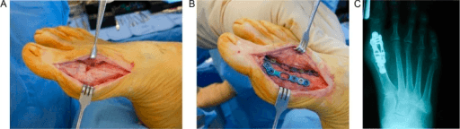
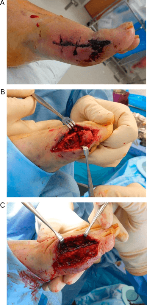
- Dorsal midline incision (preferred for dorsal-plate fixation): a straight longitudinal cut over the dorsal first MTPJ, centred between EHL and the medial border, 5 to 7 cm, extending proximal and distal to the joint line.
- Medial approach (alternative for crossing screws): a longitudinal incision over the medial border, allowing a medial capsulotomy and good plantar visualisation.
- Mark the incision before tourniquet inflation if EHL position is uncertain, and avoid skin bridges if a simultaneous lesser-ray incision is planned.
- Incise subcutaneous tissue and identify and retract EHL laterally.
- Open the dorsal capsule longitudinally and reflect full-thickness capsular flaps medially and laterally (preserve them for closure).
- Release the dorsal, medial and lateral capsule circumferentially from the metatarsal head and phalanx base.
- Protect the medial plantar digital nerve by staying dorsal to the midaxial line on the plantar-medial side.
- Bring the metatarsal head into the wound by gentle plantarflexion of the first metatarsal; expose the phalanx base by distraction and slight flexion so both articular surfaces are fully accessible.
- Goal: congruent, bleeding, cancellous bone surfaces with maximum contact area.
- Cup-and-cone (dome) reaming is preferred: graduated concave reamer on the metatarsal head, convex reamer on the phalanx base, creating matched spherical surfaces that allow multiplanar fine-tuning of position before fixation.
- Flat cut (alternative): oscillating-saw cuts perpendicular to the long axis of fixation — simpler but less forgiving of positional error.
- Remove all cartilage, including the plantar-most aspect of the metatarsal head (frequently missed); drill or burr through subchondral sclerosis to expose bleeding cancellous bone.
- Target relative to the first metatarsal shaft: 15 to 20 degrees dorsiflexion, 5 to 10 degrees valgus, neutral rotation (no pronation, no supination).
- Ground-clearance test: foot flat on a sterile back surface (or fluoroscopy) — the hallux tip should just clear the floor by 1 to 2 mm. Pressing firmly means insufficient dorsiflexion; high off the floor means excessive dorsiflexion.
- Length check: the hallux should be equal to or slightly shorter than the second toe — a significantly short hallux transfers load.
- Hold the confirmed position with provisional K-wires and confirm with AP and lateral fluoroscopy before committing.
- Rotation check: with the foot plantigrade the hallux nail should face straight up — malrotation is irreversible once the plate is applied.
- Dorsal plate plus an interfragmentary (lag) screw (evidence-based preferred construct): apply a pre-contoured low-profile locking plate spanning the fusion site, and add a compression screw (through the plate or an independent crossed lag screw) for axial compression and rotational control.
- Crossed screws (alternative, less hardware): two partially-threaded cancellous screws crossing at the fusion site — reliable, with a slightly higher non-union rate than plate constructs in comparative series.
- Confirm screw placement, compression of the fusion gap and no cortical breach on AP and lateral fluoroscopy.
- After fixation, palpate and observe the sesamoids — they should lie freely in the plantar groove and not be impinged by dorsal shift of the phalanx.
- If a sesamoid appears elevated or impinged, check that adequate plantar cartilage was removed and that dorsiflexion is not excessive.
- Repair the dorsal capsule/retinaculum over the plate as much as possible with absorbable sutures — this prevents dorsal hardware prominence and adds soft-tissue stability.
- Close the subcutaneous layer with absorbable sutures and the skin with monofilament sutures or staples.
- Apply a well-padded, non-circumferential dressing with a firm forefoot wrap to maintain position during early healing.
The medial plantar digital nerve to the hallux runs along the medial plantar surface of the proximal phalanx and first metatarsal head. It is vulnerable during medial periosteal dissection and during cup-and-cone reaming of the phalanx base. Stay dorsal to the midaxial line on the plantar-medial side and place retractors gently around the plantar-medial corner. Injury causes permanent numbness of the medial hallux and a painful neuroma. Once the dorsal plate is applied, malrotation is irreversible — so confirm all three planes (dorsiflexion, valgus, rotation) before definitive fixation.
With the foot flat on a sterile back surface (or fluoroscopy with the foot flat), the hallux tip should just clear the floor by 1 to 2 mm. Firmly pressing on the table means insufficient dorsiflexion (target 15 to 20 degrees relative to the first metatarsal shaft); a hallux high off the floor means excessive dorsiflexion. Too flat overloads the IPJ at push-off; too dorsiflexed causes dorsal footwear pressure and lesser-ray transfer metatarsalgia.
The plantar-most cartilage of the metatarsal head is the part most frequently missed, and retained cartilage or unbreached subchondral sclerosis is a leading cause of non-union. Remove all articular cartilage and drill or burr through sclerotic subchondral bone until a bleeding cancellous surface is exposed. Cup-and-cone (dome) reaming is preferred over flat cuts because it is forgiving of fine positional adjustment and maximises cancellous contact area while preserving length.
Aftercare & Complications
Rehabilitation | Phase | Timing | Immobilisation / weight-bearing | Therapy focus | |-------|--------|---------------------------------|---------------| | 1 | 0 to 2 weeks | Stiff-soled shoe or cast; non- or heel weight-bearing | Wound check and suture removal at 2 weeks | | 2 | 2 to 6 weeks | Heel weight-bearing in a stiff-soled shoe | Protect fusion until early callus on radiograph | | 3 | 6 to 12 weeks | Progressive weight-bearing as tolerated | Radiological union typically at 8 to 12 weeks | | 4 | Long term | Rocker-bottom or stiff toe-box footwear | Activity modification; reduce IPJ loading | Most patients return to desk work by about 6 weeks. Around 85 to 90 percent of well-positioned fusions unite; radiological union is typically seen at 8 to 12 weeks.
- Recognition
- Persistent fusion-site pain beyond 3 months; no bridging trabeculae on serial radiographs at 8 to 12 weeks; screw loosening; CT confirms absent bridging
- Prevention
- Complete cartilage and sclerotic-bone removal to bleeding cancellous surfaces; dorsal plate plus compression screw; smoking cessation; adequate immobilisation; optimise bone health (vitamin D, calcium)
- Management
- Asymptomatic radiological non-union — extend non-weight-bearing and observe; symptomatic — revision arthrodesis with bone graft and upgraded fixation; infected — staged debridement, antibiotics, then revision fusion
- Recognition
- IPJ pain on walking and stairs; progressive IPJ joint-space loss and osteophytes on follow-up radiographs
- Prevention
- Intraoperative ground-clearance test; target 15 to 20 degrees dorsiflexion relative to the first metatarsal shaft; fluoroscopy before final tightening
- Management
- Mild — shoe modification, rocker-bottom sole; progressive IPJ arthritis — IPJ arthrodesis; revision of the fusion rarely justified for this alone
- Recognition
- Plantar keratosis under lesser metatarsal heads 2 to 4; burning forefoot pain; markedly dorsiflexed fusion on lateral radiograph
- Prevention
- Same intraoperative checks; aim for the hallux tip to just clear the floor, not elevate more than 2 to 3 mm
- Management
- Padding, metatarsal bar, orthoses to offload lesser rays; severe cases — revision of fusion position (rare, demanding)
- Recognition
- Visible shortening; hallux significantly shorter than the second toe on AP radiograph; transfer metatarsalgia
- Prevention
- Minimal bone resection (cartilage and sclerosis only); structural bone graft in Keller's salvage; check length against the second toe before fixation
- Management
- Mild — orthotics and padding; significant symptomatic — revision with structural bone graft (high complexity)
- Recognition
- Wound edge necrosis or dehiscence at 1 to 2 weeks; exposed or infected hardware
- Prevention
- Careful handling of thin rheumatoid skin; close capsule over the plate; avoid skin tension; low-profile plates; hold footwear until healed
- Management
- Minor — local wound care; exposed hardware — early plastic surgery review, possible flap; infected — debridement and antibiotics, retain hardware until union
- Recognition
- Localised tenderness over plate or screw head; pain in footwear that resolves barefoot
- Prevention
- Low-profile anatomical plates; minimise dissection so capsule covers plate; confirm union before planning removal
- Management
- Soft wide shoes or cushioned insoles; elective hardware removal once union is confirmed (minimum 3 to 4 months)
- Recognition
- Dorsal IPJ pain and stiffness; radiographic IPJ narrowing; response to IPJ corticosteroid injection
- Prevention
- Correct fusion position; counsel patients pre-operatively that IPJ stiffness and eventual arthritis is a known long-term sequela
- Management
- Activity and shoe modification, rocker-sole footwear; corticosteroid injection; IPJ arthrodesis if refractory
Non-union work-up. Serial radiographs at 6 and 12 weeks; CT if there is doubt at 12 to 16 weeks. Exclude infection (CRP, ESR, white cell count) and identify contributors (smoking, nutrition, bone density). Revision is repeat arthrodesis with bone graft plus upgraded fixation — a dorsal plate if one was not used primarily. Keller's salvage specifics. Remove all soft-tissue interposition and periosteum from the resected proximal phalanx stump, measure the length deficit and match it with a structural bone block (iliac crest autograft remains the gold standard), and span both graft–host interfaces with a dorsal locking plate — crossed screws alone are mechanically inadequate across a graft–host interface. Reported union in specialist salvage series is lower than for primary fusion (roughly 85 to 90 percent).
Viva & Exam Focus
FUSEFUSE — Critical Steps for First MTPJ Arthrodesis
GRADEGRADE — Coughlin and Shurnas Classification
Medial plantar digital nerve to hallux. Location: runs along the medial plantar surface of the proximal phalanx, vulnerable during medial periosteal dissection and cup-and-cone reaming of the phalanx base. Protection: stay dorsal to the midaxial line during periosteal stripping and use retractors carefully around the plantar-medial corner. Injury causes permanent numbness of the medial hallux and a painful neuroma.
Position error: insufficient dorsiflexion (toe flat to floor). Target is 15 to 20 degrees dorsiflexion relative to the first metatarsal shaft. Too little dorsiflexion means the hallux lies flat and loads the interphalangeal joint during push-off, causing IPJ osteoarthritis. Too much dorsiflexion causes dorsal skin pressure in footwear. Check intraoperatively with the foot flat: the hallux tip should just clear the floor.
Position error: insufficient or excessive valgus. Target is 5 to 10 degrees valgus. Too little valgus (varus) causes the hallux to abut the second toe during gait, producing painful impingement. Too much valgus causes overlapping of the second toe and cosmetic deformity. Neutral rotation is mandatory — pronation or supination is poorly tolerated and is a common cause of revision.
Excessive bone resection causing hallux shortening. Remove cartilage and subchondral sclerosis only — do not sacrifice cortical bone unnecessarily. Shortening alters the weight-bearing platform, transfers load to lesser metatarsals, and causes cosmetic deformity. In Keller's salvage, structural bone graft is mandatory to restore length. Check length against the second toe before fixation.
Interphalangeal joint overload. If the fusion is positioned in excessive dorsiflexion (beyond 20 degrees), the ground reaction force during late stance transfers entirely to the IPJ, causing accelerated IPJ degeneration. This is the direct mechanical consequence of malposition and one of the most common late complications requiring revision. Confirm position prior to final tightening.
Clinical Decision Scenarios
Practise clinical reasoning and management decisions out loud
“You are performing a first MTPJ arthrodesis for Grade IV hallux rigidus using cup-and-cone reaming. After provisional K-wire fixation you place the foot flat on the table. The hallux tip is pressing firmly against the table surface. What does this mean and what do you do?”
“A 58-year-old woman had a Keller's resection arthroplasty for hallux rigidus at another institution 12 years ago. She now presents with severe pain, instability, and a floppy hallux. She cannot push off from the ground. Her radiographs show the classic 'floppy toe' deformity with proximal phalanx resection. How do you approach the surgical management?”
“A colleague presents you with a 47-year-old male with painful stiff first MTPJ. He has 20 degrees of dorsiflexion, bone-on-bone on radiograph, global pain, sesamoid involvement, and has failed conservative treatment including NSAIDs and orthotic insoles for 18 months. Your colleague says he would like to perform a cheilectomy. Do you agree? What would you recommend?”
Key Indications
- Grade IV hallux rigidus (Coughlin and Shurnas): bone-on-bone, rigid, global pain — primary gold standard indication
- Failed cheilectomy for advanced hallux rigidus with persistent or progressive symptoms
- Rheumatoid arthritis forefoot with first MTPJ destruction
- Salvage of failed Keller's resection arthroplasty or resection arthroplasty (requires structural bone graft to restore length)
- Neuromuscular hallux valgus with coexisting MTPJ OA; first ray hypermobility with symptomatic MTPJ arthritis
Coughlin and Shurnas Classification — Exam Essentials
- Grade I: dorsal osteophyte, motion greater than 60 degrees, dorsal pain only — cheilectomy appropriate
- Grade II: moderate restriction (40 to 60 degrees), under 25 percent joint space loss — cheilectomy appropriate
- Grade III: marked restriction (10 to 30 degrees), over 25 percent joint space loss, sesamoid involvement — cheilectomy with or without Moberg; interpositional arthroplasty debated
- Grade IV: bone-on-bone, rigid, global pain through full arc — arthrodesis is the reference standard; cheilectomy unreliable
- Threshold rule: arthrodesis for Grade IV, or Grade III with under 50 percent metatarsal head cartilage remaining at surgery
- Reference: Coughlin MJ, Shurnas PS. JBJS Am 2003;85(11):2072–2088 — landmark classification paper (the 92 percent good/excellent figure is the cheilectomy result, 86 of 93)
Critical Position Targets — Most Examinable Step
- Dorsiflexion: 15 to 20 degrees relative to first metatarsal shaft — NOT flat to floor
- Valgus: 5 to 10 degrees — too little causes hallux-second toe impingement; too much causes second toe overlap
- Rotation: neutral — pronation and supination both poorly tolerated and common causes of revision
- Ground-clearance test: foot flat on table — hallux tip should just clear the floor by 1 to 2 mm; firmly pressing means insufficient dorsiflexion
- Consequence of too flat: IPJ overload during push-off causing IPJ osteoarthritis (most common malposition complication)
Fixation — What the Evidence Actually Shows
- Dorsal plate plus an interfragmentary (lag) screw is a tension-band-style construct that resists the dorsal bending moment at push-off — favour it for revision, poor bone or graft interfaces
- No construct is decisively superior: union rates are high for both plate and crossed-screw fixation; Hyer 2008 found no difference in time-to-fusion (plate cost significantly more)
- Reference: Goucher NR, Coughlin MJ. Foot Ankle Int 2006;27(11):869–876 — dome reamers and dorsal plate with crossed lag screws, 92 percent union (4 of 54 nonunions), 96 percent satisfaction
- Cup-and-cone (dome) reaming preferred over flat cuts: forgiving of position fine-tuning, maximises cancellous contact area, preserves length
- Confirm with fluoroscopy AP and lateral before final screw tightening
Danger Zones — Five Critical Structures/Errors
- Medial plantar digital nerve: runs medial plantar surface of phalanx, injured during medial periosteal dissection — causes hallux numbness and neuroma
- Dorsiflexion malposition (flat): IPJ arthritis — most common malposition complication
- Valgus malposition (varus): hallux-second toe impingement during gait
- Shortening: from excessive bone resection; mandatory bone graft in Keller's salvage to restore length
- Wound dehiscence: thin rheumatoid or diabetic skin over prominent dorsal plate — careful tissue handling, low-profile plates
Technique Pearls
- Remove ALL articular cartilage including plantar aspect of metatarsal head — frequently missed, causes non-union
- Drill or burr subchondral sclerosis to bleeding cancellous bone — essential for union
- Intraoperative position check three planes before definitive fixation: dorsiflexion, valgus, rotation
- Check hallux length relative to second toe — hallux should be equal to or slightly shorter than second toe
- Keller's salvage: structural bone graft (iliac crest autograft) mandatory plus dorsal plate — crossed screws inadequate across graft interface
Post-operative Protocol
- 0 to 2 weeks: non-weight-bearing in stiff-soled shoe or cast; wound check at 2 weeks, suture removal
- 2 to 6 weeks: heel weight-bearing in stiff-soled shoe; protect fusion until early callus on radiograph
- 6 to 12 weeks: progressive weight-bearing as tolerated; radiological union typically 8 to 12 weeks
- Long term: rocker-bottom or stiff toe-box footwear recommended to reduce IPJ loading; sports modification
Exam Tips — High-Yield Concepts
- Know Grade IV equals arthrodesis: examiners frequently present Grade III/IV crossover cases to test whether you correctly indicate arthrodesis
- Position is the key viva topic: describe all three planes, the ground-clearance test, and consequences of malposition in each direction
- Evidence: cite Coughlin 2003 JBJS (classification/threshold), the Gibson 2005 Foot Ankle Int RCT (fusion superior to total toe replacement in pain and reoperation), and McNeil 2013 (arthrodesis the only Grade B procedure)
- Keller's salvage requires bone graft: this is a distinct procedure from primary arthrodesis and examiners often ask to differentiate
- Complications triad: non-union, malposition, IPJ arthritis — know prevention and management for each
- Against cheilectomy in Grade IV: be explicit and confident — this is a key discriminator of candidate knowledge
Background & Evidence
Pathoanatomy. Hallux rigidus is degenerative arthritis of the first MTPJ. It begins dorsally with osteophyte formation and dorsal impingement at terminal dorsiflexion, then progresses circumferentially to global cartilage loss, subchondral sclerosis and cyst formation, sesamoid involvement, and ultimately a rigid bone-on-bone joint. The Coughlin and Shurnas five-grade classification (below) captures this progression and directs treatment.
- Clinical and radiographic features
- No pain; full dorsiflexion (greater than 60 degrees); no radiographic change
- Management
- No treatment
- Clinical and radiographic features
- Mild dorsal aching; mild restriction (40 to 60 degrees); dorsal osteophyte only; no joint-space narrowing
- Management
- Cheilectomy
- Clinical and radiographic features
- Moderate pain; moderate restriction (30 to 40 degrees); dorsal and lateral osteophytes; under 25 percent joint-space loss
- Management
- Cheilectomy
- Clinical and radiographic features
- Severe pain and stiffness; marked restriction (10 to 30 degrees); extensive osteophytosis; over 25 percent joint-space loss; subchondral cysts and sclerosis; sesamoid involvement
- Management
- Cheilectomy with or without Moberg osteotomy or interpositional arthroplasty if more than half the head cartilage remains — otherwise arthrodesis
- Clinical and radiographic features
- All Grade III features plus complete joint-space loss (bone-on-bone); rigid MTPJ; possible loose bodies and sesamoid destruction
- Management
- First MTPJ arthrodesis (reference standard)
The landmark classification study. The Coughlin and Shurnas 2003 long-term series is the basis for the modern five-grade clinical-radiographic classification and for the threshold at which fusion replaces cheilectomy. A common exam trap is to misquote the headline numbers: in that paper the 92 percent good-or-excellent figure refers to the cheilectomy procedures (86 of 93), and 97 percent of patients overall had a good or excellent subjective result. The authors' actual treatment rule is that Grade IV — or Grade III with under 50 percent of the metatarsal head cartilage remaining at surgery — should be treated with arthrodesis, whereas cheilectomy was used with predictable success for Grades I, II and selected Grade III. Arthrodesis versus joint-sacrificing alternatives. The best single piece of comparative evidence is the Gibson and Thomson RCT, in which arthrodesis gave significantly greater pain relief than total toe replacement and all fusions united, while 6 of 39 phalangeal implant components required removal for loosening. Raikin and colleagues separately showed arthrodesis outperformed metallic hemiarthroplasty (24 percent hemiarthroplasty failure). The McNeil, Baumhauer and Glazebrook evidence-based review then placed this in context: arthrodesis is the only procedure for hallux rigidus that reaches fair evidence (Grade B), with cheilectomy, osteotomy, implant, resection and interpositional arthroplasty all at Grade C. Fixation and joint preparation. Goucher and Coughlin reported the benchmark dome-reamer technique (50 patients, 54 feet) using a low-profile dorsal titanium plate with crossed lag screws — 96 percent satisfaction, a 92 percent union rate (4 of 54 nonunions, 8 percent). Flavin and Stephens described an analogous low-profile contoured plate plus compression screw over a ball-and-socket preparation, with radiographic union at 6 weeks and dorsiflexion set at 20 to 25 degrees relative to the first metatarsal. The honest position on fixation is that no high-quality study has shown one construct is decisively superior: in Hyer and colleagues' comparative series, time-to-fusion did not differ between crossed screws and dorsal plate (the plate group actually had numerically more nonunions, 3 versus 1, in a small cohort) while the plate cost significantly more. The mechanical argument for a dorsal plate plus interfragmentary (lag) screw is that it is a tension-band-style construct resisting the dorsal bending moment generated at push-off; it is reasonable to prefer it for revision, poor bone stock or graft interfaces, but a candidate should not overstate the union-rate evidence. Functional and positional rationale. Brodsky and colleagues' prospective gait analysis objectively confirmed that successful fusion increases maximal ankle push-off power and single-limb support time — the biomechanical justification for fusing the joint in slight dorsiflexion rather than flat. The positional convention dates to Mann and Oates (95 percent fusion in 41 toes) and is best expressed against two reference frames: relative to the first metatarsal shaft aim for roughly 15 to 25 degrees dorsiflexion and 5 to 10 degrees valgus; relative to the floor (weight-bearing) the hallux pulp should sit only a few millimetres above the ground because of the metatarsal's natural inclination. Insufficient dorsiflexion overloads the IPJ at push-off; excessive dorsiflexion causes dorsal footwear pressure and lesser-ray transfer metatarsalgia. Relevant biomechanics. The first ray bears approximately 40 percent of forefoot load during gait, rising to 60 percent or more at push-off. Following arthrodesis the IPJ becomes the primary remaining mobile joint of the hallux, so excessive MTPJ dorsiflexion concentrates ground-reaction force at the IPJ during push-off — the mechanical basis for the IPJ arthritis that follows an over-dorsiflexed fusion. The sesamoids give flexor hallucis brevis a mechanical advantage and protect the plantar metatarsal head; after fusion they are fixed in position, so accurate position prevents dorsal sesamoid impingement.
References
Hallux rigidus: grading and long-term results of operative treatment
- Single-surgeon 19-year series: 110 of 114 patients reviewed; 93 cheilectomy procedures and 34 arthrodeses, mean follow-up 9.6 and 6.7 years respectively
- Introduced the five-grade clinical-radiographic classification still used today (Grade 0 to Grade IV)
- 97 percent of patients had a good or excellent subjective result; 92 percent (86 of 93) of cheilectomies succeeded for pain and function
- Defined the operative rule: Grade IV — or Grade III with under 50 percent metatarsal head cartilage remaining — should be treated with arthrodesis
Evidence-based analysis of the efficacy for operative treatment of hallux rigidus
- Systematic evidence-based review of 135 relevant studies graded for quality and recommendation strength
- Arthrodesis received fair evidence, Grade B recommendation — the highest of any procedure for hallux rigidus
- Cheilectomy, osteotomy, implant arthroplasty, resection arthroplasty and interpositional arthroplasty all received only Grade C (poor evidence)
- No properly powered comparative trial yet identifies a single best procedure
Arthrodesis or total replacement arthroplasty for hallux rigidus: a randomized controlled trial
- Single-surgeon RCT: 22 patients (38 toes) arthrodesis versus 27 patients (39 toes) total replacement arthroplasty
- Primary outcome was pain on VAS — improved in both groups but significantly more after arthrodesis at 24 months
- All 38 arthrodeses united (mean dorsiflexion 26 degrees); 6 of 39 phalangeal implant components required removal for loosening
- Cost ratio 2:1 in favour of arthrodesis
Hallux metatarsophalangeal joint arthrodesis using dome-shaped reamers and dorsal plate fixation: a prospective study
- Prospective series of 50 patients (54 feet) using matched male/female dome reamers and a low-profile dorsal titanium plate with crossed lag screws
- AOFAS score improved from 51 to 82; VAS pain fell from 6.3 to under 1
- Union rate 92 percent with four nonunions (8 percent); 96 percent patient satisfaction; revision rate 4 percent
- Plate is pre-set with built-in dorsiflexion and valgus; dome reaming preserves length and allows multiplanar adjustment
Comparison of arthrodesis and metallic hemiarthroplasty of the hallux metatarsophalangeal joint
- Comparative series for severe first MTPJ osteoarthritis: 21 hemiarthroplasties versus 27 arthrodeses in 46 patients
- 5 of 21 hemiarthroplasties (24 percent) failed (one revised, four converted to fusion); plantar stem cut-out seen in 8 surviving implants
- All 27 arthrodeses united with no revisions; only 2 office-based hardware removals
- AOFAS-HMI, VAS pain and satisfaction all significantly better in the arthrodesis group (mean pain 0.7 versus 2.4 of 10)
Prospective gait analysis in patients with first metatarsophalangeal joint arthrodesis for hallux rigidus
- Prospective pre- and post-operative instrumented gait analysis in 23 patients, minimum 1-year follow-up
- Significant increase in maximal ankle push-off power after fusion
- Significant increase in single-limb support time on the operated side and reduced step width
- Objectively demonstrates restored propulsion and weight-bearing function despite a fused joint
Verified citations. According to PubMed, the following references have been individually verified (title, authors, journal, year, pages and PMID). DOI links are provided where PubMed returned a DOI. 1. Coughlin MJ, Shurnas PS. Hallux rigidus: grading and long-term results of operative treatment. J Bone Joint Surg Am. 2003;85(11):2072–2088. PMID 14630834. Landmark five-grade classification; 97 percent of patients good/excellent overall and 92 percent of cheilectomies successful; arthrodesis indicated for Grade IV or Grade III with under 50 percent metatarsal head cartilage remaining. 2. McNeil DS, Baumhauer JF, Glazebrook MA. Evidence-based analysis of the efficacy for operative treatment of hallux rigidus. Foot Ankle Int. 2013;34(1):15–32. PMID 23386758. DOI. Systematic review of 135 studies; arthrodesis the only procedure reaching fair evidence (Grade B); all alternatives Grade C. 3. Gibson JNA, Thomson CE. Arthrodesis or total replacement arthroplasty for hallux rigidus: a randomized controlled trial. Foot Ankle Int. 2005;26(9):680–690. PMID 16174497. DOI. RCT (22 arthrodesis vs 27 arthroplasty); greater pain relief and all-united fusions versus 6 of 39 implant components removed; cost ratio 2:1 favouring fusion. 4. Goucher NR, Coughlin MJ. Hallux metatarsophalangeal joint arthrodesis using dome-shaped reamers and dorsal plate fixation: a prospective study. Foot Ankle Int. 2006;27(11):869–876. PMID 17144945. DOI. 50 patients/54 feet; dome reaming with low-profile dorsal titanium plate and crossed lag screws; 92 percent union (8 percent nonunion), 96 percent satisfaction. 5. Flavin R, Stephens MM. Arthrodesis of the first metatarsophalangeal joint using a dorsal titanium contoured plate. Foot Ankle Int. 2004;25(11):783–787. PMID 15574236. DOI. Prospective series; low-profile contoured plate plus compression screw over ball-and-socket preparation; union at 6 weeks, dorsiflexion 20 to 25 degrees relative to first metatarsal. 6. Mann RA, Oates JC. Arthrodesis of the first metatarsophalangeal joint. Foot Ankle. 1980;1(3):159–166. PMID 7319432. DOI. 41 toes in 28 patients; 95 percent fusion; foundational outcome series for first MTPJ arthrodesis including rheumatoid forefoot. 7. Raikin SM, Ahmad J, Pour AE, Abidi N. Comparison of arthrodesis and metallic hemiarthroplasty of the hallux metatarsophalangeal joint. J Bone Joint Surg Am. 2007;89(9):1979–1985. PMID 17768195. DOI. Severe first MTPJ OA; 24 percent hemiarthroplasty failure with plantar stem cut-out; all 27 arthrodeses united; AOFAS-HMI, pain and satisfaction superior with fusion. 8. Brodsky JW, Baum BS, Pollo FE, Mehta H. Prospective gait analysis in patients with first metatarsophalangeal joint arthrodesis for hallux rigidus. Foot Ankle Int. 2007;28(2):162–165. PMID 17296132. DOI. Instrumented gait analysis in 23 patients; significant increase in maximal ankle push-off power and single-limb support after fusion. 9. Hyer CF, Glover JP, Berlet GC, Lee TH. Cost comparison of crossed screws versus dorsal plate construct for first metatarsophalangeal joint arthrodesis. J Foot Ankle Surg. 2008;47(1):13–18. PMID 18156059. DOI. 45 fusions; overall fusion 91.1 percent; no significant difference in time-to-fusion between crossed screws and dorsal plate; plate implants cost significantly more.