Aponeurotic release or conjoined-tendon lengthening for isolated gastrocnemius contracture | intermediate
Surgical Imaging
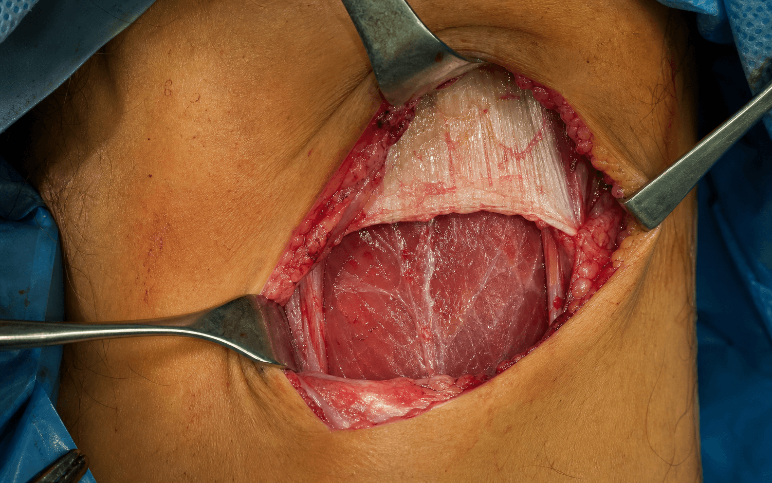
Location: The sural nerve runs in the subcutaneous tissue of the posterolateral calf, approximately 1-2 cm posterior to the lateral border of the fibula in the proximal calf. As it descends it passes posteromedial to the fibula and enters the posteromedial operative field.
Risk: The sural nerve is the most commonly injured structure during gastrocnemius recession. It may be adherent to the deep fascia or to the gastrocnemius aponeurosis in the proximal calf. Blind cutting or aggressive retraction in the posterolateral corner of the incision can transect or stretch it.
Protection: Identify the sural nerve before dividing the aponeurosis. Palpate the nerve in the subcutaneous fat posterolaterally, place a vessel loop, and keep it under direct vision throughout the release.
The trap: Releasing too much of the gastrocnemius aponeurosis, or releasing into the soleus fascia beyond the intended plane, causes excessive dorsiflexion and weakness of push-off.
Clinical consequence: Loss of concentric gastrocnemius plantar flexion power during gait (the patient cannot generate a normal heel-rise or push-off), a calcaneal gait pattern, and patient dissatisfaction despite a technically successful procedure.
The fix: Release the gastrocnemius aponeurosis incrementally. After each division, assess ankle dorsiflexion with the knee extended and flexed. Stop when dorsiflexion reaches 5-10 degrees beyond neutral with the knee extended. Intraoperative judgement is the key safeguard.
Positive Silfverskiöld (isolated gastrocnemius contracture): Dorsiflexion is limited with the knee extended but improves significantly (by at least 5 degrees) when the knee is flexed to 90 degrees. This confirms the gastrocnemius is the tight structure and the soleus is lengthened — gastrocnemius recession is the correct operation.
Negative Silfverskiöld (combined gastrocnemius-soleus contracture): Dorsiflexion is limited equally with the knee extended and flexed. The soleus or the entire tendo-Achilles is contracted. Gastrocnemius recession will under-correct — a tendo-Achilles lengthening is required.
The trap: Performing a gastrocnemius recession in a patient with a negative Silfverskiöld test. The patient will not gain meaningful dorsiflexion and will still have equinus postoperatively.
Gastrocnemius recession (Strayer): Addresses isolated gastrocnemius contracture. Preserves soleus function. Lower risk of over-lengthening. Lower wound complication rate. Preferred in diabetics and patients with forefoot pathology.
Tendo-Achilles lengthening (TAL): Required when the contracture involves the entire gastrocnemius-soleus-Achilles complex (negative Silfverskiöld). Percutaneous triple-cut TAL carries significant risks in diabetics: wound breakdown, infection, and Achilles tendon rupture.
The trap: Performing a TAL when a gastrocnemius recession would suffice — unnecessary weakening of push-off power and higher complication rate. Conversely, performing a recession when TAL is needed — residual equinus.
Why recession matters in diabetics: Gastrocnemius tightness increases forefoot pressure by approximately 20-30%. In a neuropathic diabetic foot, this elevated pressure contributes to plantar forefoot ulceration that is slow or impossible to heal despite offloading.
Indication: Recalcitrant diabetic forefoot ulceration with a positive Silfverskiöld test, after optimisation of glycaemic control, offloading, and wound care. Gastrocnemius recession reduces forefoot pressure and promotes ulcer healing.
Surgical choice: Gastrocnemius recession (not TAL) is strongly preferred in diabetics — percutaneous TAL has an unacceptably high rate of wound complications and Achilles rupture in this population.
Location: The posterior tibial neurovascular bundle (tibial nerve, posterior tibial artery and vein) runs deep to the soleus in the deep posterior compartment of the calf, between the flexor digitorum longus and flexor hallucis longus.
Risk: The bundle is deep to the operative plane in a standard Strayer release and is not routinely encountered. However, if dissection is carried too deep (past the soleus fascia into the deep posterior compartment), or if a retractor is placed aggressively against the soleus muscle belly, the tibial nerve or posterior tibial artery can be injured.
Protection: Stay in the plane between the gastrocnemius aponeurosis and the soleus fascia. Do not deepen the dissection beyond the soleus muscle belly. If exposure is inadequate, extend the incision rather than forcing deeper retraction.
S.I.L.F.V.E.RSILFVER — Gastrocnemius Recession Decision-Making
S.T.R.A.Y.E.RSTRAYER — Operative Technique Steps
Surgical Indications
Absolute Indications
- Failed non-operative treatment for a primary forefoot or midfoot pathology where isolated gastrocnemius contracture (positive Silfverskiöld test) is a demonstrated biomechanical contributor
- Recurrent or recalcitrant plantar fasciitis after a minimum of 6-12 months of structured non-operative treatment (stretching programme, orthotics, physiotherapy, night splint, at least one corticosteroid injection) with a positive Silfverskiöld test
- Diabetic forefoot ulceration with positive Silfverskiöld test, refractory to offloading and wound care optimisation
- Metatarsalgia or midfoot/forefoot overload with isolated gastrocnemius contracture, after failed non-operative management
Relative Indications
- Achilles tendinopathy (non-insertional) with isolated gastrocnemius contracture and failed non-operative treatment
- Flatfoot deformity with gastrocnemius contracture contributing to hindfoot valgus — recession may be performed as an adjunct to flatfoot reconstruction
- Equinus contracture in the paediatric population — gastrocnemius recession (Baumann or Strayer) in cerebral palsy or idiopathic toe-walking
- Chronic ankle stiffness or impingement where gastrocnemius tightness limits dorsiflexion and contributes to anterior ankle impingement symptoms
Contraindications
Absolute:
- Negative Silfverskiöld test — the contracture involves the soleus or entire tendo-Achilles; gastrocnemius recession will under-correct and a tendo-Achilles lengthening is required
- Active infection at the surgical site or in the ipsilateral extremity
- Severe peripheral vascular disease where wound healing is unlikely (absolute ABI less than 0.4, or rest pain, or tissue loss not yet optimised with vascular intervention)
Relative:
- Previous ipsilateral calf surgery or trauma — scarring may distort anatomy and increase sural nerve injury risk; approach with caution
- Marked obesity (BMI greater than 40) — impaired wound healing, deeper dissection planes, and more difficult exposure
- Active Charcot neuroarthropathy — wait until the acute phase has resolved and the foot is stabilised
- Calf weakness from neuromuscular disease (polio, stroke, Charcot-Marie-Tooth) — further weakening of the gastrocnemius may be poorly tolerated
Evidence for Non-Operative Treatment
Gastrocnemius Stretching Programme
- A structured gastrocnachemius-soleus stretching programme (wall stretches, calf stretches with the knee extended and flexed, standing on a step) is the first-line treatment for isolated gastrocnemius contracture
- Compliance is the primary determinant of success — patients must understand that stretching addresses the contracture and that the programme is long-term (months, not weeks)
- In plantar fasciitis with equinus, stretching improves symptoms in approximately 50-70% of patients when combined with orthotics and activity modification
Orthotic Management
- Heel-lift orthoses and ankle-foot orthoses (AFOs) compensate for the dorsiflexion deficit by effectively shortening the gastrocnemius-soleus demand during gait
- Forefoot rocker-bottom soles reduce forefoot pressure in diabetic patients with equinus and ulceration
- Orthotics do not correct the contracture but offload the affected structures
Injection Therapy (for Associated Pathology)
- Corticosteroid injection for plantar fasciitis: short-term pain relief in approximately 50-70% of patients, but recurrence is common
- Platelet-rich plasma (PRP) for Achilles tendinopathy: mixed evidence, not a substitute for addressing the underlying gastrocnemius contracture
- Botulinum toxin injection into the gastrocnemius: limited evidence, temporary effect (3-6 months), not a substitute for surgical recession
Evidence for Surgery
Gastrocnemius Recession for Plantar Fasciitis
Rationale: Gastrocnemius contracture increases tension on the plantar fascia and plantar forefoot structures during gait. Recession reduces this tension by lengthening the gastrocnemius component of the triceps surae, restoring more normal ankle dorsiflexion and reducing forefoot overload.
Clinical evidence:
- Multiple retrospective and prospective series demonstrate that gastrocnemius recession as an adjunct to plantar fascia release (or as a standalone procedure) improves outcomes in patients with recalcitrant plantar fasciitis and a positive Silfverskiöld test
- The Strayer procedure is the most commonly reported technique in the foot and ankle literature
Gastrocnemius Recession Techniques — Comparison
Key Evidence
Isolated gastrocnemius tightness
Gastrocnemius recession to treat isolated foot pain
Gastrocnemius recession for chronic noninsertional Achilles tendinopathy
Surgical anatomy of the gastrocnemius recession (Strayer procedure)
Proximal Medial Gastrocnemius Recession and Stretching Versus Stretching as Treatment of Chronic Plantar Heel Pain
Clinical Decision Scenarios
Practise clinical reasoning and management decisions out loud
“A 52-year-old woman presents with 18 months of recalcitrant plantar fasciitis despite structured physiotherapy, custom orthotics, two corticosteroid injections, and a night splint. On examination she has tenderness at the plantar medial calcaneal tuberosity and a positive Silfverskiöld test with dorsiflexion improving from 0 degrees (knee extended) to 10 degrees (knee flexed). How would you manage her?”
“A 65-year-old man with Type 2 diabetes (HbA1c 7.8%) has a recurrent plantar forefoot ulcer beneath the second metatarsal head that has failed to heal after 4 months of total contact casting and offloading. He has a positive Silfverskiöld test. He asks whether surgery can help his ulcer heal. How do you counsel him?”
“A 40-year-old runner presents with a 10-month history of non-insertional Achilles tendinopathy that has not responded to an eccentric calf strengthening programme, activity modification, and one PRP injection. Examination reveals a palpable tender nodular swelling in the mid-substance of the Achilles tendon, 6 cm proximal to the calcaneal insertion. Dorsiflexion is 5 degrees (knee extended) and improves to 15 degrees (knee flexed). What would you do?”
References
-
DiGiovanni CW, Kuo R, Tejwani N, Price R, Hansen ST Jr, Cziernecki J, Sangeorzan BJ (2002). Isolated gastrocnemius tightness. J Bone Joint Surg Am. 84(6):962-70. pmid: 12063330. doi: 10.2106/00004623-200206000-00010. — Landmark study establishing the prevalence of isolated gastrocnemius contracture in foot pathology and the biomechanical rationale for recession.
-
Maskill JD, Bohay DR, Anderson JG (2010). Gastrocnemius recession to treat isolated foot pain. Foot Ankle Int. 31(1):19-23. pmid: 20067718. doi: 10.3113/FAI.2010.0019. — Retrospective series of 29 patients; 93% satisfaction with isolated gastrocnemius recession for foot pain with positive Silfverskiöld test.
-
Kiewiet NJ, Holthusen SM, Bohay DR, Anderson JG (2013). Gastrocnemius recession for chronic noninsertional Achilles tendinopathy. Foot Ankle Int. 34(4):481-5. pmid: 23399888. doi: 10.1177/1071100713477620. — Case series demonstrating improved pain and function after gastrocnemius recession for non-insertional Achilles tendinopathy with gastrocnemius contracture.
-
Pinney SJ, Sangeorzan BJ, Hansen ST Jr (2004). Surgical anatomy of the gastrocnemius recession (Strayer procedure). Foot Ankle Int. 25(4):247-50. pmid: 15132933. doi: 10.1177/107110070402500409. — Cadaveric study defining the surgical anatomy of the Strayer procedure and quantifying sural nerve risk.
-
Molund M, Husebye EE, Hellesnes J, Nilsen F, Hvaal K (2018). Proximal Medial Gastrocnemius Recession and Stretching Versus Stretching as Treatment of Chronic Plantar Heel Pain. Foot Ankle Int. 39(12):1423-31. pmid: 30132688. doi: 10.1177/1071100718794659. — Level I RCT demonstrating superior outcomes with gastrocnemius recession plus stretching versus stretching alone for chronic plantar heel pain.