Stage-directed surgical treatment of lunate avascular necrosis — joint levelling, revascularisation, intercarpal fusion, and salvage | advanced
Surgical Imaging
The trap: Measuring ulnar variance on a PA wrist radiograph taken in pronation or supination — ulnar variance shifts by up to 2 mm with forearm rotation.
The fix: Ulnar variance must be measured on a standardised PA wrist radiograph taken with the shoulder abducted 90 degrees, elbow flexed 90 degrees, and the forearm in neutral rotation. Compare with the contralateral side. A difference of 2 mm or greater is considered significant. Negative variance means the ulna is shorter than the radius at the DRUJ — this is the critical measurement that determines whether radial shortening is appropriate.
The trap: Operating on a stage I patient with a salvage procedure (PRC or fusion) when joint-preserving options (joint levelling or revascularisation) are still appropriate.
The fix: Stage I (MRI signal change only, normal radiographs) and stage IIA (linear fracture, no collapse) are treated conservatively or with joint levelling/revascularisation. Stage IIIA (sclerosis without height loss) still accepts joint levelling. Only at stage IIIB (carpal collapse with fixed scaphoid rotation) does the algorithm branch to motion-sparing salvage (STT fusion or PRC). Stage IV (pancarpal arthritis) requires total wrist fusion or arthroplasty. Staging too aggressively leads to loss of motion that could have been preserved.
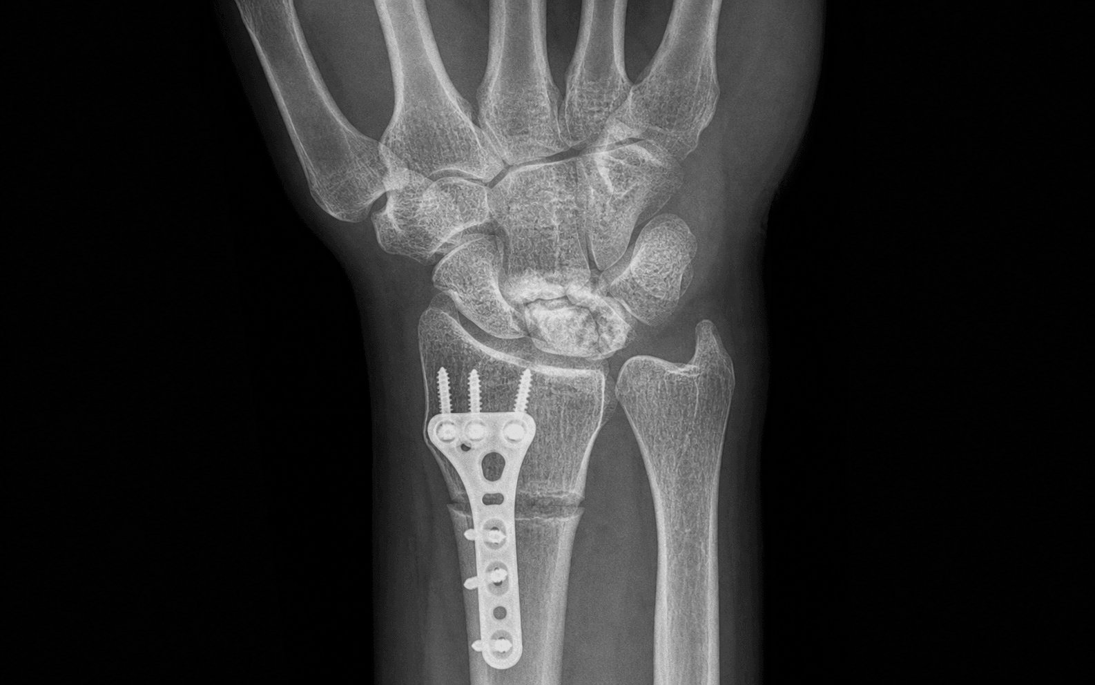
The trap: Shortening the radius by more than the measured ulnar variance plus 2 mm — this overloads the DRUJ and causes ulnar-sided wrist pain, radioulnar impingement, and progressive DRUJ arthritis.
The fix: Calculate the exact amount of shortening preoperatively from the PA wrist film. The target shortening equals the ulnar variance (for a neutral wrist) or the ulnar variance minus 2 mm (for a slightly positive postoperative variance). Never shorten by more than 4 mm total. Intraoperatively confirm the achieved shortening with fluoroscopy before final plate fixation. If the DRUJ is already symptomatic or arthritic, radial shortening is contraindicated — use capitate shortening instead.
The trap: Performing a proximal row carpectomy when the capitate head or the lunate fossa of the radius has chondrosis or degenerative change — the capitate-radius articulation will fail and the patient develops rapid postoperative arthritis.
The fix: MRI is mandatory before PRC. If the capitate head cartilage is intact on MRI (no marrow oedema, no subchondral cysts, no cartilage loss), PRC gives reliable outcomes. If capitate head wear is present, perform total wrist fusion or arthroplasty instead. An intraoperative arthroscopy or direct visual inspection of the capitate articular surface during the dorsal approach confirms the MRI finding — if chondrosis is seen, abandon PRC.
The trap: The posterior interosseous nerve (PIN) lies on the floor of the fourth extensor compartment (EDM tunnel) during the dorsal wrist approach. It can be inadvertently transected, crushed, or entrapped in hardware, causing a painful neuroma or loss of PIN-innervated extension (though this is clinically silent as the PIN is purely motor to EDM and EPL beyond the supinator).
The fix: Identify the PIN in the fourth extensor compartment as the dorsal retinaculum is elevated. It runs with the posterior interosseous artery on the dorsal capsule. Either protect it with a vessel loop and retract it, or perform a prophylactic PIN neurectomy at the level of the distal radius (which provides pain relief without detectable motor deficit). Never leave it as an unidentified structure crossing the operative field where it can be caught by plates or screws.
The trap: Choosing STT fusion for a patient whose primary complaint is pain at the radiocarpal joint rather than the midcarpal joint — STT fusion stabilises the proximal carpal row but does not address radiocarpal arthritis.
The fix: STT fusion (scaphotrapeziotrapezoid) and scaphocapitate fusion are indicated in Lichtman IIIB when the patient has isolated carpal collapse without radiocarpal arthritis. They preserve carpal height and some radiocarpal motion while eliminating midcarpal instability. PRC is preferred when the patient has midcarpal arthritis or when the surgeon wants a simpler procedure that avoids nonunion risk. The decision is nuanced: PRC gives more flexion-extension arc (approximately 100 degrees vs approximately 60 degrees for STT fusion) but loses carpal height; STT fusion preserves height but reduces radial deviation.
V.A.R.I.A.N.C.EVARIANCE — Surgical Decision Framework
L.I.C.H.T.M.A.NLICHTMAN — Staging and Treatment Algorithm
Surgical Indications
Indications by Lichtman Stage and Ulnar Variance
The surgical management of Kienböck disease is guided by two independent variables: the Lichtman radiographic stage (which reflects the degree of lunate collapse and carpal instability) and the ulnar variance (which determines which joint-levelling procedure is biomechanically appropriate).
- Ulnar Negative
- Observe; immobilise; consider radial shortening if progressive pain
- Ulnar Neutral / Positive
- Observe; immobilise; consider capitate shortening or core decompression if progressive
- Ulnar Negative
- Radial shortening osteotomy (2-4 mm)
- Ulnar Neutral / Positive
- Capitate shortening osteotomy or revascularisation
- Ulnar Negative
- Radial shortening osteotomy (2-4 mm)
- Ulnar Neutral / Positive
- Capitate shortening, revascularisation, or core decompression
- Ulnar Negative
- STT fusion, scaphocapitate fusion, or PRC
- Ulnar Neutral / Positive
- STT fusion, scaphocapitate fusion, or PRC
- Ulnar Negative
- Total wrist fusion or arthroplasty
- Ulnar Neutral / Positive
- Total wrist fusion or arthroplasty
Absolute Indications for Surgery
- Lichtman stage IIA or greater with persistent pain despite 3-6 months of conservative treatment (immobilisation, NSAIDs, activity modification)
- Progressive lunate collapse on serial radiographs
- Loss of carpal height or fixed scaphoid flexion (stage IIIB)
- Pancarpal arthritis (stage IV)
Relative Indications
- Stage I with persistent symptoms greater than 6 months despite immobilisation (some surgeons offer earlier joint levelling in manual labourers)
- Bilateral Kienböck disease (surgical treatment of the more symptomatic side)
- Patient preference for definitive surgical management over prolonged immobilisation
Contraindications to Specific Procedures
Radial shortening osteotomy:
- Ulnar-neutral or positive variance (shortening the radius would worsen the imbalance)
- Pre-existing DRUJ arthritis or instability
- Active infection at the surgical site
- Uncorrected smoking (significantly increases nonunion risk)
Proximal row carpectomy:
- Capitate head chondrosis on MRI (PRC relies on capitate-radius articulation)
- Radiocarpal arthritis (stage IV)
- Active infection
STT or scaphocapitate fusion:
- Midcarpal arthritis beyond the targeted fusion site
- Advanced pancarpal arthritis (stage IV)
- Poor bone quality precluding reliable fixation
Evidence for Conservative Treatment
Immobilisation and Observation
- Stage I and early stage IIA disease may be managed with short-arm thumb spica cast or splint immobilisation for 4-12 weeks to reduce mechanical load on the lunate
- Activity modification and avoidance of repetitive axial loading are recommended regardless of stage
- NSAIDs provide symptomatic pain relief but do not alter disease progression
- Approximately 20-30 percent of stage I patients improve with immobilisation alone; however, many progress to surgical intervention
Core Decompression
- Percutaneous drilling of the lunate to stimulate revascularisation by creating vascular channels
- Suitable for early stages (I-IIA) regardless of ulnar variance
- Limited evidence base — small case series report symptom improvement in 60-80 percent of patients
- Low morbidity; can be combined with other procedures
Evidence for Joint-Levelling Procedures
Radial Shortening Osteotomy
- The most studied surgical procedure for Kienböck disease with the largest evidence base
- Shortens the distal radius by 2-4 mm, effectively converting a ulnar-negative wrist to neutral or slightly positive variance
- Offloads the lunate by redistributing axial load from the radial side of the wrist to the ulnar side
- Multiple long-term series report pain relief in 70-90 percent of patients with Lichtman stages I-IIIA
- Preserves wrist motion — most patients maintain 80-90 percent of preoperative range
- Union rate greater than 95 percent with plate fixation in non-smokers
- Key landmark study: Weiss et al. (1991) reported 94 percent good-to-excellent results with radial shortening in a series of 36 wrists with average 5-year follow-up
Capitate Shortening Osteotomy
- Shortens the capitate by 2-3 mm, achieving the same lunate offloading effect as radial shortening but without altering the DRUJ relationship
- Indicated when ulnar variance is neutral or positive (radial shortening would worsen DRUJ mechanics)
- Fixation with an intramedullary headless compression screw (Acutrak or Herbert-type) or mini-fragment plate
- Almquist (1982) described the original technique; subsequent series report 70-85 percent satisfactory results
- Nonunion rate approximately 5-10 percent — higher than radial shortening because the capitate osteotomy site is smaller
Radial Shortening vs Capitate Shortening Osteotomy
Evidence for Revascularisation
Vascularised Bone Grafting
- Pedicled or free vascularised bone grafts (most commonly the 4th and 5th extensor compartment artery-based grafts from the distal radius, or the medial femoral condyle free flap) are used to restore blood supply to the lunate
- Most commonly combined with a joint-levelling procedure (radial shortening or capitate shortening) to simultaneously offload and revascularise the lunate
- Indicated in stage IIA-IIIA with a viable (non-collapsed) lunate shape — revascularisation cannot restore a collapsed lunate architecture
- The Iliac crest bone graft based on the vascularised pedicle was described by Hori (1979); more recent work uses the medial femoral condyle free flap with good structural support
- Series report pain relief in 65-85 percent and radiographic evidence of revascularisation (restored MRI signal) in approximately 60-75 percent
Evidence for Motion-Sparing Salvage
Intercarpal Fusion (STT or Scaphocapitate)
- STT fusion (scaphotrapeziotrapezoid) stabilises the proximal carpal row by preventing scaphoid flexion, which is the primary collapse pattern in Kienböck disease
- Indicated at Lichtman stage IIIB when carpal height is reduced and the scaphoid has rotated into fixed flexion but pancarpal arthritis has not yet developed
- Watson and Ryu (1986) described the technique; series report 70-85 percent good-to-excellent results
- Nonunion rate approximately 5-15 percent — the most common complication; requires careful surface preparation and rigid fixation
- Scaphocapitate fusion is an alternative that preserves more radial deviation than STT fusion
Proximal Row Carpectomy
- Removes the entire proximal carpal row (scaphoid, lunate, triquetrum), allowing the capitate to articulate with the lunate fossa of the radius
- Advantages: no nonunion risk (no fusion), simpler procedure, predictable pain relief, faster rehabilitation
- Disadvantages: loss of carpal height, reduced grip strength (approximately 60-70 percent of contralateral), potential for capitate-radius arthritis over time (long-term follow-up shows radiographic arthritis in 15-25 percent of patients at 10 years, though many remain asymptomatic)
- Contraindication: capitate head chondrosis or midcarpal arthritis — preoperative MRI is mandatory
- Culp (2010) reported 85 percent good-to-excellent results in a series of PRCs for Kienböck disease at stage IIIB
Evidence for Total Wrist Salvage
Total Wrist Fusion
- The gold standard salvage for Lichtman stage IV with pancarpal arthritis
- Provides reliable pain relief in greater than 90 percent of patients
- Eliminates wrist motion but restores grip strength to approximately 80-90 percent of the contralateral side
- Plate-and-screw fixation (dorsal wrist fusion plate) is the most common technique
- Union rate greater than 95 percent with modern plating systems
Total Wrist Arthroplasty
- Motion-preserving alternative to fusion for low-demand patients with stage IV disease
- Relatively newer implants (fourth-generation designs) have improved survival but long-term data remain limited
- Contraindicated in high-demand manual labourers, active infection, or poor bone stock
- Revision to fusion is possible but complex — arthroplasty is generally reserved for older or lower-demand patients
Clinical Decision Scenarios
Practise clinical reasoning and management decisions out loud
“A 32-year-old right-handed male carpenter presents with a 12-month history of insidious right wrist pain. He reports pain with heavy gripping and pushing. Examination reveals tenderness over the dorsum of the wrist, reduced grip strength, and pain on axial loading. PA radiograph in neutral rotation shows a sclerotic lunate with a linear fracture line, carpal height preserved, scapholunate angle 48 degrees, and ulnar variance minus 2 mm. MRI shows low T1 signal in the lunate with preserved lunate morphology. What is the diagnosis, stage, and surgical management?”
“A 45-year-old woman presents with long-standing right wrist pain. PA radiograph shows a collapsed and fragmented lunate, carpal height ratio 0.48, scapholunate angle 68 degrees, and mild narrowing of the radioscaphoid joint. Ulnar variance is neutral. MRI shows low T1 signal throughout the lunate with capitate head cartilage intact. What is the Lichtman stage and what are the surgical options?”
“A 28-year-old woman with Kienbock disease underwent a radial shortening osteotomy 18 months ago. The osteotomy united uneventfully. She now presents with worsening wrist pain, reduced grip, and stiffness. Radiographs show the lunate has collapsed further, the carpal height ratio is 0.50, and the scapholunate angle is 65 degrees. What is happening and what is the next step?”