Osteochondritis Dissecans of the Knee — Fixation

ArthroplastyAdvancedCore Procedure

Osteochondritis Dissecans of the Knee — Fixation

Surgical technique guide for internal fixation of osteochondritis dissecans (OCD) lesions of the knee — arthroscopic and open approaches, headless compression screws, bioabsorbable fixation, subchondral drilling, fragment salvage in juvenile and adult patients

High-yield overview

Arthroscopic or open fixation of unstable OCD lesions with headless compression screws or bioabsorbable devices | advanced

Surgical Imaging

OCD medial femoral condyle screw fixation
Osteochondritis dissecans of the medial femoral condyle fixed with two headless compression screws, securing the osteochondral fragment for healing.Credit: AI-generated medical illustration · OrthoVellum
Critical Danger Structures and Exam Traps
Juvenile vs Adult OCD — The Skeletal Maturity Trap

The trap: Treating all OCD lesions as adult OCD — operating too early on a stable juvenile lesion or using rigid implants that cross an open physis and risk premature physeal closure.

The fix: Check skeletal maturity with a full-leg radiograph assessing physes and a hand radiograph for bone age. Juvenile OCD (open physes) with a stable lesion: protected weight-bearing and activity modification for 3-6 months before considering surgery. If fixation is needed in a juvenile patient, use bioabsorbable devices that do not cross the physis where possible, or place screws entirely within the epiphysis.

Fragment Viability — Preserving the Native Piece

The trap: Discarding a displaced osteochondral fragment and proceeding directly to microfracture or marrow stimulation in a young patient. The native fragment, if viable, provides hyaline cartilage that is biomechanically superior to fibrocartilage from marrow stimulation.

The fix: Always assess the fragment at arthroscopy. A viable fragment has intact subchondral bone attached to the articular cartilage. If the fragment is in-situ or hinged but viable, fix it. Only debride to marrow stimulation when the fragment is fragmented, necrotic, or unsalvageable. In patients under 25 with a viable fragment, fixation should always be attempted before considering salvage procedures.

Hardware Prominence — Chondral Damage from proud Screws

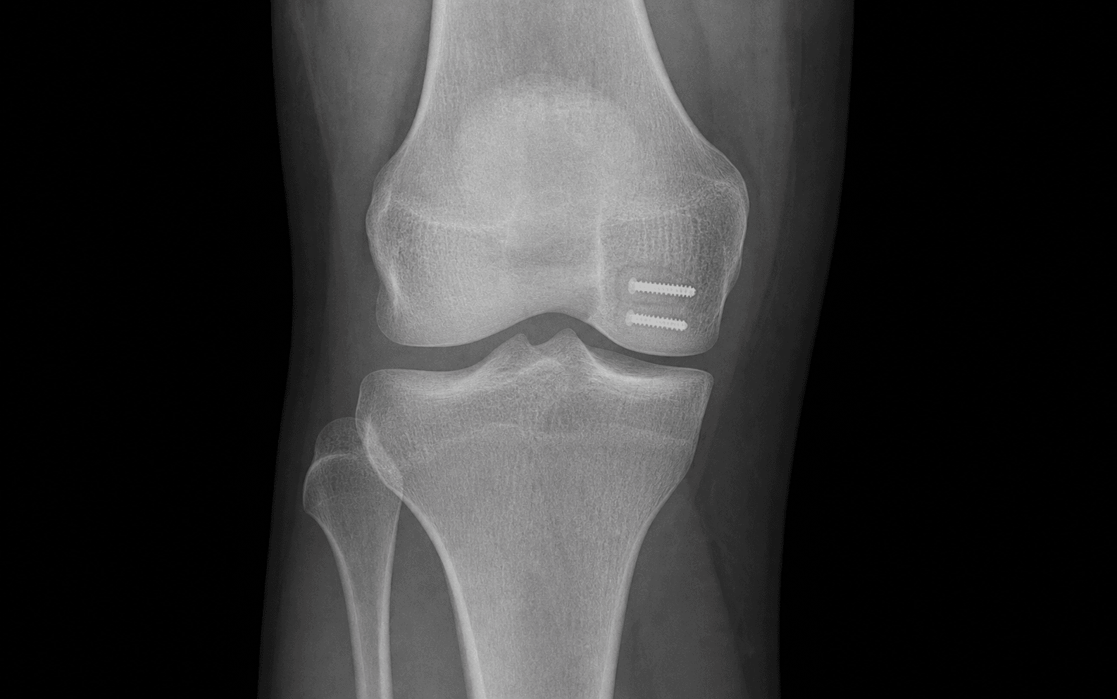
The trap: Leaving headless compression screws proud to the articular surface, causing chondral damage, catching, and early osteoarthritis. Even 1 mm of proud hardware can shred the opposing tibial articular cartilage during knee flexion.

The fix: Use headless compression screws (Herbert-type, Acutrak, MMP) countersunk fully beneath the articular surface. Confirm screw burial by direct visualisation and fluoroscopy. Check that no screw head protrudes by running a probe over the fixation site at full arthroscopic visualisation. If the bone is too soft to hold a countersunk screw, use bioabsorbable pins.

Lesion Stability Assessment — MRI vs Arthroscopy

The trap: Relying solely on MRI to determine lesion stability and planning surgery without arthroscopic probing. MRI has a sensitivity of approximately 75-85% for detecting instability — some unstable lesions appear stable on MRI.

The fix: Diagnostic arthroscopy with probe testing is the definitive assessment. An unstable fragment lifts away from the bed with probe pressure, has a fluid-filled interface, and may have a hinge of attached cartilage. An absolutely stable lesion is firmly anchored with no gap. Plan for fixation of unstable lesions and observe stable lesions non-operatively.

Physeal Sparing in Juvenile OCD

The trap: Placing metal compression screws across an open physis during OCD fixation, risking premature physeal arrest, limb-length discrepancy, or angular deformity in a growing child.

The fix: In juvenile patients with open physes, direct the screws within the epiphysis, parallel to but not crossing the physis. Alternatively, use bioabsorbable pins or nails (PLLA, PGA) placed entirely within the epiphyseal segment. If the lesion is large and fixation requires a screw trajectory that crosses the physis, consider bioabsorbable devices that resorb without permanent physeal tethering, and monitor with serial radiographs.

Missed Procedural Indication — When Not to Fix

The trap: Fixing a small, stable, healed lesion in a skeletally immature patient who would have healed with protected loading alone. Fixation carries surgical risks (hardware complications, arthrofibrosis, infection) and should be reserved for genuinely unstable or detachable lesions.

The fix: The treatment algorithm is driven by two axes — skeletal maturity and lesion stability. A stable juvenile lesion: non-operative. An unstable juvenile lesion: fixation. An unstable adult lesion: fixation or salvage. A stable adult lesion with closed physes: activity modification and monitoring, with surgery if it destabilises. Do not operate without a clear instability indication.

Mnemonic

S.T.A.B.L.ES.T.A.B.L.E — OCD Lesion Stability Assessment

Mnemonic

F.I.X.A.T.EF.I.X.A.T.E — OCD Fixation Principles

Mnemonic

D.R.I.L.LD.R.I.L.L — Subchondral Drilling and Healing Stimulation

Surgical Indications

Absolute Indications

  • Unstable or detached OCD lesion confirmed on arthroscopic probing (Dipaola grade II-IV) in a skeletally mature or immature patient with symptoms
  • Symptomatic loose body from a displaced OCD fragment with mechanical locking or catching
  • Failed non-operative management after 3-6 months of protected weight-bearing and activity modification in a juvenile patient with an unstable lesion
  • Progressive lesion enlargement or instability on serial MRI despite non-operative treatment

Relative Indications

  • Symptomatic stable lesion (Dipaola grade I) in a skeletally mature adult (closed physes) with persistent pain despite 3-6 months of non-operative management
  • Lesion greater than 2 square cm with high T2 signal but intact cartilage — fixation may prevent detachment
  • Patient approaching skeletal maturity with a juvenile lesion that has not healed — fixation before physeal closure may improve healing potential
  • Adult OCD with a viable fragment and a contained bed — fixation preferred over marrow stimulation when the fragment is salvageable

Contraindications

Absolute:

  • Active knee infection (septic arthritis — arthroscopy and fixation deferred until infection resolved)
  • Fragment completely necrotic with no attached subchondral bone and no articular cartilage integrity — fixation will fail, proceed to salvage (microfracture, OATS, or allograft)

Relative:

  • Advanced degenerative changes (Outerbridge grade IV changes elsewhere in the knee) — fixation of an OCD lesion will not alter the degenerative cascade
  • Patient with poor compliance with postoperative weight-bearing restrictions
  • Very small lesion (less than 1 square cm) that has healed with non-operative management

Evidence for Non-Operative Treatment

Juvenile OCD (Open Physes)

  • Juvenile OCD with open physes has a significantly higher healing rate with non-operative management compared to adult OCD — reported healing rates range from 50-60% with protected weight-bearing and activity modification over 3-6 months
  • The key principle is that the open physis provides ongoing growth and remodelling capacity, and the juvenile subchondral bone has better vascularity
  • Immobilisation is generally not recommended — hinged knee brace with protected weight-bearing (touch-down or partial weight-bearing) and activity restriction (no competitive sports) is the standard approach
  • Serial MRI every 3-4 months to monitor lesion healing: look for progressive resolution of the T2 signal line, reconstitution of the subchondral bone, and cartilage integrity
  • The most common reason non-operative management fails: ongoing high-impact activity that repeatedly stresses the fragment — patient education and compliance are critical

Adult OCD (Closed Physes)

  • Non-operative success in adult OCD is substantially lower than in juvenile OCD — healing rates of approximately 20-30% with protected loading in stable lesions
  • Adult OCD is generally considered for non-operative management only when the lesion is stable (Dipaola grade I) and the patient has low physical demands
  • Many adult stable lesions are managed non-operatively initially, but have a higher rate of eventual surgery compared to juvenile lesions

Evidence for Surgical Fixation

Arthroscopic vs Open Fixation

Arthroscopic fixation (preferred when feasible):

  • Minimally invasive — avoids arthrotomy, reduces postoperative pain and scarring
  • Direct visualisation of the articular surface for precise fragment reduction
  • Access to the most common lesion site (lateral medial femoral condyle) is generally good through standard anteromedial and anterolateral portals
  • Limitations: difficult for very large lesions, lesions on the posterior femoral condyle, or lesions requiring extensive bone grafting of the bed

Open fixation (through medial parapatellar or limited approach):

  • Required for very large lesions (greater than 3 square cm), lesions with significant bone loss requiring grafting, or lesions in difficult arthroscopic locations
  • Allows direct inspection of the fragment and bed, bone grafting of the crater, and placement of multiple fixation devices under direct vision
  • Higher morbidity than arthroscopy: larger incision, longer recovery, risk of arthrofibrosis

Arthroscopic vs Open OCD Fixation


Key Evidence

Evidence

Management of osteochondritis dissecans of the knee: current concepts review

Level III
Kocher MS, Tucker R, Ganley TJ, Flynn JMAm J Sports Med (2006)
Clinical implication: Always determine skeletal maturity first — this single factor drives the entire treatment pathway for OCD of the knee.
Evidence

Internal fixation of juvenile osteochondritis dissecans lesions of the knee

Level IV
Kocher MS, Czarnecki JJ, Andersen JS, Micheli LJAm J Sports Med (2007)
Clinical implication: Juvenile OCD fixation with bioabsorbable pins provides reliable healing without hardware removal — use physeal-sparing technique to protect growth.
Evidence

Internal Fixation of Osteochondritis Dissecans of the Knee Leads to Good Long-Term Outcomes and High Degree of Healing without Differences between Fixation Devices

Level III
Perelli S, Molina Romoli AR, Costa-Paz M, Erquicia JI, Gelber PE, Monllau JCJ Clin Med (2019)
Clinical implication: Both metal and bioabsorbable fixation produce reliable healing — choose the device based on fragment size, bone quality, and physeal status.

Clinical Decision Scenarios

Practise clinical reasoning and management decisions out loud

Viva scenarioAdvanced
Clinical prompt

A 13-year-old male footballer presents with 6 weeks of activity-related right knee pain. MRI shows a 15 mm OCD lesion on the lateral aspect of the medial femoral condyle with an intact articular cartilage surface and a thin T2 high-signal line at the fragment-bone interface. His physes are open. How do you manage him?

Practical approach
This is a classic presentation of juvenile OCD: skeletally immature male athlete, lateral medial femoral condyle lesion, MRI shows an intact cartilage surface with a high-signal line at the interface. The intact cartilage suggests the fragment is still in situ and may be stable. **Skeletal maturity is the first and most important factor** — his open physes mean this is juvenile OCD, which has a substantially higher rate of healing with non-operative management compared to adult OCD (up to 50-60% heal with protected loading alone). **Management plan**: Non-operative management is appropriate as the first line. I would place him in a hinged knee brace with protected weight-bearing (touch-down weight-bearing initially, progressing to partial weight-bearing as tolerated). He must stop all impact and pivoting activities, including football. I would prescribe gentle range-of-motion exercises within the brace and quadriceps isometric exercises. I would repeat the MRI at 3 months to assess for healing (look for progressive resolution of the T2 signal line and reconstitution of the subchondral bone). If the MRI shows healing at 3 months, I gradually increase weight-bearing and activities. If the lesion is unchanged or has progressed to instability at 3 months, I proceed to diagnostic arthroscopy with probe testing. If the fragment is unstable on probing, I proceed to arthroscopic fixation with bioabsorbable pins (to avoid the open physis) and subchondral drilling. If the fragment is stable on arthroscopy despite the MRI findings, I continue non-operative management. **Key counselling points**: Healing may take 3-6 months. Compliance with activity restriction is essential — continued impact loading is the most common reason non-operative treatment fails. Football may not be possible for this season. The prognosis is good with juvenile OCD and appropriate treatment.
Viva scenarioAdvanced
Clinical prompt

A 17-year-old female gymnast with closed physes presents after an acute twisting injury. MRI shows a 20 mm OCD lesion on the lateral medial femoral condyle with a displaced osteochondral fragment sitting in the intercondylar notch. The articular cartilage on the fragment appears intact. Talk me through your management.

Practical approach
This patient has adult OCD (closed physes at age 17) with a displaced fragment but an intact articular cartilage surface on the fragment. The fragment is viable and should be salvaged — the native hyaline cartilage is biomechanically superior to any salvage procedure. **Immediate management**: Diagnostic arthroscopy. At arthroscopy I assess the fragment in the intercondylar notch and the empty crater on the medial femoral condyle. I classify this as Dipaola grade IV (displaced loose body with empty crater). **Fragment assessment**: I examine the loose fragment — intact articular cartilage, viable subchondral bone attached. The fragment is viable and suitable for fixation. **Fixation plan**: I retrieve the fragment with a grasper and bring it to the crater. I debride the crater bed to bleeding cancellous bone, removing all fibrous tissue. I reduce the fragment anatomically into the crater, confirming congruent reduction by direct visualisation from the anterolateral viewing portal. I place two 1.6 mm K-wires percutaneously for temporary fixation. Under fluoroscopic guidance, I place two or three 3.0 mm headless compression screws from the medial femoral cortex, directed through the fragment into the subchondral bed. I countersink each screw fully beneath the articular cartilage and confirm no hardware is proud with a probe. **Additional healing stimulation**: I drill 1.5 mm K-wires through the fixed fragment into the bed to create vascular channels. **Rehabilitation**: Because the physes are closed, I use metal headless compression screws for rigid fixation. Toe-touch weight-bearing for 2 weeks, progressing to partial weight-bearing at 4 weeks, full weight-bearing by 6-8 weeks. Return to gymnastics at 6-9 months, guided by MRI confirmation of healing. **Counselling**: The fragment is displaced, which means the blood supply to the fragment has been disrupted — there is a risk of avascular necrosis and nonunion of approximately 10-15%. The prognosis is less favourable than for an in-situ fixation. A second arthroscopy for screw removal may be needed at 6-12 months.
Viva scenarioAdvanced
Clinical prompt

You are asked to see a 22-year-old man with a 3-year history of medial knee pain. Previous MRI showed a 25 mm OCD lesion on the lateral medial femoral condyle. He was managed non-operatively and his symptoms persisted. Repeat MRI now shows an empty crater on the medial condyle with no identifiable loose body and full-thickness cartilage loss. How do you proceed?

Practical approach
This patient has chronic OCD with a lost fragment, an empty crater, and full-thickness cartilage loss. This is an unsalvageable lesion — there is no native fragment to fix. The treatment strategy shifts from fixation to salvage. **Assessment**: I would get a standing AP, lateral, and Rosenberg view radiographs to assess for any associated degenerative change in the medial compartment. I would check alignment with a full-leg weight-bearing radiograph — if there is a varus deformity contributing to medial compartment overload, a tibial osteotomy may be needed as part of the treatment plan. **Salvage options for a 25 mm (2.5 square cm) defect in a 22-year-old**: - **Microfracture** is an option for lesions less than 2-3 square cm and is the simplest procedure, but it produces fibrocartilage which has limited durability — usually acceptable for 5-7 years. Given this patient is young (22), fibrocartilage may not last his lifetime and a more durable option is preferred. - **Osteochondral autograft transfer (OATS)**: a 25 mm defect can be filled with a single large-diameter autograft plug or multiple smaller plugs in a mosaicplasty pattern. This provides hyaline cartilage. The donor-site morbidity is the main limitation. - **Autologous chondrocyte implantation (ACI)**: suitable for defects of this size. Two-stage procedure — arthroscopic biopsy of healthy cartilage, cell culture, then open implantation at a second stage. Produces hyaline-like cartilage but requires 12-18 month rehabilitation and is expensive. **My recommendation**: For a 22-year-old with a 25 mm defect and no significant degenerative change, I would recommend OATS as the primary salvage option. It provides durable hyaline cartilage with a single-stage procedure. If the defect is too large for a single autograft plug or the geometry does not suit a cylindrical plug, ACI would be the alternative. If there is associated varus malalignment, I would perform a medial closing-wedge high tibial osteotomy at the same time or in a staged fashion to offload the medial compartment and protect the graft.
Exam day cheat sheet
OCD Knee Fixation — Exam Day Summary

References

Evidence

Management of osteochondritis dissecans of the knee: current concepts review

Kocher MS, Tucker R, Ganley TJ, Flynn JMAm J Sports Med (2006)
Clinical implication: Always determine skeletal maturity before planning treatment — it drives the entire treatment pathway.
Evidence

Internal fixation of juvenile osteochondritis dissecans lesions of the knee

Kocher MS, Czarnecki JJ, Andersen JS, Micheli LJAm J Sports Med (2007)
Clinical implication: Juvenile OCD fixation with bioabsorbable pins provides reliable healing without hardware removal — use physeal-sparing technique to protect growth.
Evidence

Functional and radiographic outcomes of unstable juvenile osteochondritis dissecans of the knee treated with lesion fixation using bioabsorbable pins

Adachi N, Deie M, Nakamae A, Okuhara A, Kamei G, Ochi MJ Pediatr Orthop (2015)
Clinical implication: Bioabsorbable pin fixation is effective for unstable juvenile OCD and avoids hardware removal surgery — but only use when instability is confirmed.
Evidence

Internal Fixation of Unstable Osteochondritis Dissecans in the Skeletally Mature Knee with Metal Screws

Barrett I, King AH, Riester S, van Wijnen A, Levy BA, Stuart MJ, Krych AJCartilage (2016)
Clinical implication: Metal headless compression screws provide reliable fixation for adult OCD — countersink fully and confirm no hardware prominence to avoid a second procedure.
Evidence

Internal Fixation of Osteochondritis Dissecans of the Knee Leads to Good Long-Term Outcomes and High Degree of Healing without Differences between Fixation Devices

Perelli S, Molina Romoli AR, Costa-Paz M, Erquicia JI, Gelber PE, Monllau JCJ Clin Med (2019)
Clinical implication: Both metal and bioabsorbable fixation produce reliable healing — choose the device based on fragment size, bone quality, and physeal status.
Evidence

Juvenile Osteochondritis Dissecans of the Knee Joint: Midterm Clinical and MRI Outcomes of Arthroscopic Retrograde Drilling and Internal Fixation with Bioabsorbable Pins

Komnos G, Iosifidis M, Papageorgiou F, Melas I, Metaxiotis D, Hantes MCartilage (2021)
Clinical implication: Combined arthroscopic retrograde drilling and bioabsorbable fixation is an effective physeal-sparing approach for juvenile OCD.
Editorially reviewed — transparent references and correction processPublished by OrthoVellum Medical Education TeamEditorial boardMethodologyReview policy
Educational disclosure

Educational content is reviewed for source visibility, editorial coherence, and correction readiness.

No individual clinician credential is claimed unless a named person is shown.

Verify before clinical use; this is not medical advice or a substitute for local guidance.