Open or arthroscopic release of a contracted lateral retinaculum for isolated excessive lateral patellar tilt without instability | intermediate
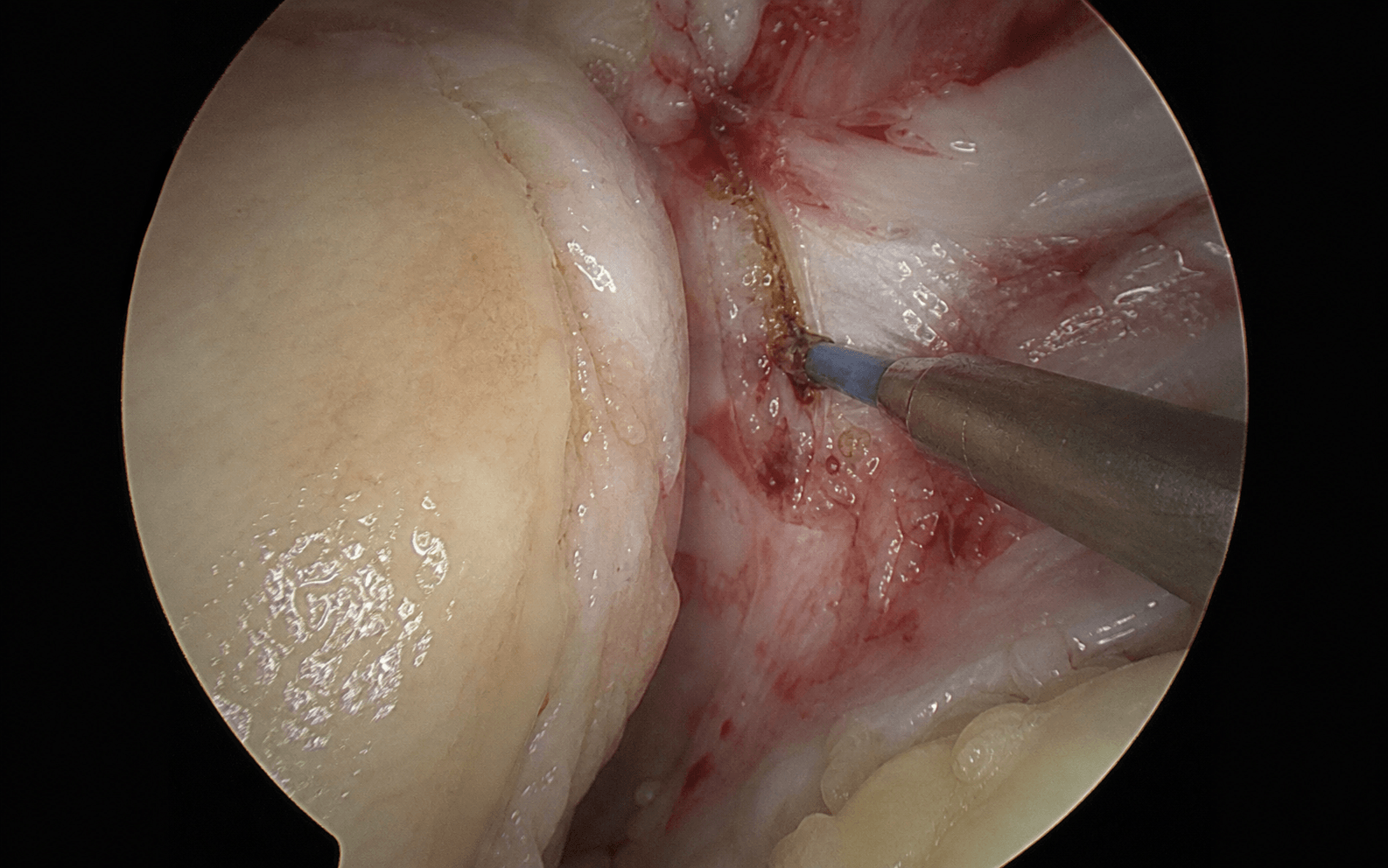
Surgical Imaging
The trap: applying a lateral release broadly for patellofemoral pain or instability — the historical error that produced inconsistent, often poor results.
The fix: release only for OBJECTIVE excessive lateral tilt with a tight lateral retinaculum (reduced medial glide, cannot evert) and a NON-subluxating patella with a negative lateral glide and no apprehension.
The trap: performing an isolated lateral release for recurrent patellar dislocation. Instability is MPFL deficiency and/or bony malalignment — a release corrects neither.
The fix: for instability, perform MPFL reconstruction and/or tibial tubercle transfer. An isolated release will not stabilise the knee and risks making it worse.
The mechanism: over-extensive release removes the lateral retinaculum's role as a secondary lateral stabiliser, allowing the patella to sublux medially — a difficult, painful problem.
The prevention: keep the indication narrow, prefer a lengthening over a complete release, spare the vastus lateralis tendon, and confirm intra-operatively that the patella cannot be dislocated medially.
The trap: dividing the superior lateral geniculate artery and vein at the superolateral corner without coagulation produces a tense post-operative haemarthrosis — the most common early complication.
The fix: identify and coagulate the geniculate leash at the proximal extent of the release, then re-confirm haemostasis under arthroscopic vision before closing.
The trap: cutting the vastus lateralis tendon at the superolateral corner of the patella weakens the extensor mechanism and removes a dynamic lateral stabiliser.
The fix: identify the white glistening vastus lateralis tendon first and keep the release anterior and inferior to it; carry the division distally from the superior pole along the lateral patellar border.
The trap: a complete release where a lengthening would do — leaving the patella without lateral restraint in a patient who may have any element of laxity.
The fix: where an isolated lateral procedure is needed, prefer a Z-lengthening of the lateral retinaculum: it corrects the tilt while preserving a continuous lateral restraint, protecting against medial instability.
L.A.T.E.R.A.LLATERAL — Indication and Assessment
D.A.N.G.E.RDANGER — Operative Dangers and Pitfalls
The Modern Indication — Narrow and Objective
The One Clear Indication
Lateral retinacular release (LRR) is indicated for the isolated, objectively tight lateral retinaculum producing excessive lateral patellar tilt without frank instability — historically termed excessive lateral pressure syndrome (ELPS) or lateral patellar compression syndrome (Ficat). A contracted lateral retinaculum holds the patella in lateral tilt, increasing lateral-facet contact pressure and producing lateral retropatellar pain and, over time, lateral-facet chondrosis.
Diagnostic Criteria (all should be present)
- Excessive lateral patellar tilt on axial imaging (Merchant view or CT): the patella is tilted laterally with a tight lateral facet against the lateral femoral condyle
- Objectively tight lateral retinaculum on examination: a reduced or absent medial glide (the patella cannot be translated medially the normal one to two quadrants) and a positive patellar tilt test (the lateral border cannot be lifted/everted to a normal angle)
- No lateral instability: a non-subluxating patella with a negative lateral glide and no apprehension — the patella does not translate laterally or dislocate
- Lateral-facet overload on MRI (lateral-facet subchondral bone-marrow oedema, lateral-facet chondral loss) consistent with chronic lateral compression
- Normal bony alignment: no significant trochlear dysplasia, a normal TT-TG distance, and normal patellar height — that is, instability morphology is excluded
What LRR Does NOT Treat
- Frank lateral patellar instability (recurrent dislocation or subluxation). The defining pathology is a deficient medial restraint (the MPFL) and/or bony malalignment, neither of which a lateral release corrects. The correct operation is MPFL reconstruction and, where the tibial tubercle is laterally positioned (increased TT-TG) or there is patella alta, a tibial tubercle transfer (osteotomy).
- Nonspecific anterior knee pain without objective lateral tilt. The historical broad use of LRR for anterior knee pain produced inconsistent, often poor results and is not recommended.
Indications
Absolute Indications
- Symptomatic excessive lateral pressure syndrome with documented lateral tilt, an objectively tight lateral retinaculum, and no instability, that has failed an adequate non-operative programme (activity modification, quadriceps and vastus medialis obliquus rehabilitation, iliotibial band stretching, patellar taping or bracing over three to six months)
Relative Indications
- An adjunct to a primary instability procedure (MPFL reconstruction or tibial tubercle transfer) where genuine objective lateral tightness would otherwise prevent central patellar tracking — increasingly performed as a lengthening rather than a complete release
- Lateral retinacular pain with documented tilt in the setting of lateral-facet patellofemoral arthrosis
Contraindications
Absolute:
- Patellar instability as the primary problem treated by an isolated LRR — the classic error. Release does not stabilise the knee and can create iatrogenic medial instability
- Normal patellar tilt with no objective lateral tightness
- Active septic arthritis
Relative:
- Generalised ligamentous laxity or multidirectional patellar laxity — high risk of over-release and medial instability
- Complex regional pain syndrome tendency or hypersensitivity conditions
- Inflammatory arthropathy in an active flare (optimise medical management first)
Diagnostic Workup
Clinical Examination
- Patellar tilt test: with the knee extended and the quadriceps relaxed, attempt to lift the lateral border of the patella. Normally the patella can be tilted so the lateral border rises to near or above the horizontal. A fixed lateral tilt that cannot be corrected indicates a tight lateral retinaculum
- Medial glide test: translate the patella medially. Normal medial translation is about one to two quadrants of patellar width. A markedly reduced or absent medial glide confirms lateral tightness
- Lateral glide and apprehension: translate the patella laterally. Excessive lateral translation with apprehension indicates instability — a contraindication to isolated release
- Compression or grind (Clarke's) test: reproduces retropatellar pain in patellofemoral overload
- Ober's test: assesses for iliotibial band tightness, which contributes to lateral retinacular tension
Imaging
- Merchant (axial) view at 45 degrees flexion: assess tilt and subluxation, and the sulcus and congruence angles
- Laurin lateral patellofemoral angle: a line along the lateral patellar facet and a line across the anterior femoral condyles. The angle normally opens (diverges) laterally; parallel lines, or an angle opening medially, indicate pathological lateral tilt
- CT patellar tracking at 0, 15, 30 and 45 degrees flexion to confirm persistent tilt through a flexion arc
- MRI: lateral-facet subchondral oedema and chondral loss consistent with overload, and critically to assess the MPFL, trochlear morphology and sulcus to exclude instability causes
Excluding Instability Morphology
The whole premise of an isolated release is that instability is absent, so the following should be normal:
- TT-TG distance: less than 20 mm (a value greater than 20 mm is abnormal and points toward instability)
- Patellar height: Insall-Salvati ratio less than 1.2 (a ratio greater than 1.2 indicates patella alta, an instability factor)
- Trochlear morphology: no high-grade dysplasia (sulcus angle less than 145 degrees; a crossing sign indicates dysplasia)
- Q angle: less than 15 degrees in men and less than 20 degrees in women
Evidence
Non-Operative Treatment
- Most patellofemoral pain — including mild tilt — responds to a structured programme of quadriceps and hip-abductor strengthening, vastus medialis obliquus retraining, iliotibial band stretching, activity modification and patellar taping or bracing
- LRR should be reserved for the small subgroup with objective lateral tilt and a tight lateral retinaculum who fail a documented three- to six-month non-operative programme
Surgical Results and the Shift Away From Broad LRR
- Early enthusiasm for LRR across a broad range of patellofemoral complaints gave way to recognition that results are unpredictable unless the indication is restricted to true excessive lateral pressure syndrome
- For instability specifically, isolated LRR has been associated with high recurrence and inferior outcomes compared with medial-side reconstruction; modern algorithms treat instability with MPFL reconstruction and/or tibial tubercle transfer
- Over-release producing iatrogenic medial patellar instability is now a recognised and difficult complication, driving the trend toward lateral retinacular lengthening over complete release
Patellofemoral Problem → Correct Operation
Clinical Decision Scenarios
Practise clinical reasoning and management decisions out loud
“A 24-year-old woman is referred requesting a lateral release for recurrent patellar dislocation — she has dislocated her right patella three times, most recently six weeks ago. Her MRI shows a deficient medial patellofemoral ligament and a TT-TG distance of 22 mm. How do you counsel her about the lateral release she is asking for?”
“A 38-year-old runner has a two-year history of lateral retropatellar pain. Examination shows a reduced medial glide and a fixed lateral tilt you cannot evert; there is no apprehension. Her axial radiograph shows lateral patellar tilt with a Laurin angle that opens medially; MRI shows lateral-facet subchondral oedema with an intact MPFL and a normal TT-TG. How do you manage her?”
“A 30-year-old woman is referred six months after a lateral retinacular release done elsewhere for patellofemoral pain. She now describes a new sensation of the patella falling inward, medial-sided knee pain and giving way. Examination shows the patella can be subluxed medially with little resistance. What has happened and how do you manage her?”