Dorsal intermetatarsal approach · central metatarsals (2nd–4th) · intermediate
- Operate for displacement greater than 3-4mm in any plane, angulation greater than 10 degrees (especially plantar), multiple metatarsal fractures with loss of transverse arch stability, open fractures, irreducible fractures with soft-tissue interposition, or an associated Lisfranc component.
- The dorsal intermetatarsal approach exploits the interval between metatarsals — it avoids the extensor tendons overlying each shaft and gives access to both adjacent metatarsals through one incision.
- The dorsalis pedis artery runs between the 1st and 2nd metatarsals, 2-3cm deep to the skin — identify and protect it before any dissection in that interspace. Superficial peroneal nerve branches cross the dorsum with variable anatomy, 1-2cm deep.
- Rotational assessment is critical and easily missed on X-ray: when reduced, all toes must point in the same direction and the metatarsal head must align with the base — compare to the contralateral foot.
- Under ground-reaction load the metatarsal bends apex-dorsal, so the plantar cortex is the tension side. A dorsal plate works best as a neutralization or bridge plate over a reconstructed plantar cortex — restore plantar cortical contact to resist collapse.
When & Why
Indication. A displaced fracture of a central metatarsal (2nd, 3rd or 4th) shaft, neck or head that meets an operative threshold — displacement greater than 3-4mm in any plane, angulation greater than 10 degrees (plantar angulation is the least tolerated), multiple metatarsal fractures with loss of transverse arch stability, open fractures requiring debridement and stabilization, irreducible fractures with soft-tissue interposition, or a fracture with an associated Lisfranc (tarsometatarsal) component. Minimally displaced fractures that meet none of these thresholds are managed non-operatively. Assess the whole forefoot, not just the one line on the X-ray. Before committing, obtain AP, lateral and oblique foot radiographs — all three are essential — and on them measure displacement, angulation, rotation and look for adjacent metatarsal and tarsometatarsal injury:
- Weight-bearing views (if the patient can stand) reveal true displacement and arch disruption that non-standing films underestimate.
- CT for base fractures — a central-ray base fracture must be screened for a Lisfranc injury pattern; weight-bearing CT improves detection of subtle tarsometatarsal injuries.
- Compare rotation to the contralateral foot — rotational malreduction is invisible on plain X-ray but causes crossed toes. The fixation decision. The exposure is the same regardless of implant; the choice of fixation follows the fracture pattern and the soft tissues:
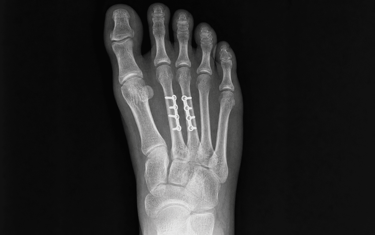
Low-profile 2.0-2.4mm dorsal locking plate over a reconstructed plantar cortex, 6-8 cortices minimum. The benchmark for displaced shaft and neck fractures — reliable union and a low symptomatic-removal rate.
Best for minimally comminuted 2nd-4th shaft fractures wanting buried hardware. A 2.7-3.5mm screw (or elastic nail) suits the slender central-ray canal; minimal dissection, no removal, but limited rotational control.
When the soft tissues preclude a plate, for temporary fixation in polytrauma, or in children. Crossed 1.6-2.0mm wires give less stability, need pin-site care and are removed at 4-6 weeks.
Consent specifically for nonunion, malunion (particularly rotational malunion and transfer metatarsalgia from plantar angulation), hardware prominence and shoe irritation, wound infection, superficial peroneal nerve injury with dorsal numbness or a painful neuroma, MTP stiffness, and — in high-energy patterns — compartment syndrome. Setup. Supine with a bump under the ipsilateral hip, thigh tourniquet (280-300mmHg), foot at the end of the table or on a radiolucent arm board so fluoroscopy reaches the foot easily. Loupe magnification helps identify and protect the dorsal cutaneous nerves.
The Operation
The goal is to restore length, alignment and rotation of the central metatarsal through the dorsal intermetatarsal interval — protecting the dorsalis pedis artery, the superficial and deep peroneal nerves and the extensor tendons — and to hold that reduction with a low-profile dorsal plate (or an intramedullary screw / K-wires where the pattern demands) so the metatarsal parabola is preserved and metatarsalgia avoided. The exposure is laid out in full below (and in depth on the dorsal approach to the lesser metatarsals page).
Operative sequence
- Supine, ipsilateral hip bump, thigh tourniquet at 280-300mmHg, foot at the end of the table on a radiolucent board for fluoroscopy.
- Palpate and mark the metatarsal shafts, the fracture site, the intermetatarsal intervals and the dorsalis pedis pulse (between the 1st and 2nd metatarsal bases).
- Confirm AP, lateral and oblique foot views; measure displacement (greater than 3-4mm = operative), angulation (greater than 10 degrees = operative) and rotation, and look for multiple-ray and Lisfranc involvement.
- Weight-bearing films show true displacement and arch disruption; CT a base fracture to rule out a Lisfranc injury.
- Plan fixation by pattern: transverse or comminuted shaft (plate), long oblique (lag screw plus neutralization plate), minimally comminuted shaft (intramedullary screw), soft-tissue compromise (K-wires).
- A dorsal longitudinal incision in the intermetatarsal space, 4-6cm long, centered over the fracture. It sits between the extensor tendons rather than over a shaft, so it avoids them and can reach two adjacent metatarsals.
- By ray: 2nd metatarsal — between 1st and 2nd (protect dorsalis pedis); 3rd — between 2nd and 3rd, or 3rd and 4th; 4th — between 3rd and 4th (preferred).
- For multiple fractures, keep skin bridges of at least 3-4cm between parallel incisions to prevent necrosis; a single midline incision can fix simultaneous 2nd and 3rd metatarsal fractures through subperiosteal dissection.
- Incise skin sharply to subcutaneous tissue; identify and protect the superficial peroneal nerve branches crossing the dorsum with variable anatomy, 1-2cm deep. Cut ends retract and form painful neuromas — use loupe magnification and retract gently (or vessel-loop them).
- Identify the extensor digitorum longus (EDL) tendons overlying each metatarsal and retract them laterally — do not transect.
- Elevate the dorsal interosseous muscles subperiosteally off the fracture site. Minimal periosteal stripping preserves blood supply and prevents nonunion.
- Between the 1st and 2nd metatarsal, the deep peroneal nerve runs with the dorsalis pedis artery at 2-3cm depth — protect them during deep dissection in that interval. The plantar neurovascular bundle lies 8-10mm plantar to the shaft and is protected by keeping the dissection on the dorsal surface and maintaining the plantar periosteal sleeve.
- Complete exposure of the fracture by subperiosteal dissection; evacuate the fracture hematoma.
- Identify the fracture pattern — transverse, short oblique, long oblique or comminuted — because it drives fixation: simple two-part patterns differ from comminuted patterns needing bridge plating.
- Restore length with pointed reduction clamps, temporary K-wires or longitudinal traction for impacted/shortened fractures.
- Restore alignment in sagittal and coronal planes: excessive plantar angulation causes metatarsalgia under that head; dorsal angulation transfers load to the adjacent metatarsal.
- Rotation is critical and easily missed on X-ray. Align the metatarsal head with the base orientation so all toes point the same direction, and compare to the contralateral foot — rotational malunion causes crossed toes and does not remodel in adults.
- A 2.0mm or 2.4mm low-profile dorsal locking plate (mini-fragment 2.7mm system also usable), contoured to the shaft, with 3-4 screws (6-8 cortices) each side of the fracture.
- The plantar cortex is the tension side during gait, so the dorsal plate is a neutralization plate over a lag screw in simple oblique patterns, or a bridge plate spanning comminution (do not compress comminuted fragments). Restoring plantar cortical contact is what resists collapse.
- For simple oblique patterns place the interfragmentary lag screw first, then the neutralization plate. Use locking screws in osteoporotic bone; drill perpendicular to bone and check depth so screws do not penetrate the adjacent interspace.
- Intramedullary screw for minimally comminuted 2nd-4th shaft fractures wanting buried hardware: achieve reduction first (closed or mini-open), enter at the base (antegrade) or head (retrograde), match the slender canal with a 2.7-3.5mm screw or elastic titanium nail (reserve larger 4.0-4.5mm screws for the broader fifth metatarsal/Jones), bury the head beneath the cortex, confirm on fluoroscopy. Advantages: minimal dissection, buried hardware, no removal. Disadvantages: limited rotational control, technically demanding, oversized screws risk cortical blow-out.
- K-wires when soft tissues preclude a plate or for temporary fixation: 1.6-2.0mm wires crossed (two or three) for rotational stability, inserted antegrade from the fracture site or retrograde from the head; bend and cut outside the skin for later removal at 4-6 weeks. Disadvantages: pin-site infection risk, less stable than a plate, loss of reduction before removal.
- Multiple metatarsals: fix the central rays (2nd/3rd) first — they are the keystone of the transverse arch and have the least mobility — then the more mobile lateral rays (4th/5th). For three or more fractures with severe soft-tissue injury, consider damage control with an external fixator acutely and staged ORIF once soft tissues recover; maintain a high index of suspicion for compartment syndrome.
- Neck fractures often have apex-plantar angulation causing pseudo-shortening and metatarsalgia; options are plantar-to-dorsal lag screws, a dorsal plate, or K-wires. Head fractures are often intra-articular and need anatomic reduction (mini-plate, headless compression screws buried beneath cartilage, or K-wires) to prevent MTP arthritis; the head has a limited blood supply, so minimize stripping (AVN risk).
- Check reduction on AP, lateral and oblique views: no rotation (all heads aligned, toes parallel), alignment restored in all planes, no intra-articular screw penetration into the TMT or MTP joints, and an acceptable (not prominent) plate profile.
- If the patient is awake with an ankle block, simulate weight-bearing under fluoroscopy to assess stability.
- Irrigate copiously; release the tourniquet and achieve meticulous hemostasis with bipolar cautery, confirming no vascular compromise to the toes.
- Close in layers: re-approximate the dorsal interossei if elevated (2-0 absorbable), subcutaneous layer (3-0 absorbable), skin (4-0 monofilament or subcuticular).
- Apply a soft dressing with gentle compression or a posterior splint in neutral for comfort, and elevate strictly post-operatively to minimize swelling and wound complications.
Before any deep dissection in the 1st-2nd interspace, identify the dorsalis pedis artery (palpable on the dorsum, running between the 1st and 2nd metatarsals, 2-3cm deep) and the deep peroneal nerve that accompanies it, and protect them. The superficial peroneal nerve branches cross the whole dorsum with variable anatomy, only 1-2cm deep, and their cut ends retract and form painful neuromas — use loupe magnification, identify and retract them, and never transect. Keep dissection on the dorsal surface to leave the plantar neurovascular bundle (8-10mm plantar to the shaft) protected within its periosteal sleeve.
Rotation is the reduction that is invisible on X-ray and the one examiners will ask about. Align the metatarsal head with the base so all toes point the same direction, and compare to the contralateral foot. Rotational malunion causes crossed toes, does not remodel in adults, and demands revision — prevent it with careful intra-operative clinical alignment, not fluoroscopy alone.
Plantar angulation greater than 10 degrees functionally shortens the ray (the head sits lower and more prominent) and drives metatarsalgia under that head; dorsal angulation shortens the ray radiographically and transfers load to the adjacent metatarsal (a transfer lesion). Either way the metatarsal parabola — 2nd longest, then 1st and 3rd progressively shorter — is disrupted. Restore sagittal alignment and length to preserve the parabola.
Aftercare & Complications
Rehabilitation | Phase | Timing | Immobilisation | Therapy | |-------|--------|-----------------|---------| | 1 | Day 0-1 | Soft dressing or posterior splint | Neurovascular check; strict elevation; begin gentle toe range of motion immediately | | 2 | Weeks 0-6 | Non-weight-bearing in a CAM boot or cast | Continue toe ROM; wound check and suture removal at 2 weeks; serial X-rays at 2, 4 and 6 weeks | | 3 | Weeks 6-8 | Transition to protected weight-bearing | Progressive weight-bearing as radiographic healing allows; physiotherapy for gait re-education | | 4 | Weeks 8-12 | Supportive athletic shoe | Advance to full weight-bearing; return to normal activities as tolerated | Hardware removal is rarely needed unless prominent and symptomatic, and then only after 12-16 weeks. Intramedullary screws and buried hardware can remain permanently. Most patients return to normal activities around 10-12 weeks and to sport at 3-4 months. Complications
- Recognition
- Crossed toes, toe overlap, pain with shoe wear; easily missed on X-ray — compare to the contralateral foot
- Prevention
- Careful intra-operative rotational assessment; all toes point the same direction; compare to the contralateral foot
- Management
- Early: revision ORIF within 6 weeks. Late: corrective osteotomy once malunion is established at 3-6 months
- Recognition
- Pain under adjacent metatarsal heads during gait with plantar callus, worse walking
- Prevention
- Avoid plantar angulation greater than 10 degrees; restore metatarsal length and sagittal alignment
- Management
- Conservative first — metatarsal pad, rocker-bottom shoe; surgical — corrective osteotomy to restore the parabola
- Recognition
- Persistent pain at 3-6 months with no bridging callus on X-ray; possible hardware failure
- Prevention
- Adequate fixation (6-8 cortices); minimal periosteal stripping; avoid excessive soft-tissue dissection
- Management
- Revision ORIF with bone graft; consider IM screw or plate augmentation; rule out infection first
- Recognition
- Shoe irritation over the dorsal plate, pain with tight shoes, localized tenderness
- Prevention
- Low-profile 2.0-2.4mm plates; bury screw heads; consider an IM screw for buried fixation
- Management
- Symptomatic — remove after union (12-16 weeks). Asymptomatic — leave in situ
- Recognition
- Dorsal foot numbness, painful neuroma, positive Tinel sign, hypersensitivity
- Prevention
- Identify and protect branches under loupe magnification; avoid transection; gentle retraction only
- Management
- Observation for neuropraxia (3-6 months); neuroma excision and burial if symptomatic; gabapentin for neuropathic pain
- Recognition
- Wound erythema, drainage, fever, raised inflammatory markers, purulent discharge
- Prevention
- Meticulous sterile technique; prophylactic antibiotics; minimize soft-tissue trauma; tension-free closure
- Management
- Superficial — antibiotics and wound care. Deep — washout and debridement, retain hardware if stable, remove if loose
- Recognition
- Reduced toe motion (especially dorsiflexion), pain with passive motion
- Prevention
- Early toe ROM exercises from day 1-2; avoid immobilizing the MTP joints; physiotherapy
- Management
- Gentle manipulation and physiotherapy; rarely MTP arthrolysis; usually well tolerated
Viva & Exam Focus
DORSALDORSAL — indications for ORIF of a central metatarsal
PIMPPIMP — fixation options by pattern
Between the 1st and 2nd metatarsals, palpable on the dorsum, 2-3cm from the skin, running with the deep peroneal nerve — injury causes vascular compromise. Identify and protect before deep dissection in that interspace.
Dorsal branches cross the metatarsals with variable anatomy, 1-2cm deep to the skin — injury causes dorsal foot numbness and a painful neuroma. Identify under loupe magnification and retract gently.
Runs with the dorsalis pedis between the 1st and 2nd metatarsals at 2-3cm depth — injury weakens the toe extensors. Protect during deep dissection in the 1st-2nd interspace.
Tendons overlie each metatarsal shaft and must be retracted laterally, not transected — injury causes toe drop (extension weakness).
Runs 8-10mm plantar to the metatarsal shaft — protected by staying on the dorsal surface and maintaining the plantar periosteal sleeve.
Clinical Decision Scenarios
Practise clinical reasoning and management decisions out loud
“A 35-year-old construction worker sustains a displaced 2nd metatarsal shaft fracture with 5mm displacement and 15 degrees plantar angulation after a heavy object fell on his foot. Walk me through your management.”
“You are fixing a 3rd metatarsal fracture and notice that despite anatomic reduction on AP and lateral views, the 3rd toe is crossing over the 2nd toe. What is the problem and how do you fix it?”
“A 42-year-old presents with pain under the 2nd metatarsal head 3 months after ORIF of a 3rd metatarsal shaft fracture that has united. What is the likely cause and how do you prevent it?”
Indications
- Displacement greater than 3-4mm in any plane
- Angulation greater than 10 degrees (especially plantar)
- Multiple metatarsal fractures with arch instability
- Open fractures requiring debridement and stabilization
- Irreducible fractures with soft-tissue interposition
- Fractures with an associated Lisfranc component
Key anatomy
- Superficial peroneal nerve — dorsal branches cross the metatarsals, 1-2cm deep, variable anatomy
- Dorsalis pedis artery — between 1st and 2nd metatarsal, 2-3cm deep, palpable on the dorsum
- Deep peroneal nerve — accompanies dorsalis pedis, supplies EDB and toe extensors
- Extensor tendons — EDL overlies each metatarsal, retract do not transect
- Plantar neurovascular bundle — 8-10mm plantar to the shaft, protected by the periosteal sleeve
- Central rays (2nd/3rd) — keystone of the arch, least mobile, fixed at the TMT joints
Critical steps
- Three-view imaging — AP, lateral, oblique; assess displacement, angulation, rotation
- Dorsal intermetatarsal approach — between metatarsals, protects extensor tendons
- Protect dorsalis pedis (1st-2nd, 2-3cm deep) and the superficial peroneal nerve
- Anatomic reduction — restore length, alignment, rotation; all toes point the same direction
- Dorsal plate fixation — 2.0-2.4mm low-profile, 6-8 cortices minimum
- Alternatives — IM screw (buried, shaft) or K-wires (temporary)
- Fluoroscopic verification — three views, confirm rotation, no intra-articular screws
Danger zones
- Dorsalis pedis artery (1st-2nd, 2-3cm deep) — palpate and protect
- Superficial peroneal nerve (dorsal branches, variable, 1-2cm deep) — identify and retract
- Deep peroneal nerve (with dorsalis pedis) — protect in deep dissection
- Extensor tendons (overlie each metatarsal) — retract laterally, do not transect
- Plantar neurovascular bundle (8-10mm plantar) — maintain the periosteal sleeve
Technique pearls
- ROTATION is critical — all toes point the same direction, compare to the contralateral
- Plantar cortex is the tension side — dorsal plate acts as a neutralization plate
- Plantar angulation greater than 10 degrees causes metatarsalgia from pseudo-shortening
- Multiple metatarsals — fix central rays (2nd/3rd) first, then lateral rays
- Skin bridges greater than 3-4cm between parallel incisions prevent necrosis
- 6-8 cortices minimum for stable shaft fixation
- IM screw for shaft fractures — buried, minimal dissection
Complications
- Rotational malunion — crossed toes, easily missed on X-ray, requires revision
- Transfer metatarsalgia — from plantar angulation or shortening, disrupts the parabola
- Nonunion — inadequate fixation or excessive stripping, needs revision with graft
- Hardware prominence — low-profile plates, consider IM screw, remove if symptomatic
- Nerve injury — superficial peroneal causes neuroma, identify and protect
- Infection — superficial vs deep, retain stable hardware, remove if loose
- MTP stiffness — early ROM exercises, usually well tolerated
Post-op protocol
- Immediate — elevation, neurovascular check, toe ROM from day 1
- Week 0-6 — NWB in CAM boot, serial X-rays at 2, 4, 6 weeks
- Week 6-8 — protected weight-bearing based on radiographic healing
- Week 8-12 — progress to full weight-bearing, supportive shoe
- Hardware removal — rarely needed unless prominent, after 12-16 weeks minimum
- Return to activity — 10-12 weeks normal activities, 3-4 months for sport
Exam tips
- Emphasize ROTATION assessment — the examiner will ask about it
- Demonstrate dorsalis pedis location and protection in the 1st-2nd interspace
- Explain why plantar angulation is problematic (metatarsalgia from pseudo-shortening)
- Discuss fixation options: plate (standard), IM screw (buried), K-wire (temporary)
- Know the sequence for multiple metatarsals: central rays first (2nd/3rd keystone)
- High-yield complications: rotational malunion and transfer metatarsalgia
- Describe the metatarsal parabola: 2nd longest, then 1st and 3rd progressively shorter
Background & Evidence
Epidemiology. Metatarsal fractures are common, and the central rays are frequently injured together. In a prospective series of 355 patients with 411 metatarsal fractures (mean age 42 years), the fifth metatarsal was the most commonly fractured ray with a female predominance in older age groups — critically, multiple fractures occur in contiguous rays: 63 percent of third metatarsal fractures had an associated second or fourth metatarsal fracture. The practical implication is that a central-ray fracture should prompt a deliberate search for adjacent metatarsal injury, because contiguous multi-ray patterns are the rule rather than the exception and they change fixation sequencing. Pathoanatomy — the metatarsal parabola. The metatarsals form a parabola in which the 2nd metatarsal is the longest, then the 1st and 3rd are progressively shorter. During gait the metatarsal bends apex-dorsal under ground-reaction load, so the plantar cortex is the tension side and the dorsal cortex is loaded in compression. This is why a dorsally applied plate functions as a neutralization or bridge plate over a reconstructed plantar cortex, and why plantar angulation is so poorly tolerated: it functionally shortens the ray (pseudo-shortening), drives the head plantarward and produces metatarsalgia under that head, while dorsal angulation radiographically shortens the ray and transfers load to the adjacent metatarsal (a transfer lesion). Restoring length and sagittal alignment preserves the parabola and is the whole point of fixation. Key evidence. Low-profile dorsal plating is the well-supported default: in 45 patients with 75 first-to-fourth metatarsal shaft and neck fractures treated with dorsal plates (Bryant, 2018), union was 100 percent at a mean 10.9 weeks with full weight-bearing at 7.5 weeks, low residual angulation, and no patient requesting plate removal. Minimally invasive intramedullary fixation is a valid alternative for short shaft and neck fractures — titanium elastic nailing of 22 such fractures (Hettchen, 2015) gave a mean AOFAS midfoot score of 93.9 with no nonunion, secondary displacement or nail breakage. Contemporary technique (Kamin, 2021) emphasises restoration of length, axis, rotation and joint position with 2.0-2.4mm interlocking plates and lag-screw compression where the pattern permits, preserving the metatarsal index. Finally, because base fractures accompany Lisfranc injuries, they must be screened with weight-bearing imaging or CT: a missed tarsometatarsal injury and non-anatomic reduction are the dominant drivers of poor outcome (Ahluwalia, 2022).
References
The epidemiology of metatarsal fractures
- Prospective series of 355 patients with 411 metatarsal fractures over 1 year; mean age 42 years
- Fifth metatarsal was the most commonly fractured ray; female predominance in older age groups
- Multiple fractures occur in contiguous rays — 63% of third metatarsal fractures had an associated second or fourth metatarsal fracture
Union Rate and Rate of Hardware Removal Following Plate Fixation of Metatarsal Shaft and Neck Fractures
- 45 patients, 75 first-to-fourth metatarsal shaft and neck fractures treated with dorsal plate fixation
- 100% union with mean time to union 10.9 weeks and full weight-bearing at 7.5 weeks; residual coronal 3.9 and sagittal 2.2 degrees angulation
- No patient underwent plate removal and 26 of 27 surveyed did not want the plate removed
Fixation of displaced fifth metatarsal shaft and neck fractures
- Operative goal is restoration of length, axis, rotation and joint position while preserving the metatarsal index (Maestro curve)
- Interlocking plates with 2.0-2.4mm screws are preferred to minimise later soft-tissue irritation; interfragmentary lag screws used where fragments allow
- Reduction performed under longitudinal traction with reduction clamps and temporary K-wires; partial weight-bearing in orthosis for 6 weeks
Elastic Stable Intramedullary Nailing (ESIN) of Metatarsal Fractures
- 22 patients treated with antegrade titanium elastic nailing for short metatarsal shaft and neck fractures; 19 followed for mean 25.6 months
- Mean AOFAS midfoot score 93.9; no nonunion, secondary displacement or nail breakage
- Single case of skin irritation; no refracture after nail removal
Surgical controversies and current concepts in Lisfranc injuries
- Narrative evidence-based review; prognosis correlates with the number of foot columns injured and with quality of anatomical reduction
- Weight-bearing CT improves identification of subtle tarsometatarsal injuries that accompany base fractures
- Recent evidence favours dorsal plate fixation over trans-articular screws for better reduction quality; primary arthrodesis is a safe alternative in selected patients