Mature hyaline cartilage transferred in a single stage — mosaicplasty
- True hyaline cartilage transfer — Type II collagen is maintained, unlike the fibrocartilage (Type I) produced by microfracture
- Single-stage procedure — no cell culture and no second surgery, unlike ACI/MACI
- Donor site morbidity is the limiting factor — a maximum of 3-4 plugs, which caps the technique near 4cm squared
- Perpendicular placement of every plug is critical for survival and congruity
- Ideal size 1-4cm squared — larger than microfracture, smaller than the lesions suited to ACI or allograft
When & Why
OATS (osteochondral autograft transfer system), or mosaicplasty, transfers cylindrical osteochondral plugs — subchondral bone with its intact overlying hyaline cartilage — from a low-load donor area of the same knee into a focal chondral or osteochondral defect. It occupies the middle of the cartilage-restoration algorithm: it delivers the durability of true hyaline cartilage in a single stage for lesions too large for microfracture but still within donor-site capacity. Understanding where it sits relative to microfracture, ACI/MACI and osteochondral allograft is essential for exam success and for selecting the right patient.
A focal, full-thickness chondral or osteochondral defect of 1-4cm squared on the femoral condyle or trochlea — typically a single, contained lesion in a young, active patient, including defects that have failed microfracture.
Lesions over 4cm squared (donor limitation), diffuse osteoarthritis, bipolar (kissing) lesions, inflammatory arthropathy, uncorrected malalignment, and — relatively — patellofemoral defects.
Establish the mechanism (trauma versus insidious), the symptoms (mechanical catching or locking, pain), any previous cartilage treatment, the patient's activity demands and sport goals, and the lesion duration (acute versus chronic).
Look for an effusion, compartment tenderness and crepitus suggesting cartilage pathology; assess alignment; and rule out associated ligamentous instability.
Malalignment is a major risk factor for OATS failure. Always assess alignment clinically and with long-leg standing films if concerned. Consider a concomitant or staged osteotomy if significant varus or valgus is present — any resurfacing performed into an uncorrected mechanical axis will overload and fail.
Imaging protocol
Weight-bearing AP, lateral, Rosenberg (45 degree PA) and skyline views. Assess joint space, alignment and established osteoarthritic change. Chronic lesions may show subchondral change or cysts.
Assess lesion size, location, depth and containment. Evaluate subchondral bone integrity. Identify associated meniscal or ligamentous pathology. Assess donor-site availability.
Helpful for precise lesion sizing and bone-stock assessment, particularly in revision cases or complex geometries.
On MRI, evaluate lesion dimensions measured in two planes, depth (partial versus full-thickness), the subchondral bone (cysts, oedema), containment (stable shoulders) and donor-site availability. MRI can underestimate lesion size — always confirm the true dimensions at diagnostic arthroscopy before committing.
The Operation
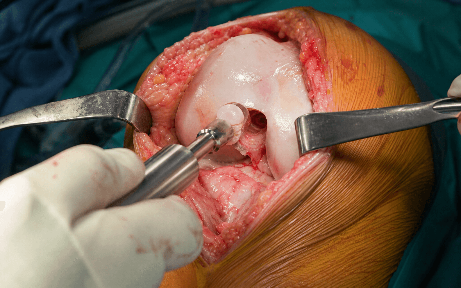
The goal is to fill a focal, contained chondral or osteochondral defect with one or more mature hyaline-cartilage plugs that sit flush, perpendicular and press-fit stable, restoring a congruent articular surface while keeping donor-site harvest within safe limits. The exposure is the rate-limiting step, so the operation is planned around achieving perpendicular access — laid out in the steps below.
The most important intra-operative decision is made at diagnostic arthroscopy: measure the defect with a calibrated probe in two planes, confirm it is focal (1-4cm squared), contained with stable shoulders, and not bipolar (kissing). If the lesion is larger than expected or bipolar, abandon OATS and convert to a different strategy (osteochondral allograft, ACI/MACI, or a staged plan with osteotomy) rather than over-harvesting the donor.
Operative sequence
- Position — supine, thigh tourniquet, leg in a holder or over a post so the knee can be flexed through a full range and the lesion brought into view.
- Anaesthesia/analgesia — GA or regional; the donor site is a recognised source of postoperative pain, so plan multimodal analgesia.
- Diagnostic arthroscopy first — probe and measure the lesion, assess the rest of the joint, and rule out bipolar disease and uncorrected instability or malalignment.
- Measure the defect with a calibrated probe in two planes and confirm it is focal (1-4cm squared), contained with stable shoulders, and not bipolar.
- If larger than expected or bipolar, abandon OATS and convert (osteochondral allograft, ACI/MACI, or a staged plan with osteotomy) rather than over-harvest the donor.
- Arthroscopic — feasible for small, well-positioned condylar lesions where a perpendicular trajectory can be achieved through a portal.
- Mini-arthrotomy (most common) — a limited medial or lateral parapatellar arthrotomy gives perpendicular access and the most reliable plug seating.
- Access is the rate-limiting step: choose the exposure that lets the recipient harvester sit perpendicular to the defect. The single reason to convert from arthroscopic to open is inability to keep the harvester perpendicular — a non-perpendicular tunnel produces an oblique plug, surface step-off and point loading. Plan the approach around perpendicular access, not cosmesis.
- Prepare the recipient socket first (per instrument system), drilling to a defined depth (commonly 15-20mm) with the tube harvester held strictly perpendicular.
- Create vertical walls and a fresh bleeding cancellous base; debride unstable cartilage back to a stable rim. Measure socket depth precisely.
- Donor sites (low-load, non-articulating) — the superolateral and superomedial margins of the trochlea (just above the sulcus terminalis, at the periphery of the patellofemoral contact zone) and the lateral wall of the intercondylar notch.
- Harvest perpendicular to the donor articular surface and slightly longer than the recipient socket so the plug can be trimmed and seated flush.
- Limit harvest to 3-4 plugs maximum — donor availability and morbidity, not the lesion alone, cap the technique near 4cm squared.
- Match donor curvature to recipient convexity and orient the plug's cartilage cap to recreate the local surface contour; a flatter trochlear plug placed into a more convex condyle creates a proud edge or a step.
- Depth — donor plug depth must equal recipient socket depth. Trim bone from the base if too long; never trim the cartilage cap.
- Height — seat flush or up to 1mm proud, never recessed. Recessed plugs are load-shielded and integrate poorly; excessively proud plugs (more than 1mm) cause peak contact stress and graft overload.
- Press-fit — an interference (press) fit provides primary stability without screws or supplemental fixation. Deliver with gentle, axial tamping; avoid repeated heavy impaction, which kills surface chondrocytes.
- Place the largest central plug first, then fill the periphery; aim for the highest practical fill.
- Residual 1-2mm gaps between plugs (commonly 10-20 percent of the defect) fill with fibrocartilage from the marrow — this is acceptable. Complete hyaline coverage is not required; aim for maximal practical hyaline fill while keeping every plug flush and perpendicular.
- Take the knee through a range of motion to confirm the plugs are stable, congruent and do not catch.
- Standard layered arthrotomy or portal closure.
- Document defect size, donor sites, number and diameter of plugs, and the final seating (flush versus proud).
Match donor curvature to recipient. A plug harvested from a flatter trochlear margin and placed into a more convex condyle creates a proud edge or a step. Harvest from a donor region whose convexity approximates the recipient and orient the plug's cartilage cap to recreate the local surface contour.
OATS needs no screws, wires or glue — an accurately sized interference (press) fit is the fixation. That makes gentle, axial delivery essential: repeated heavy impaction fractures the subchondral bone and kills the surface chondrocytes that make the graft worth transferring.
Aftercare & Complications
Rehabilitation Rehabilitation is built around protecting the bone-to-bone junction while it heals, then restoring motion and strength. OATS progresses faster than ACI because the mature bone-to-bone interface heals more predictably than grafted cells.
- Weight-bearing
- Toe-touch only (crutches)
- Rehabilitation
- CPM or early ROM; quad sets and straight-leg raises
- Goal
- Protect the bone-to-bone junction; gain motion
- Weight-bearing
- Progress to 50 percent, wean crutches by week 8
- Rehabilitation
- Progressive closed-chain strengthening; stationary bike when ROM allows; pool therapy
- Goal
- Full ROM; protected daily function
- Weight-bearing
- Full weight-bearing, no impact
- Rehabilitation
- Continue closed-chain strengthening; avoid open-chain loading
- Goal
- Return to normal gait and daily activity
- Weight-bearing
- Full
- Rehabilitation
- Jogging progression, agility drills, sport-specific training; no cutting or pivoting
- Goal
- Sport preparation
- Weight-bearing
- Full
- Rehabilitation
- Progressive return to cutting and pivoting sport if functional criteria met
- Goal
- Full return to sport (often faster than ACI)
OATS rehabilitation is generally faster than ACI because the mature bone-to-bone junction heals more predictably than ACI cell integration. Full weight-bearing is typically achieved by 8-12 weeks and full return to high-level sport is expected at 9-12 months.
Outcomes
- Good/excellent
- 90-95%
- Failure rate
- Under 5%
- Comment
- Early outcomes excellent
- Good/excellent
- 85-90%
- Failure rate
- 5-10%
- Comment
- Durability maintained
- Good/excellent
- 85%
- Failure rate
- 10-15%
- Comment
- Superior to microfracture
Unlike microfracture — which deteriorates at 2-5 years as its fibrocartilage repair degrades — OATS outcomes are durable to 10 years and beyond. This is attributed to the transfer of true hyaline cartilage with its mature Type II collagen matrix.
Complications
- Incidence
- 10-15%
- Risk factors
- Multiple plugs, large harvest
- Prevention and management
- Limit to 3-4 plugs; counsel preoperatively
- Incidence
- 5-10%
- Risk factors
- Poor press-fit, early loading
- Prevention and management
- Proper sizing, protected weight-bearing
- Incidence
- 5-10%
- Risk factors
- Malalignment, poor technique
- Prevention and management
- Address alignment; meticulous perpendicular placement
- Incidence
- 5-8%
- Risk factors
- Prolonged immobilisation
- Prevention and management
- Early ROM protocol
- Incidence
- Variable
- Risk factors
- Curvature mismatch
- Prevention and management
- Careful donor-recipient matching
The most common complaint after OATS is donor-site symptoms — anterior knee pain, crepitus, or pain on stair descent. This is why limiting harvest to 3-4 plugs maximum is essential. Counsel patients preoperatively that this is the expected, accepted morbidity of the technique.
Viva & Exam Focus
OATSOATS — key principles
Hook:OATS gives you breakfast in one serving — everything included in a single-stage procedure.
PLUGPLUG — placement principles
Hook:Put the PLUG in right — perpendicular, level and uniform.
Exam viva scenarios
Practise clinical reasoning and management decisions out loud
“A 32-year-old male athlete presents 18 months after microfracture for a 2cm squared medial femoral condyle lesion. He has persistent pain and MRI shows incomplete fill. What are your options?”
“During OATS for a 1.5cm squared medial femoral condyle lesion, you are about to insert the osteochondral plug. What are the critical technical points for plug placement?”
“A 28-year-old female has a 3.5cm squared lateral femoral condyle lesion with associated valgus malalignment. How do you approach this?”
Definition
- Transfer of bone plus hyaline cartilage plugs
- Same patient (autograft)
- Single-stage procedure
- True Type II collagen preserved
Key numbers
- 1-4cm squared = optimal lesion size
- 3-4 plugs = maximum safe harvest
- 6-10mm = common plug diameters
- 10-15% = donor-site morbidity
- 85-90% = good results at 10 years
Donor sites (TIN)
- Trochlear margins (supero-lateral/medial)
- Intercondylar notch walls
- Non-weight-bearing areas only
Plug placement (PLUG)
- Perpendicular to surface
- Level or 1mm proud (never recessed)
- Uniform depth matching
- Gaps fill with fibrocartilage
Advantages over microfracture
- Type II collagen (hyaline)
- Superior biomechanics
- Durable to 10+ years
- Better sport return rates
Limitations
- Donor-site morbidity (10-15%)
- Limited to lesions under 4cm squared
- Curvature matching required
- Patellar lesions challenging
Background & Evidence
Why hyaline matters. OATS transfers mature hyaline cartilage containing Type II collagen, proteoglycans and organised chondrocytes. This is biomechanically superior to the fibrocartilage (Type I collagen) produced by microfracture. Integration studies show 90-95 percent Type II collagen at one year with OATS, and the mature matrix is why outcomes remain durable at a decade where microfracture has typically failed.
- OATS (hyaline)
- Type II
- Microfracture (fibrocartilage)
- Type I
- OATS (hyaline)
- High (native-like)
- Microfracture (fibrocartilage)
- 50-80% of normal
- OATS (hyaline)
- Bone-to-bone plus cartilage
- Microfracture (fibrocartilage)
- Fibrous integration
- OATS (hyaline)
- Maintained at 10+ years
- Microfracture (fibrocartilage)
- Declines 2-5 years
- OATS (hyaline)
- Normal (65-80%)
- Microfracture (fibrocartilage)
- Reduced
- OATS
- Hyaline (Type II)
- Microfracture
- Fibrocartilage (Type I)
- ACI/MACI
- Hyaline-like
- OCA
- Hyaline
- OATS
- 1-4cm squared
- Microfracture
- Under 2cm squared
- ACI/MACI
- Over 2cm squared
- OCA
- Over 4cm squared
- OATS
- Single
- Microfracture
- Single
- ACI/MACI
- Two
- OCA
- Single
- OATS
- Yes (10-15%)
- Microfracture
- None
- ACI/MACI
- None (biopsy only)
- OCA
- None (allograft)
- OATS
- Low-moderate
- Microfracture
- Low
- ACI/MACI
- High
- OCA
- High
Classification by lesion size. Size, with the donor-site ceiling it imposes, is the single biggest driver of suitability.
- Plugs needed
- Single 6-8mm plug
- Suitability
- Excellent for OATS
- Plugs needed
- 1-2 plugs (8-10mm)
- Suitability
- Ideal for OATS
- Plugs needed
- 3-4 plugs (mosaicplasty)
- Suitability
- OATS acceptable, consider ACI
- Plugs needed
- Exceeds donor capacity
- Suitability
- ACI or allograft preferred
- OATS suitability
- Excellent
- Technical considerations
- Most common, easy access
- OATS suitability
- Excellent
- Technical considerations
- Good access, match curvature
- OATS suitability
- Good
- Technical considerations
- Requires careful contouring
- OATS suitability
- Moderate-poor
- Technical considerations
- Difficult access, thin bone
- OATS suitability
- Moderate
- Technical considerations
- Consider if contained
Plugs are taken from non-weight-bearing zones: the superolateral and superomedial trochlear margins (accessible, low-load), and the walls of the intercondylar notch (where larger plugs are possible).
Bone-to-bone healing takes 6-8 weeks; residual gaps fill with fibrocartilage; chondrocyte viability exceeds 90 percent; and the subchondral bone remodels by one year.
Guidelines, registries and global practice
ICRS cartilage consensus uses a size-based algorithm favouring OAT for small-to-medium focal defects (roughly 1-4cm squared), directing larger or bipolar lesions to allograft or ACI/MACI. NICE/NHS (UK) recommends ACI for larger eligible defects in specific pathways, with OAT a standard option for small focal lesions. AAOS (US) emphasises addressing malalignment, meniscal status and instability alongside any resurfacing. Practice converges globally: lesion size, location, alignment and prior treatment drive technique selection more than geography.
Document lesion size measured at arthroscopy (two planes), containment and bipolar status, donor site(s) used with number and diameter of plugs, final seating (flush versus proud), alignment and instability assessment, and consent including donor-site morbidity (anterior knee pain or crepitus).
The main international variation is access to fresh osteochondral allograft (OCA). Where established tissue banks exist (much of North America), OCA is a single-stage option for lesions over 4cm squared and for revisions; where allograft supply is limited (many European, Asian and Australasian centres), surgeons rely more on staged ACI/MACI or push mosaicplasty toward its upper limit. Availability and reimbursement of cell-based therapies (ACI/MACI) also vary by health system, but the cartilage-restoration algorithm itself is broadly shared.
References
Autologous Osteochondral Mosaicplasty - 10-Year Experience
- 831 patients; good/excellent results in 92% of femoral condylar, 87% tibial, 79% patellofemoral, 94% talar mosaicplasties
- Long-term donor-site morbidity (Bandi score) was 3%
- Second-look arthroscopy: congruent gliding surface with surviving hyaline cartilage and fibrocartilage filling of donor sites
- Complications: 4 deep infections and 36 painful postoperative haemarthroses
OAT vs Microfracture in Young Athletes - RCT
- 60 athletes (mean age 24.3y) randomised to OAT vs microfracture; 95% followed at mean 37 months
- Good/excellent (modified HSS and ICRS) in 96% OAT vs 52% microfracture (p less than 0.001)
- Return to preinjury sport: 93% (26/28) OAT vs 52% (15/29) microfracture
- 1 failure in the OAT group vs 9 in the microfracture group; biopsy showed better repair histology with OAT
OAT vs Microfracture - 10-Year RCT Follow-Up
- Same RCT cohort followed to mean 10.4 years (range 9-11)
- Failure at 10 years: 14% (4/28) OAT vs 38% (11/29) microfracture (p less than 0.05)
- OAT maintained significantly better ICRS and Tegner scores at 10 years
- Radiographic OA (Kellgren-Lawrence I): 25% OAT vs 48% microfracture (not significant)
ACI vs Osteochondral Cylinder Transplantation - Histology
- 40 patients with femoral condyle defects randomised to osteochondral cylinder transplantation vs ACI
- Osteochondral transplants retained their hyaline character on histology; ACI defects filled mainly with fibrocartilage
- Recovery (Lysholm) was faster after osteochondral transplantation than after ACI at 6, 12 and 24 months
- A persistent interface (gap) remained between transplant and host cartilage in all biopsied plugs
Return to Sport After Cartilage Surgery - Meta-Analysis
- 44 studies, 2549 athletes; overall return to sport 76% across all techniques
- Highest return after OAT (93%), then OCA (88%), ACI (82%), microfracture (58%)
- OAT had the fastest return to sport (5.2 months) vs microfracture (9.1), OCA (9.6) and ACI (11.8) (p less than 0.001)
- Age, lesion size and preoperative Tegner score were not significant determinants of return rate