Open or tubular minimally invasive posterior nerve root decompression without fusion | advanced
Surgical Imaging
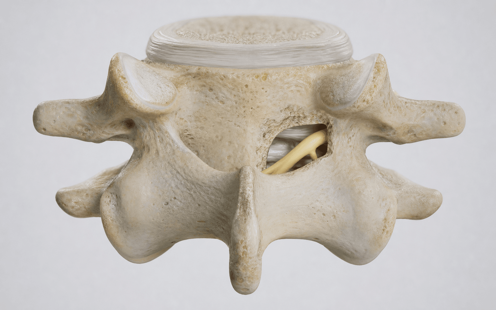
The trap: Drilling too aggressively through the medial facet during keyhole creation, removing more than 50 percent of the facet mass. This is the single most consequential error in posterior cervical foraminotomy because it produces a painful segmental instability that often requires unplanned posterior instrumentation and fusion.
The fix: Repeatedly palpate and visualise the lateral facet margin during drilling. Use a high-speed diamond burr and remove bone incrementally. Stop when the exiting nerve root is decompressed — do NOT continue drilling to 'clean up' the facet. If more than 50 percent of the facet must be removed to achieve decompression, abandon the foraminotomy and plan a fusion procedure instead.
The trap: The cervical dura is thin and tightly applied to the nerve root and posterior longitudinal ligament in the lateral recess. Aggressive Kerrison rongeur use against the dura, or drilling too deeply through the ligamentum flavum, can tear the dura and create a CSF leak.
The fix: Identify the ligamentum flavum and remove it carefully with Kerrison rongeurs under microscopic or loupe magnification before exposing the dura. Never advance a burr or rongeur blindly. If a durotomy occurs, primary repair with 6-0 Prolene, cover with a fat graft or fibrin sealant, and place the patient flat postoperatively. Consult neurosurgery for large tears.
The trap: The exiting nerve root lies directly in the surgical corridor. Over-retraction, thermal injury from the burr, or direct compression with instruments can cause a root injury ranging from neuropraxia to permanent radiculopathy. The spinal cord lies medial to the working area and is vulnerable to medial migration of instruments.
The fix: Perform all bone work under microscopic magnification. Irrigate continuously during burr use to prevent thermal necrosis. Never retract the nerve root forcefully — use gentle medial retraction with a Penfield dissector. Keep all instruments oriented laterally. Monitor somatosensory and motor evoked potentials throughout the procedure.
The trap: The posterior epidural venous plexus is prominent in the cervical spine. Bleeding from these veins obscures the surgical field, risks nerve root compression from a haematoma, and can be difficult to control in the confined keyhole corridor.
The fix: Apply bipolar diathermy to bleeding epidural veins before bone removal begins, where possible. Use thrombin-soaked Gelfoam and cottonoid patties. Work methodically — achieve haemostasis before each incremental step of bone removal. Maintain a clear field at all times. Have a Valsalva manoeuvre performed by the anaesthetist before closure to check for occult bleeding.
The trap: Unilateral foraminotomy is performed on the symptomatic side. Confusion about laterality — especially with patients referred with imaging labelled from a different institution or with non-standard radiological conventions — leads to wrong-side surgery.
The fix: Perform a formal time-out. Correlate symptoms (dermatomal distribution of pain and numbness, specific myotomal weakness) with imaging. Confirm the laterality with the patient: ask them to point to the side of their arm pain. Mark the surgical site before positioning and draping. Verify once more after the patient is prone, before incision.
The trap: Offering a posterior keyhole foraminotomy for a patient with central canal stenosis, myelopathy, or predominantly anterior compression. The keyhole approach decompresses only the lateral foramen and exiting root — it cannot safely address central canal narrowing or ventral cord compression.
The fix: Recognise the limitations of the approach. Central stenosis or myelopathy requires laminectomy with or without fusion, or an anterior corpectomy/discectomy depending on the pathology. Kyphotic deformity worsens with posterior-only surgery. Always assess the canal diameter, cord signal change, and sagittal alignment on preoperative MRI before selecting this approach.
K.E.Y.H.O.L.EKEYHOLE — Posterior Cervical Foraminotomy Principles
F.A.C.E.T.SFACETS — Patient Selection and Contraindications
R.O.O.T.SROOTS — Intraoperative Dangers
Surgical Indications
Absolute Indications
- Unilateral cervical radiculopathy from a posterolateral soft disc herniation with concordant symptoms and imaging, failing at least 6-12 weeks of conservative treatment (physiotherapy, NSAIDs, activity modification)
- Unilateral foraminal stenosis from an uncovertebral joint (Luschka) osteophyte compressing the exiting nerve root, with dermatomal pain and/or motor weakness
- Recurrent radiculopathy after prior ACDF at an adjacent level, where further anterior surgery is undesirable and the recurrent compression is posterolateral
Relative Indications
- Patient preference for motion-preserving surgery over fusion (avoids adjacent segment disease, preserves cervical range of motion, no graft or plate-related complications)
- Young patient (under 50) with a soft lateral disc where long-term fusion-related adjacent segment degeneration is a concern
- Prior anterior cervical surgery (scarring makes repeat anterior approach harder) where the new pathology is posterolateral and accessible posteriorly
- Contraindication to anterior surgery (prior irradiation, tracheostomy, severe cervical kyphosis precluding anterior exposure)
Contraindications
Absolute:
- Myelopathy from central canal stenosis or anterior cord compression — posterior foraminotomy decompresses only the lateral foramen and cannot address ventral cord compression
- Significant cervical kyphosis — posterior decompression removes posterior tension band elements and can worsen the kyphotic deformity
- Segmental instability on dynamic flexion-extension radiographs (greater than 3.5 mm translation or greater than 11 degrees angular motion)
- Pathology requiring resection of more than 50 percent of the facet joint — proceed to laminectomy with fusion or ACDF instead
Relative:
- Central disc herniation — ACDF provides direct anterior decompression and disc space restoration
- Bilateral radiculopathy — requires bilateral foraminotomies or alternative approach
- Predominant axial neck pain without radiculopathy — posterior foraminotomy targets the nerve root, not the disc or facet joint as pain generators; facet injection or medial branch blocks should be considered first
- Obesity with a thick posterior cervical soft tissue envelope — limits visualisation and increases retraction pressure in the minimally invasive tubular approach
- Prior posterior cervical surgery with scar tissue — increases difficulty of dissection and nerve root identification
Pathology and Rationale
Why Posterior and Not Anterior?
Posterior cervical foraminotomy accesses the nerve root from behind, drilling the lamina-facet junction to decompress the lateral foramen and remove a posterolateral disc fragment or foraminal osteophyte. The key advantage is that NO fusion is performed — the facet joint, disc, and motion segment are preserved.
This matters because:
- Adjacent segment disease: ACDF eliminates motion at the operated level and transfers biomechanical stress to adjacent segments, accelerating degeneration above and below the fusion over 10-20 years
- Fusion morbidity: ACDF carries risks of graft subsidence, plate/screw complications, dysphagia, dysphonia (recurrent laryngeal nerve injury), and pseudarthrosis
- Recovery: Foraminotomy patients typically return to work faster than ACDF patients because no bone graft healing or fusion is required
The posterior approach is ideal when the compressive pathology is posterolateral (lateral to the cord) and the compression can be reached by removing bone at the lamina-facet junction without violating the disc space anteriorly.
Best Pathology for Keyhole Foraminotomy
- Soft posterolateral disc herniation: the most common indication. A herniated nucleus pulposus fragment that has migrated postero-laterally into the neural foramen, compressing the exiting root. The fragment is accessible through the keyhole corridor and can be removed with pituitary forceps after bone decompression.
- Foraminal osteophyte: an uncovertebral joint (Luschka) osteophyte encroaching on the neural foramen from the anterior-lateral direction. These are harder than soft discs and may require more bone removal, increasing the risk of facet over-resection.
Pathology Better Suited to ACDF
- Central or paracentral disc herniation with cord compression or myelopathy
- Disc space collapse with loss of height and foraminal narrowing anteriorly
- Reversal of cervical lordosis or fixed kyphosis
- Discogenic axial neck pain (discography-positive) — foraminotomy does not address the disc
Evidence
Outcomes of Posterior Cervical Foraminotomy
Multiple case series and comparative studies report excellent outcomes for posterior cervical foraminotomy in appropriately selected patients:
- Clinical success rate: 85-97 percent good-to-excellent outcomes at short-to-medium term follow-up in appropriately selected patients with unilateral radiculopathy
- Arm pain relief: complete or near-complete resolution of radiculopathy in 85-95 percent of patients
- Nerve root recovery: motor strength recovery in patients with preoperative weakness is seen in 80-90 percent of cases within 6-12 months
- Return to work: median return to work 2-6 weeks (compared with 6-12 weeks after ACDF in most series)
- Reoperation rate: 5-10 percent at 5-10 years (primarily for recurrent disc herniation or persistent symptoms)
- Adjacent segment disease: NOT a concern (no fusion performed) — this is a long-term advantage over ACDF
Posterior Cervical Foraminotomy vs ACDF — Key Differences
Key Evidence
Surgical management of cervical soft disc herniation. A comparison between the anterior and posterior approach
Same-segment and adjacent-segment disease following posterior cervical foraminotomy
Minimally invasive cervical microendoscopic foraminotomy: an initial clinical experience
Complications, outcomes, and need for fusion after minimally invasive posterior cervical foraminotomy and microdiscectomy
Comparing mid-term outcomes between ACDF and minimally invasive posterior cervical foraminotomy in the treatment of cervical radiculopathy
Clinical Decision Scenarios
Practise clinical reasoning and management decisions out loud
“A 42-year-old man presents with a 3-month history of right C7 radiculopathy — shooting pain down the lateral forearm into the middle finger, with objective triceps weakness (4/5) and a diminished right triceps reflex. MRI shows a posterolateral soft disc herniation at C6-7 compressing the right C7 root in the foramen. His cervical alignment is lordotic. How do you counsel him and what operation would you offer?”
“During a posterior cervical keyhole foraminotomy at C5-6 for a left C6 radiculopathy, you have removed the lateral ligamentum flavum and identified the C6 root. As you continue drilling the medial facet to decompress the foramen, you notice a small amount of clear fluid welling up from the epidural space. What do you do?”
“You are performing a posterior cervical keyhole foraminotomy at C6-7. After initial bone removal, you realise that the foraminal osteophyte is larger than expected and achieving adequate root decompression will require removing approximately 60 percent of the facet joint. What do you do?”
References
-
Herkowitz HN, Kurz LT, Overholt DP (1990). Surgical management of cervical soft disc herniation: comparison between the anterior and posterior approach. Spine. — Comparative series demonstrating equivalent outcomes between posterior foraminotomy and ACDF for unilateral radiculopathy.
-
Fessler RG, Khoo LT (2002). Minimally invasive cervical foraminotomy. Spine. — Early series describing the tubular retractor technique for posterior cervical foraminotomy with excellent short-term outcomes.
-
Rao RD, David KS, Wang M (2011). Posterior cervical foraminotomy: a review of long-term outcomes. J Bone Joint Surg Am. — Long-term follow-up study with minimum 10-year data confirming durability of posterior foraminotomy when facet integrity is maintained.
-
Wimberley DW, Parent A, Balyan R, et al. (2013). Posterior foraminotomy versus anterior discectomy with fusion for cervical radiculopathy. J Spinal Disord Tech. — Retrospective comparative study confirming no significant outcome difference between the two approaches for unilateral radiculopathy.
-
Clarke MJ, Appropriateness study group (2007). Cervical foraminotomy: a review of indications and outcomes. Neurosurgery. — Systematic review identifying facet preservation (greater than 50 percent) as the critical technical principle and reporting 85-97 percent success rates in selected patients.