Arthroscopic reconstruction through revised tunnels, with optional staged bone grafting and anterolateral augmentation · advanced
- FIRST find out WHY it failed. Failure is usually multifactorial: technical error (above all femoral tunnel malposition), biological non-incorporation, traumatic re-injury, or unaddressed pathology (posterolateral corner, meniscal deficiency, malalignment). Re-operating without a cause dooms the revision to repeat failure.
- A CT scan is MANDATORY before every revision — assess tunnel position, widening and confluence with the proposed new tunnel. Reported two-stage thresholds cluster at 10 to 14 mm; bone-graft when no acceptable new aperture can be made.
- STAGE the operation: one-stage if the tunnels are well-positioned and small; two-stage bone grafting if massively widened, confluent, or infected.
- CHOOSE THE GRAFT by the patient. Autograft (contralateral bone-patellar tendon-bone, then quadriceps tendon) is preferred in young, high-demand patients — the MARS cohort shows allograft reruptures roughly twice as often in the under-25s.
When & Why
Indication. Revision ACL reconstruction is for the symptomatic failed primary reconstruction — recurrent instability with a positive Lachman and pivot shift, or graft rupture (traumatic or atraumatic) — where the patient has persistent giving-way that limits function. Before offering surgery you must explain why the first reconstruction failed, because re-operating without a diagnosis is the surest route to a second failure. Failure is multifactorial — diagnose the mechanism. Technical error, chiefly tunnel malposition, is repeatedly the single largest contributor in revision series, followed by traumatic re-injury and biological or unaddressed causes. Treat the relative frequencies below as approximate, not fixed.
- Cause
- Femoral tunnel malposition
- Relative frequency
- Leading technical cause
- Key features
- Too anterior or too vertical — a vertical graft that impinges
- Cause
- Tibial tunnel malposition
- Relative frequency
- Moderate
- Key features
- Too anterior causes extension impingement; too posterior causes flexion loss
- Cause
- Inadequate notchplasty
- Relative frequency
- Less common
- Key features
- Graft impingement on the notch roof
- Cause
- Failed incorporation
- Relative frequency
- Moderate
- Key features
- Graft intact but incompetent at 6 to 12 months
- Cause
- Re-injury
- Relative frequency
- Common
- Key features
- New trauma to a healed graft, often contact sport
- Cause
- Missed posterolateral corner injury
- Relative frequency
- Moderate
- Key features
- Chronic posterolateral instability and varus thrust
- Cause
- Untreated meniscal pathology
- Relative frequency
- Moderate
- Key features
- Meniscal deficiency increases graft load
- Cause
- Uncorrected malalignment
- Relative frequency
- Less common but high-impact
- Key features
- Varus alignment with posterolateral corner deficiency
Investigation protocol. Imaging and examination define the failure mechanism and the operative plan: - CT (mandatory): tunnel position, widening, and confluence risk with the proposed new tunnel — 3D reconstruction is invaluable for planning.
- MRI: graft integrity (competent versus failed), meniscal status, cartilage grade, and associated ligament injury.
- Stress radiographs: compare to the contralateral side for objective laxity.
- Clinical examination: pivot shift grade, varus and valgus opening, and the dial test for the posterolateral corner. The staging decision — read it off the CT. Whether the revision is one- or two-stage is decided before the incision, from the tunnels:
- Threshold
- Less than 12 mm
- Plan
- One-stage — use the existing tunnels
- Threshold
- Less than 14 mm with more than 2 mm rim
- Plan
- One-stage — drill new anatomic tunnels
- Threshold
- More than 14 to 16 mm, or less than 2 mm rim between old and new
- Plan
- Two-stage — bone graft, reconstruct at 3 to 6 months
- Threshold
- Any
- Plan
- Staged debridement, then delayed reconstruction
Graft selection — match the graft to the patient. The graft hierarchy runs from the strongest autograft to allograft, chosen against age, demand, and what has already been harvested.
- Advantages
- Gold-standard autograft; bone-to-bone healing
- Disadvantages
- Donor morbidity, anterior knee pain
- Best indication
- Young athlete, failed ipsilateral BPTB
- Advantages
- Strong, thick graft; low morbidity
- Disadvantages
- Learning curve, variable bone plug
- Best indication
- Failed hamstring primary, intact quad
- Advantages
- Familiar technique
- Disadvantages
- Less stiff than BPTB; donor morbidity
- Best indication
- Failed ipsilateral hamstring
- Advantages
- No donor morbidity; large size
- Disadvantages
- Higher failure in the young (MARS)
- Best indication
- Older, lower-demand, or multiligament knee
- Advantages
- Very strong; large
- Disadvantages
- Slower incorporation
- Best indication
- Massive tunnels, multiligament
MARS (Level 2): autograft was 2.78x less likely to rerupture than allograft at 2 years; at 6 years BTB autograft was 4.2x less likely than BTB allograft (3.5% vs 8.4%). Default to autograft in young, high-demand patients and reserve allograft for the older or lower-demand knee.
Consent for outcomes that are genuinely inferior to the primary reconstruction — higher re-failure, lower return to pre-injury sport, and a fall-off in activity as most patients down-regulate their sport. Counsel specifically on donor-site morbidity (anterior knee pain, patellar fracture), nerve injury (saphenous, peroneal), infection (1 to 2%), and stiffness. Setup. Supine with a thigh-mounted leg holder and the tourniquet high on the thigh, positioned so the knee can reach full flexion for anteromedial-portal drilling. Have the CT on screen, fluoroscopy available, revision ACL instrumentation to hand, bone-graft materials prepared, and a back-up graft option in case the primary choice is unavailable.
The Operation
The goal is to expose the joint arthroscopically, remove the failed graft, restore anatomic tunnels in sound bone, pass a new graft under correct tension, and address every contributor to the original failure — all while protecting the neurovascular structures that lie immediately behind the knee. The access and diagnostic steps come first; they are the foundation on which the whole revision rests.
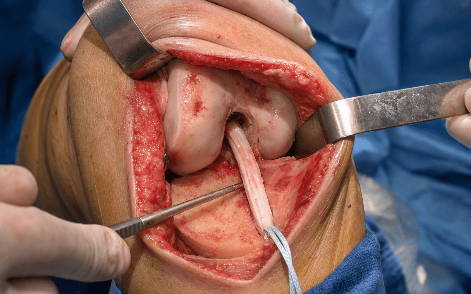
Operative sequence
- Supine with a thigh-mounted leg holder and a high thigh tourniquet; flex and support the hip so the knee reaches the full range needed for arthroscopic work.
- Establish standard anterolateral and anteromedial portals. In the scarred, revised knee the portals may need to sit more laterally or be modified to clear dense scar — plan them around the proposed new tunnel positions.
- CT on screen and fluoroscopy ready to correlate tunnel position throughout.
- Systematic evaluation: probe the graft remnant for competence, grade the menisci and articular cartilage, and confirm PCL integrity.
- If the graft is intact but incompetent (soft, stretched) at 6 to 12 months, this is biological failure — it may be augmented rather than simply discarded.
- Confirm the pre-operative failure mechanism against what you actually find inside the joint.
- Use the shaver and radiofrequency ablator to clear scar; preserve the notch roof.
- Identify the old tunnel apertures and assess them for sclerosis and widening.
- Take care with posterior debridement — the popliteal artery lies immediately behind the posterior capsule.
- Remove interference screws that protrude into the tunnel or obstruct the new position. Bioabsorbable screws may be expanded or fragmented and need curetting out.
- Metal screws may need a trephine or a reverse threader; the femoral screw occasionally requires an outside-in or mini-open lateral approach for access.
- Leave well-positioned, non-interfering hardware alone.
- Follow the graft hierarchy decided pre-operatively: contralateral BPTB (gold-standard autograft, bone-to-bone healing), ipsilateral quadriceps tendon (thick and strong, low morbidity if the hamstring is already harvested), or allograft for the older, lower-demand or multiligament knee.
- Prepare the graft on the back table to the measured size and mark its orientation; always have a back-up plan.
- Protect the saphenous nerve in hamstring harvest and the patella in BPTB harvest.
- If the tunnels are well-positioned and less than 12 mm, reuse them.
- If they are malpositioned but less than 14 mm and non-confluent (more than 2 mm rim), drill new anatomic tunnels in one stage.
- If they are widened beyond 14 to 16 mm or confluent (less than 2 mm rim), this becomes a TWO-STAGE procedure — pack both tunnels with bone graft and reconstruct at 3 to 6 months (Stage 1 ends here).
- Confirm every tunnel position with fluoroscopy before reaming.
- Femoral tunnel via the anteromedial portal (preferred) at 1:30 to 2:00 on the right knee (10:00 to 10:30 on the left), low on the lateral wall — the anatomic footprint.
- Tibial tunnel with a standard ACL guide at 55 to 60 degrees, placed in the posterior half of the native footprint.
- Knee at 110 to 120 degrees flexion for anteromedial-portal drilling; start with a smaller reamer if the bone is soft or the tunnel widened.
- Protect the posterior femoral wall — aim for a 2 mm posterior cortical rim, confirmed on fluoroscopy before final reaming.
- Pass the graft with correct orientation and tension; fix the femoral side first — interference screw, suspensory button, or hybrid.
- Cycle the graft 20 to 30 times to remove creep; fix the tibial side at 20 to 30 degrees flexion with a posterior drawer to set tension.
- Suspensory fixation (EndoButton, TightRope) is useful in revision where bone stock is poor; an interference screw needs an adequate bony rim; hybrid fixation gives maximum security.
- Aim for 1 to 2 mm of laxity at full extension — over-tensioning causes a flexion contracture, under-tensioning persistent laxity.
- Meniscus: repair if possible; root repair is critical to prevent extrusion and osteoarthritis; consider meniscal allograft transplantation for symptomatic meniscal deficiency.
- Posterolateral corner and alignment: a varus knee with posterolateral deficiency (the triple-varus knee) needs a high tibial osteotomy before or with the ACL — uncorrected varus will destroy the graft.
- Anterolateral augmentation: a lateral extra-articular tenodesis or anterolateral ligament reconstruction for a high-grade pivot shift, the young high-risk athlete, or the revision setting.
- Arthroscopic confirmation of graft position, tension and absence of impingement; perform a notchplasty if the graft catches on the notch roof.
- Check the full range — the knee must achieve full extension and at least 120 degrees flexion, with negative Lachman and pivot shift.
- Achieve haemostasis, photo-document the graft, close the portals in the standard fashion, and brace in extension.
The popliteal artery lies directly behind the posterior capsule, separated from the posterior tibial cortex by only a few millimetres; the gap is smallest in extension and increases with flexion. It is most at risk during tibial tunnel over-reaming, posterior debridement, and any deep femoral or transtibial drilling that breaches the posterior wall. Flex the knee to displace the artery posteriorly, never over-drill the femoral tunnel posteriorly, confirm an intact 2 mm posterior cortical rim on fluoroscopy, and use a calibrated tibial guide that stops at the far cortex. If pulsatile bleeding occurs, inflate the tourniquet and obtain immediate vascular surgery review.
Danger structures. Five structures are injured repeatedly in revision ACL surgery — know where each lies and how to protect it before you ream a tunnel.
At risk: tibial and femoral tunnel over-reaming, posterior debridement, and deep transtibial drilling. Protection: flex the knee to drop the artery posteriorly; never over-drill the femoral tunnel posteriorly; confirm a 2 mm posterior cortical rim on fluoroscopy; use a calibrated tibial guide and stop at the far cortex. Pulsatile bleeding means tourniquet up and vascular surgery now.
At risk: hamstring harvest, medial portal placement, and tibial tunnel reaming. Protection: oblique hamstring incision about 3 cm below the joint line, blunt dissection to sartorius, stay anterior to the gracilis insertion; place the posteromedial portal under direct vision.
At risk: the lateral portal, posterolateral corner work, and anterolateral reconstruction — it wraps the fibular neck only 1 to 2 cm distal to the joint line and sits about 10 mm from the posterolateral capsule. Protection: keep the lateral portal within 1 cm of the joint line, flex the knee, palpate the fibular neck, and identify the nerve directly during anterolateral work. A new foot drop is urgent.
At risk: femoral tunnel drilling, especially in soft widened revision bone. Protection: fluoroscopy before reaming, aim for a 2 mm posterior rim, anteromedial-portal drilling visualises better than transtibial, and use a smaller reamer if the tunnel is widened.
At risk: too horizontal a tibial tunnel or over-reaming of a widened tunnel — the neurovascular bundle lies directly posterior. Protection: set the tibial guide at 55 to 60 degrees, confirm with fluoroscopy before over-reaming, and stage with bone grafting if the widening is massive.
One-staging a knee with inadequate bone or tunnel confluence invites fixation failure and tunnel communication. If the CT shows widening beyond 14 to 16 mm or a less than 2 mm rim between old and new tunnels, stop, bone-graft both tunnels, and reconstruct at 3 to 6 months once a CT confirms incorporation.
Anteromedial-portal drilling at 110 to 120 degrees flexion places the femoral tunnel independently in the anatomic footprint, which transtibial drilling cannot reliably do. If the posterior wall worries you, start with a smaller reamer or switch to an outside-in technique under fluoroscopy.
The STABILITY RCT (Level 1) showed adding a lateral extra-articular tenodesis to hamstring ACLR cut graft rupture from 11% to 4% in high-risk young patients (NNT 14.3); the SANTI cohort showed hamstring plus ALL failed far less than BPTB or quadrupled hamstring in young pivoting athletes. Anterolateral augmentation is now a strong consideration in the high-risk revision.
Aftercare & Complications
Rehabilitation. Rehabilitation after revision is slower and more guarded than after a primary reconstruction, with return to sport driven by objective criteria rather than time alone. | Phase | Timing | Brace & weight-bearing | Focus | |-------|--------|------------------------|-------| | 1 | 0 to 2 weeks | Extension brace, touch weight-bearing | Full extension from day 1 (critical), quad activation, CPM if available | | 2 | 2 to 6 weeks | Hinged brace, partial weight-bearing | Progressive flexion, closed-chain quad, patellar mobility | | 3 | 6 to 12 weeks | Brace weaned, full weight-bearing | Proprioception, normalise gait, light strengthening | | 4 | 3 to 6 months | None | Agility, neuromuscular retraining, sport-specific drills | | 5 | 6 to 9+ months | None | Return-to-sport testing and psychological readiness | Return-to-sport criteria (typical minimum, often longer in revision): - At least 9 to 12 months — longer for revision and high-risk sport.
- Isokinetic strength greater than 90% limb symmetry index.
- Hop testing greater than 90% limb symmetry index.
- Negative pivot shift and a stable Lachman.
- Psychological readiness (for example an ACL-RSI score).
- Counsel realistic expectations — most patients down-regulate their sport after revision. Complications
- Recognition
- Recurrent instability, positive pivot shift and Lachman; may be gradual (biological) or acute (traumatic)
- Prevention
- Address all causes of primary failure; autograft in the young; anterolateral augmentation in high-risk cases; correct alignment
- Management
- Identify the cause — technical: correct the tunnels; biological: augmentation or a different graft; traumatic: re-revision possible
- Recognition
- Loss of motion (extension loss most common), anterior knee pain, pain with forced range, palpable scar
- Prevention
- Optimise pre-operative motion, atraumatic technique, aggressive early extension, avoid inflammation
- Management
- Intensify therapy; arthroscopic lysis of adhesions if no progress by 3 to 6 months; manipulation under anaesthesia for focal contracture
- Recognition
- Communication between tunnels on CT or arthroscopy, loss of fixation, unstable graft, failed incorporation
- Prevention
- Careful pre-operative CT planning and 3D reconstruction; keep at least 2 mm between tunnels; stage if needed
- Management
- Two-stage reconstruction with bone grafting — pack both tunnels and wait 3 to 6 months for incorporation
- Recognition
- Fever, effusion, wound erythema, raised inflammatory markers (CRP, ESR), positive aspiration
- Prevention
- Prophylactic antibiotics, meticulous sterile technique, minimise operative time, optimal skin closure
- Management
- Aspirate and culture; arthroscopic lavage with graft retention if early; two-stage with graft removal if established
- Recognition
- Saphenous: medial numbness. Peroneal: foot drop, lateral numbness. Infrapatellar branch: anterior numbness
- Prevention
- Careful portal placement, protect nerves in hamstring harvest, avoid lateral structures in anterolateral work
- Management
- Observation for neurapraxia (3 to 6 months typical); EMG if no recovery; exploration if complete palsy
- Recognition
- Anterior knee pain (BPTB), hamstring or quad weakness, fracture of the patella or tibial tubercle
- Prevention
- Consider allograft in low-demand patients, meticulous harvest technique, avoid an excessive bone plug
- Management
- Therapy for weakness; fracture fixation if displaced; counsel on the expected recovery timeline
- Recognition
- Screw prominence or migration, bioabsorbable screw expansion, interference with new tunnels
- Prevention
- Appropriate screw sizing, avoid proud placement, use suspensory fixation for poor bone
- Management
- Symptomatic removal once healed; may need a staged approach if it interferes with the revision
Viva & Exam Focus
FAILUREFAILURE — causes of ACL reconstruction failure
STAGEDSTAGED — two-stage revision indications
Clinical Decision Scenarios
Practise clinical reasoning and management decisions out loud
“A 22-year-old male professional footballer presents 18 months after primary ACL reconstruction with a hamstring autograft. He describes persistent instability with pivoting movements. Clinical exam shows Grade 2 Lachman and Grade 2 pivot shift. How would you investigate and manage this patient?”
“The CT scan shows tunnel widening greater than 16 mm on both the tibial and femoral sides, with significant bone loss. What is your two-stage revision strategy?”
“A 28-year-old woman presents 3 years after ACL reconstruction. She has a Grade 3 pivot shift, a varus thrust gait, and a dial test positive at 30 degrees. MRI shows an intact but incompetent ACL graft. What is your comprehensive management plan?”
Indications
- Failed primary ACLR with recurrent instability
- Graft rupture (traumatic or atraumatic)
- Technical failure — tunnel malposition is the most common cause
- Biological failure — graft intact but incompetent at 6 to 12 months
- Unaddressed pathology causing ongoing instability (PLC, meniscal, malalignment)
Key anatomy
- Femoral tunnel: 1:30 to 2:00 (right) or 10:00 to 10:30 (left), low on the lateral wall
- Tibial tunnel: posterior half of the native footprint, 55 to 60 degree angle
- Popliteal artery: just behind the posterior capsule — avoid posterior wall breach and deep over-reaming; flex the knee for posterior work
- Saphenous nerve: posteromedial, at risk with the medial portal and hamstring harvest
- Peroneal nerve: 1 to 2 cm distal to the fibular neck, at risk with lateral procedures and anterolateral work
Critical steps
- 1. CT scan — tunnel position, widening and confluence assessment
- 2. Identify the cause of failure (MARS framework)
- 3. Staging decision — one-stage if tunnels less than 14 mm, two-stage if greater than 16 mm
- 4. Graft selection — autograft preferred in the young (MARS evidence)
- 5. Anatomic tunnel placement — anteromedial-portal drilling preferred
Danger zones
- Posterior wall: a 2 mm rim is required — fluoroscopy before reaming
- Popliteal artery: directly posterior to the knee — avoid over-drilling the femoral tunnel
- Peroneal nerve: at risk with anterolateral reconstruction — identify it directly
- Tunnel convergence: less than 2 mm between tunnels means communication risk
Technique pearls
- Anteromedial-portal drilling at 110 to 120 degrees flexion for an anatomic femoral position
- Suspensory fixation is useful with poor bone stock from tunnel widening
- Cycle the graft 20 to 30 times before final fixation to remove creep
- Fix the tibial side at 20 to 30 degrees flexion with a posterior drawer to set tension
Complications
- Re-failure: rerupture about 3% at 2 years and about 6% at 6 years (MARS) — higher with allograft in the young
- Arthrofibrosis: prevented with aggressive early extension exercises
- Tunnel convergence: requires a two-stage procedure with bone grafting
- Nerve injury: saphenous (medial) and peroneal (lateral) — identify and protect
Post-op protocol
- Brace in extension; immediate CPM if available
- Full extension exercises from day 1 (critical)
- Progressive weight-bearing over 6 weeks
- Return to sport: minimum 9 to 12 months, greater than 90% limb symmetry index, hop tests, psychological readiness
Exam tips
- MARS cohort (Level 2): autograft 2.78x less rerupture at 2 years; BTB autograft 4.2x less at 6 years (3.5% vs 8.4%)
- STABILITY RCT: adding LET cut graft rupture from 11% to 4% in high-risk young patients (NNT 14.3)
- Tunnel thresholds: reported two-stage thresholds cluster at 10 to 14 mm; bone-graft when no acceptable new aperture can be made
- Triple varus: bone varus plus PLC plus thrust means you MUST correct alignment before or with the ACL
- Always identify the cause of primary failure before revising
Background & Evidence
Why revision differs from primary. Revision ACL reconstruction is common and rising as primary reconstruction volume grows, and outcomes are consistently inferior to the primary procedure — higher rerupture, lower patient-reported function, and a fall-off in activity as most patients down-regulate their sport. Meniscal loss and high-grade chondral damage independently predict a worse result, which is why meniscal preservation and chondral status weigh so heavily in planning and consent. Outcomes (MARS cohort and related data).
- Rate or finding
- 3.3% overall — 24 allograft and 12 autograft of 1112 followed
- Notes
- Autograft 2.78x less likely to rerupture
- Rate or finding
- 5.8% overall; 3.5% autograft vs 8.4% allograft
- Notes
- BTB autograft 4.2x less likely than BTB allograft
- Rate or finding
- Significantly improved vs baseline
- Notes
- But the Marx activity score falls — patients down-regulate sport
- Rate or finding
- Lower than after primary reconstruction
- Notes
- Counsel realistic expectations
- Rate or finding
- Inferior to primary ACLR
- Notes
- Higher rerupture, lower function and activity
Risk factors for revision failure: - Younger age and high-demand pivoting sport.
- Allograft use, especially in the young.
- A previous revision (a second revision fares worse than a first).
- Meniscal deficiency and high-grade chondral damage.
- Uncorrected alignment or posterolateral corner deficiency. The evidence base. The MARS cohort underpins graft choice, demonstrating a widening autograft survival advantage out to 6 years. The STABILITY randomised trial and the SANTI cohort underpin anterolateral augmentation in high-risk young knees. The systematic review by Gopinath and colleagues codifies two-stage practice — consistent indications despite variable technique, with the tunnel-diameter threshold for two-stage clustering at 10 to 14 mm.
References
Effect of graft choice on the outcome of revision ACL reconstruction in the MARS cohort
- Prospective multicentre cohort of 1205 revision ACL reconstructions (median age 26 years), 82% questionnaire follow-up at 2 years
- Autograft was a significant predictor of better 2-year IKDC and KOOS sport/QoL scores than allograft
- Graft rerupture occurred in 3.3% (37/1112) by 2 years; use of autograft made patients 2.78x less likely to rerupture than allograft
Association between graft choice and 6-year outcomes of revision ACL reconstruction in the MARS cohort
- 1234 revision patients followed to 6 years; overall graft rerupture 5.8% (55/949)
- BTB autograft rerupture 3.5% vs BTB allograft 8.4% — BTB autograft 4.2x less likely to rerupture than BTB allograft
- Autograft predicted fewer reoperations within 6 years (OR 0.56) and higher activity level than BTB allograft
Lateral extra-articular tenodesis reduces failure of hamstring autograft ACL reconstruction: 2-year STABILITY RCT
- Multicentre RCT, 618 high-risk patients aged 25 or younger randomised to hamstring ACLR with or without iliotibial-band LET
- Clinical failure (rotatory laxity or rupture) 25% with LET vs 40% without; graft rupture 4% (11/291) vs 11% (34/298)
- Number needed to treat with LET to prevent one graft rupture over 2 years was 14.3
Anterolateral ligament reconstruction is associated with significantly reduced ACL graft rupture rates at minimum 2-year follow-up (SANTI Study Group)
- Prospective comparative cohort of 502 young pivoting-sport athletes (mean age 22.4 years)
- Graft rupture: HT plus ALL 4.13% vs BPTB 16.77% vs quadrupled hamstring 10.77%
- HT plus ALL graft failure was 2.5x lower than BPTB and 3.1x lower than 4HT; age 25 or younger and preoperative laxity over 7 mm were independent risk factors
Consistent indications and good outcomes despite high variability in techniques for two-stage revision ACL reconstruction: a systematic review
- Systematic review of 13 studies, 355 two-stage revision ACL patients
- Most common indications were tunnel malposition and tunnel widening; reported tunnel-diameter thresholds for two-stage clustered at 10 to 14 mm
- Most common bone grafts were iliac crest autograft and allograft bone chips or dowels; PROs (Lysholm, Tegner, IKDC) improved from pre- to post-operative
Meniscal and articular cartilage predictors of clinical outcome after revision ACL reconstruction
- MARS cohort analysis of meniscal and articular cartilage status against two-year clinical outcome after revision ACL reconstruction
- Meniscal loss and high-grade chondral damage independently predicted worse patient-reported outcome
Rehabilitation predictors of clinical outcome following revision ACL reconstruction in the MARS cohort
- Prognostic analysis of the prospective MARS cohort identifying rehabilitation factors associated with two-year clinical outcome after revision ACL reconstruction
Revision anterior cruciate ligament reconstruction: report of 11-year experience and results in 114 consecutive patients
- Foundational single-surgeon series reporting revision ACLR experience in 114 consecutive patients over 11 years