Glenohumeral fusion for brachial plexus palsy, deltoid loss and failed arthroplasty — a salvage operation powered by the scapular rotators
- The OPTIMAL FUSION POSITION is 15–30° abduction, 15–25° forward flexion, 40–50° internal rotation — so the hand can reach the mouth and the perineum. Malposition causes severe disability and may need revision.
- Brachial plexus palsy is the MOST COMMON indication worldwide — the hallmark is a non-functioning deltoid and rotator cuff with no prospect of recovery. Arthrodesis transfers shoulder positioning to the scapular rotators.
- FUNCTIONING SCAPULAR MUSCLES are the absolute prerequisite — serratus anterior (long thoracic nerve, C5–C7) and trapezius (accessory nerve) must be intact to rotate the scapula and achieve elevation after fusion. Without them, fusion achieves nothing.
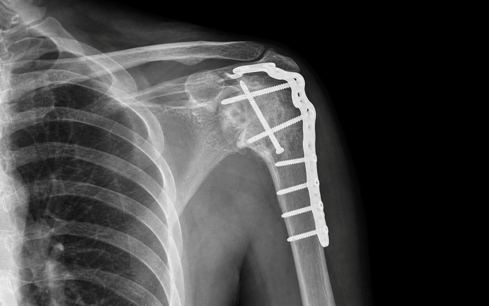
- NON-UNION is the most common complication (10–15%) — risk factors are poor bone contact, inadequate fixation, smoking, prior radiation and infection. A long contoured plate from the scapular spine to the proximal humerus, plus iliac crest graft, is essential.
- Bilateral shoulder disease is a contraindication — the patient needs one functional upper limb for activities of daily living and crutch use.
When & Why
Shoulder arthrodesis is a SALVAGE operation. It is offered when the glenohumeral joint is painful, unstable or flail AND reconstruction by arthroplasty or soft-tissue surgery is impossible — most often because the deltoid and rotator cuff are both lost. The aim is not to restore a moving shoulder, but to give a stable, painless platform that the scapulothoracic muscles can position in space so the hand becomes useful again. The six indications
The most common indication worldwide. A complete, non-recovering injury leaves a flail, painful glenohumeral joint with a paralysed deltoid and cuff. Best when elbow flexion and hand function are preserved.
Failed anatomic or reverse TSA with irreparable glenoid bone loss, periprosthetic infection, or multiple revision failures. Often needs structural allograft for bone-stock deficiency.
Loss of BOTH the deltoid and rotator cuff. RSA needs a functioning deltoid and TSA needs an intact cuff — when both are gone, arthrodesis is the only reconstructive option.
Uncontrolled septic glenohumeral arthritis where arthroplasty is contraindicated. Performed as a salvage once infection is eradicated; higher non-union risk in irradiated or infected tissue.
Tumour resection of the proximal humerus or scapula with glenohumeral involvement, paralytic conditions (poliomyelitis, upper motor neuron lesion), and severe post-traumatic bone and soft-tissue loss.
Bipolar (humeral and glenoid) bone loss where instability surgery is not feasible. Rare — most instability is managed by soft-tissue or bone procedures.
Before you operate — the non-negotiable prerequisites. These decide whether the operation will work at all: - Functioning serratus anterior and trapezius — the motor for elevation after fusion. Test clinically (ask the patient to attempt elevation and watch the scapula rotate) and confirm with EMG if there is any doubt. Scapular winging (serratus palsy) means arthrodesis will not achieve functional elevation.
- A normal contralateral shoulder — the patient must have one functional limb for hygiene, dressing and crutch use.
- Preserved elbow and hand function (especially in brachial plexus cases) — otherwise a positioned shoulder adds little.
- Realistic counselling — this is salvage, not restoration; elevation will be limited to roughly 90° via scapulothoracic motion.
Arthrodesis is contraindicated when it cannot work or will harm the patient. Absolute contraindications: bilateral shoulder disease (the patient cannot use crutches or perform perineal hygiene if both shoulders are fused), non-functioning scapular muscles (fusion is futile without scapulothoracic rotation), and active uncontrolled infection at the operative site. Relative contraindications: significant contralateral shoulder disease, severe osteoporosis (fixation may fail), Charcot arthropathy (high non-union risk), and a patient who cannot comply with prolonged immobilisation. The classic trap: proposing arthrodesis for bilateral brachial plexus palsy — the patient would be left with no functional upper limbs.
Setup. Beach chair or lateral decubitus with the arm draped free so it can be manipulated to set and confirm the fusion position. Image intensifier (fluoroscopy) must be available throughout, and loupe magnification helps nerve and surface identification.
The Operation
The goal: through a posterior approach, prepare broad bleeding cancellous surfaces on the humeral head, glenoid and acromion; set the arm in the precise fusion position; pack iliac crest bone graft; and lock the construct with a long contoured reconstruction plate spanning from the scapular spine to the proximal humeral shaft. The exposure is laid out in full as the first steps below.
Operative sequence
- Beach chair or lateral decubitus, arm draped free so it can be moved to set and check position.
- Fluoroscopy positioned to confirm abduction on the AP view before and during fixation.
- Decide the target position before draping: 15–30° abduction, 15–25° forward flexion, 40–50° internal rotation.
- Longitudinal incision along the spine of the scapula, curving laterally over the posterior deltoid toward the proximal humerus.
- The posterior approach is preferred: gravity assists arm positioning and the surface for plating (the posterior-superior shoulder) is exposed directly.
- Elevate the posterior deltoid from the spine of the scapula to expose the posterior scapula and proximal humerus.
- Develop the internervous plane between teres minor (axillary nerve) and infraspinatus (suprascapular nerve). In brachial plexus palsy these muscles are denervated and simply swept aside — the interval is still used.
- Identify and protect the AXILLARY NERVE as it exits the quadrangular space and winds around the surgical neck with the posterior circumflex humeral artery. The suprascapular nerve is at risk from a deep posterior retractor in the spinoglenoid notch.
- Expose the posterior glenohumeral capsule and open it to enter the joint.
- In plexus cases the denervated infraspinatus, teres minor and supraspinatus are reflected to broaden the exposure; the deltoid muscle belly is preserved as a biological sleeve and padding over the plate even though it is denervated.
- Denude the humeral head of all articular cartilage down to bleeding cancellous bone with a curette, osteotome or burr.
- Denude the glenoid articular surface completely to bleeding cancellous bone, and prepare the undersurface of the acromion similarly — the humeral head will be brought up against the acromion to add surface area to the fusion mass.
- Create flat opposing surfaces to maximise bone contact, and drill multiple holes into both surfaces to encourage vascular ingrowth.
- Maximum bone-contact surface area is the single most important technical factor for union; inadequate preparation is the most preventable cause of non-union.
- Abduction 15–30°, forward flexion 15–25°, internal rotation 40–50°.
- With the elbow at 90°, the forearm should point toward the face (confirms abduction and flexion) and be able to swing down toward the perineum (confirms internal rotation). The hand should reach the mouth and the top of the head, and the lower back.
- Hold the position with a temporary K-wire and confirm with fluoroscopy before definitive fixation.
- Iliac crest autograft is the gold standard — harvest cancellous and corticocancellous bone and pack it into any gaps between the humeral head and glenoid, and around the acromial contact superiorly.
- A massive subacromial corticocancellous graft dramatically lowers the non-union rate (Atlan 2012, PMID 22464233: pseudarthrosis fell from 43% with cancellous-only graft to 4% with a structural corticocancellous graft).
- For failed arthroplasty or tumour with major bone loss, plan for a structural femoral-head allograft.
- Contour a 3.5mm or 4.5mm pelvic reconstruction plate (or a dedicated arthrodesis plate) onto the posterior-superior shoulder, spanning from the scapular spine or acromion, across the glenohumeral joint, down to the proximal-to-mid humerus.
- Proximal fixation: 3–4 screws into the scapular spine or base of the acromion. Across the joint: 2–3 compression screws through the prepared surfaces. Distal fixation: 3–4 screws into the proximal humeral shaft.
- A SHORT plate will fail — the weight of the arm generates an enormous bending moment at the fusion site, so the plate must be a long-moment-arm device anchored both proximally and distally. Supplementary screws from the humerus into the acromion may be added.
- Confirm the abduction angle on the AP fluoroscopic view.
- Check the arm hangs comfortably at the side (not excessively abducted) and that, with the elbow at 90°, the forearm sits in appropriate internal rotation pointing toward the face and perineum.
- Repair the posterior deltoid back to the scapular spine fascia; layered closure over a deep drain; compressive dressing.
- Apply a shoulder spica cast or a custom thoracohumeral orthosis in the fusion position for 6–12 weeks.
First, protect the AXILLARY NERVE in the quadrangular space and at the surgical neck throughout the posterior dissection — in a brachial plexus case document any pre-existing deficit. Second, use a LONG plate: a short plate spanning only the glenohumeral joint cannot resist the arm's bending moment and is a leading cause of fixation failure and non-union.
The whole fusion position reduces to one test: with the elbow flexed to 90°, the forearm must point toward the face and be able to reach down to the perineum. If it does both, the position is correct. Combined with about 60° of scapulothoracic rotation, a well-positioned fusion gives roughly 90° of functional forward elevation.
Older series (Cofield averaged about 45° abduction) found patients could not sleep comfortably, could not use crutches, and the arm protruded awkwardly. Rowe (1974, PMID 4847239) argued against the high angles then in vogue, and modern teaching favours a lower-profile position — often summarised as the 'rule of 30s' — measured from the side of the body.
Aftercare & Complications
Rehabilitation External immobilisation in the fusion position is mandatory until there is radiological evidence of early union. A custom thoracohumeral orthosis is better tolerated than a spica and allows hygiene, and is preferred in most modern centres; some units (Atlan 2012) immobilise selectively. | Phase | Timing | Immobilisation | Focus | |-------|--------|----------------|-------| | Immobilisation | 0–12 weeks | Spica cast or custom orthosis in the fusion position | X-ray at 6 and 12 weeks for bridging callus; CT at 3–6 months if union is doubtful | | Scapular re-education | 12 weeks onward | Orthosis removed once early union is confirmed | Train serratus anterior and trapezius to position the arm through scapulothoracic motion | | Return to function | 3–6 months | None | Light activities of daily living at 3–4 months; heavy manual work and driving at around 6 months | Expected function | Measure | Expected after arthrodesis | |---------|----------------------------| | Forward elevation | 60–90° via scapulothoracic rotation | | Abduction | 60–80° via scapulothoracic rotation | | Hand to mouth | Usually achievable | | Perineal hygiene | Usually achievable with 40–50° internal rotation | | Pain relief | Significant in most patients | | Satisfaction | 75–90% in brachial plexus palsy (Chammas 2004) | | Lifting strength | About 50–70% of the contralateral arm | Complications
- Rate
- 10–15%
- Mechanism / recognition
- Persistent pain beyond 6 months; CT shows a radiolucent line at the fusion site. Distinguish fibrous union (may still consolidate) from true non-union.
- Prevention / management
- Maximum bone contact at surgery, rigid long-plate fixation, iliac crest graft, smoking cessation. Revision: refresh surfaces to bleeding bone, bone graft, longer or better-positioned plate; electrical stimulation as an adjunct. Smoking is the strongest modifiable risk factor.
- Rate
- 5–10%
- Mechanism / recognition
- Excessive abduction (arm sticks out, cannot use crutches), insufficient internal rotation (cannot reach the perineum), or excessive internal rotation (cannot reach forward).
- Prevention / management
- Set and check all three planes with fluoroscopy before definitive fixation. Early (before consolidation): revise. After solid fusion: corrective humeral osteotomy — a major undertaking.
- Rate
- 5–10%
- Mechanism / recognition
- Plate fracture or screw loosening from the arm's bending moment; on X-ray a broken plate or loose screws, usually with non-union.
- Prevention / management
- Use a long plate with adequate proximal and distal fixation; protect in a spica until early union. Revision: longer plate plus bone graft.
- Rate
- 5–8%
- Mechanism / recognition
- Humeral head set too superior (too much abduction) or prominent hardware; altered mechanics stress the acromion.
- Prevention / management
- Keep abduction at or under 30°; countersink screw heads. Symptomatic hardware is removed after union; a displaced acromial fracture is managed in a sling, or fixed if non-union occurs.
- Rate
- 2–5%
- Mechanism / recognition
- Axillary nerve from retraction, suprascapular nerve from a posterior retractor, or brachial plexus from traction. In plexus cases document pre-existing deficits.
- Prevention / management
- Identify and protect the axillary nerve; gentle retraction. Neuropraxia: observe; neurotmesis: explore and repair if possible.
- Rate
- 1–3%
- Mechanism / recognition
- Wound or deep infection around hardware; higher in irradiated tissue, the immunocompromised, and revisions.
- Prevention / management
- Prophylactic antibiotics, meticulous closure, optimise comorbidities. Acute: washout, retain metalwork if not yet consolidated, IV antibiotics. Chronic: remove metalwork after union is confirmed and manage the infection.
- Rate
- 10–20%
- Mechanism / recognition
- Failure to achieve expected function, usually from non-functioning scapular muscles missed preoperatively, bilateral disease, or unrealistic expectations.
- Prevention / management
- Thorough preoperative scapular-muscle assessment and realistic counselling that this is a salvage procedure with elevation limited to about 90°.
Viva & Exam Focus
FUSIONFUSION — the key elements of shoulder arthrodesis
BRACHIALBRACHIAL — patient selection for arthrodesis in plexus palsy
Clinical Decision Scenarios
Practise clinical reasoning and management decisions out loud
“A 28-year-old man sustained a right brachial plexus injury in a motorcycle accident 18 months ago. He has a completely flail shoulder with no deltoid or rotator cuff function but intact elbow flexion and hand function. EMG confirms no reinnervation at the shoulder. He is right-handed. What are his surgical options and what is your recommended management?”
“What is the optimal fusion position for shoulder arthrodesis and how do you justify each component biomechanically?”
“A patient develops non-union of a shoulder arthrodesis 8 months post-operatively. CT confirms a fibrous union with no bridging bone at the glenohumeral site. The plate appears intact. What is your management?”
Indications
- Brachial plexus palsy — most common; flail shoulder with functioning serratus and trapezius
- Failed arthroplasty — irreparable bone loss, repeated revision failures
- Irreparable rotator-cuff tear PLUS deltoid loss — RSA impossible (needs deltoid) and TSA impossible (needs cuff)
- Infection / chronic osteomyelitis — after eradication
- Flail shoulder from tumour resection or paralytic conditions
Contraindications
- Bilateral shoulder disease — cannot use crutches or perform perineal hygiene bilaterally
- Non-functioning serratus anterior or trapezius — fusion achieves nothing without scapular rotation
- Active uncontrolled infection at the operative site
- Significant contralateral shoulder disease — relative contraindication
- Charcot arthropathy — very high non-union risk
Fusion position
- Abduction: 15–30° (NOT the older figure of about 45° — too much, outdated)
- Forward flexion: 15–25°
- Internal rotation: 40–50° — the most important; enables perineal hygiene and hand-to-mouth
- Practical test: elbow at 90°, the forearm points to the face AND the perineum
- Malposition is the second most common cause of failure after non-union
Prerequisites (confirm preoperatively)
- Serratus anterior intact — scapular winging test; EMG if uncertain
- Trapezius intact — clinical testing; accessory-nerve function
- Contralateral shoulder NORMAL
- Elbow and hand function ideally preserved (for brachial plexus cases)
- Infection excluded (inflammatory markers, aspiration if required)
Fixation principles
- Long contoured plate from the scapular spine/acromion to the proximal humeral shaft
- A short plate WILL fail — the bending moment from arm weight is enormous
- Glenohumeral compression screws across the prepared surfaces
- Iliac crest autograft mandatory — packs all gaps
- Spica cast or custom orthosis for 6–12 weeks post-op
Complications
- Non-union (10–15%): most common — bone graft plus revision fixation with a longer plate
- Malposition (5–10%): second most common cause of failure — confirm position intraoperatively
- Hardware failure (5–10%): short plate plus non-union — replace with a longer plate
- Subacromial impingement / acromial fracture: altered mechanics
- Functional dissatisfaction (10–20%): mainly from non-functioning scapular muscles missed preoperatively
Key references
- Rowe 1974 (PMID 4847239): re-evaluated arm position; argued against high abduction angles
- Cofield and Briggs 1979 (PMID 457712): 71 shoulders, about 45°/25°/25°, internal fixation, position had little effect on outcome
- Chammas 2004 (PMID 15274265): 27 BPI patients; function improves even with a flail hand if elbow flexion is restored
- Atlan 2012 (PMID 22464233): 54 cases; subacromial corticocancellous graft cut pseudarthrosis from 43% to 4%
Examiner favourites
- Fusion position → 15–30° abduction, 15–25° flexion, 40–50° internal rotation, with justification
- Why serratus anterior is essential → scapular rotation is the motor for post-fusion elevation
- Why bilateral disease is a contraindication → crutches and perineal hygiene become impossible
- Most common complication → non-union (10–15%), managed by bone graft plus revision plate
- Most common indication globally → brachial plexus palsy (Chammas 2004)
Background & Evidence
Who needs it. Brachial plexus palsy is the most common indication for shoulder arthrodesis worldwide. The other indications cluster around a common theme — a glenohumeral joint that cannot be salvaged by arthroplasty: failed anatomic or reverse TSA with bone loss, combined deltoid and cuff loss, uncontrolled infection, tumour resection, and paralytic flail shoulders. Why arthrodesis works. When the deltoid and rotator cuff are both lost, neither an anatomic total shoulder (which needs an intact cuff) nor a reverse replacement (which needs a functioning deltoid) is possible. Fusing the glenohumeral joint converts the scapulothoracic articulation into the only remaining shoulder motor. After fusion, serratus anterior (long thoracic nerve, C5–C7, protracting and upwardly rotating the scapula) and trapezius (accessory nerve) rotate the glenoid upward so the fused arm can be positioned in space. This is why functioning scapular muscles are the absolute prerequisite — without them the operation is futile. The denervated deltoid is preserved as a biological soft-tissue sleeve over the hardware even though it cannot act as a motor. The fusion-position debate. The recommended position has evolved away from the high-abduction postures of the mid-20th century toward a lower, more functional position. The principle — not the precise degree — is what matters: enough abduction and flexion for the hand to reach the face via scapular rotation, and enough internal rotation to reach the perineum and midline.
- Recommended position
- Argued against high abduction; measure from the side of the body
- Key message
- Moved practice away from extreme abduction toward a functional, lower-profile position
- Recommended position
- Mean achieved about 45° abduction, 25° flexion, 25° rotation
- Key message
- 71 shoulders with rigid fixation; the exact position had little effect on the functional result
- Recommended position
- 15–30° abduction, 15–25° flexion, 40–50° internal rotation (the 'rule of 30s')
- Key message
- Enough abduction/flexion to reach the face via scapular rotation, and enough internal rotation to reach the perineum and midline
Key evidence. The modern technical lesson comes from Atlan (2012, PMID 22464233): a structural subacromial corticocancellous graft cut the pseudarthrosis rate from 43% to 4% — the strongest evidence that graft strategy drives union. Chammas (2004, PMID 15274265) clarified patient selection in plexus palsy: restore elbow flexion first, and a flail hand is not in itself a contraindication. Cofield and Briggs (1979, PMID 457712) established durable pain relief with rigid single-stage fixation, and Scalise and Iannotti (2008, PMID 18171959) defined the high revision and non-union burden in the failed-arthroplasty setting.
References
Re-evaluation of the position of the arm in arthrodesis of the shoulder in the adult
- Re-evaluated the ideal arm position for shoulder arthrodesis in adults
- Argued against the historically high abduction angles then in use, which left the arm protruding and impaired comfort and crutch use
- Established the principle of measuring abduction from the side of the body and using a comparatively modest, functional position
Glenohumeral arthrodesis. Operative and long-term functional results
- 71 shoulders in 70 patients fused with internal fixation, mean follow-up 9.5 years
- Mean achieved position approximately 45° abduction, 25° flexion, 25° rotation
- Solid fusion after one operation in 68 of 71 shoulders; the remaining 3 required a second arthrodesis
- Pain relief adequate in three-quarters; 82% felt they had benefited and results did not deteriorate over time
- The exact position of fusion had little effect on the functional result
Glenohumeral arthrodesis in upper and total brachial plexus palsy. A comparison of functional results
- 27 adults compared: 11 upper palsy with a functional hand versus 16 total palsy with a flail hand, mean follow-up 70 months
- All patients had recovered active elbow flexion against resistance BEFORE shoulder fusion
- Both groups gained functional capability; a flail hand did not reduce post-operative active range of movement
- Pectoralis major strength was a significant prognostic factor for hand excursion and shoulder strength
Functional outcome of glenohumeral fusion in brachial plexus palsy: a report of 54 cases
- 54 patients, mean follow-up 37 months; overall fusion 76% after the first procedure, 94% at final follow-up
- A massive subacromial corticocancellous autograft reduced pseudarthrosis to 4% versus 43% with cancellous-only graft (p less than 0.001)
- Mean scapulothoracic motion 59° abduction and 48° rotation; abduction over 45° achieved in more than 75% of cases
- 48 of 54 patients had no post-operative immobilisation
Glenohumeral arthrodesis after failed prosthetic shoulder arthroplasty
- 7 consecutive patients fused after failed shoulder arthroplasty with severe bone loss and deltoid/cuff insufficiency
- Penn Shoulder Score improved from a mean of 17 to 58 points (p = 0.008)
- Fusion achieved in 5 of 7; 4 of 7 needed additional bone-grafting, and 2 ended with persistent non-union
- Delayed union requiring further surgery was the most common complication