Thoracic Disc Herniation — Discectomy

SpineAdvancedCore Procedure

Thoracic Disc Herniation — Discectomy

Surgical technique guide for discectomy in thoracic disc herniation — approach selection by herniation location, posterolateral (transpedicular/costotransversectomy), lateral extracavitary and transthoracic approaches, cord protection, level localisation, and management of calcified discs

High-yield overview

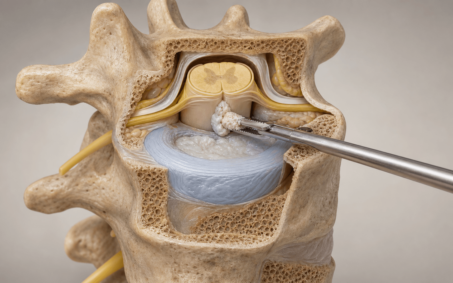
Surgical discectomy via approach tailored to herniation location | cord must NEVER be retracted | advanced

Surgical Imaging

Thoracic discectomy decompressing the cord
Thoracic disc herniation compressing the spinal cord; the herniated fragment is removed to decompress the cord.Credit: AI-generated medical illustration · OrthoVellum
Critical Danger Structures and Exam Traps
Wrong-Level Surgery — Thoracic Counting

The trap: Counting ribs or vertebral bodies from surface anatomy (C7 prominence, T12 rib) is unreliable — up to 15% of thoracic spine procedures have incorrect level localisation in some series. The T2 vertebra is difficult to identify on an intraoperative lateral because the shoulders overlap.

The fix: Use a preoperative full-length localiser radiograph. Place a localising needle, k-wire or towel clip in a spinous process at the intended level and confirm with AP + lateral X-ray or CT before incision. Cross-reference with preoperative MRI by counting from T1 (which articulates with the first rib) on a sagittal localiser. Never rely on a single radiographic view alone.

Spinal Cord Manipulation — Paraplegia

Mechanism: The thoracic spinal cord has a watershed blood supply in the mid-thoracic region (T4-T9), where the anterior spinal artery is narrowest and receives the fewest radicular feeders. Even gentle retraction can cause cord ischaemia and permanent motor loss.

Prevention: Choose an approach that allows the herniation to be resected without any contact with the cord. For lateral discs, a transpedicular or costotransversectomy approach reaches the disc lateral to the cord. For central/broad-based discs, an anterior (transthoracic) or lateral extracavitary approach provides direct visualisation without cord retraction.

Artery of Adamkiewicz (Great Anterior Radicular Artery)

Location: Most commonly arises from a left posterior intercostal artery between T9 and T11 (75% of individuals), but can arise from T5-L4 and from the right in up to 25%. It joins the anterior spinal artery and supplies the anterior two-thirds of the lower thoracic and upper lumbar cord.

Risk: Sacrifice of the segmental vessel feeding the artery of Adamkiewicz during a transthoracic or lateral approach risks anterior spinal artery syndrome — flaccid paraplegia with preserved posterior column function (vibration and proprioception). Prevention: Preoperative CT angiography or MRA in all patients undergoing anterior or lateral approaches to the lower thoracic spine. If the artery arises at or adjacent to the herniation level, consider a different approach or temporary clamping with neuromonitoring.

Dural Tear / CSF Leak — Calcified Discs

Risk: Calcified thoracic discs adhere to the ventral dura in a significant proportion of cases. In the series by Stillerman, up to 30% of calcified discs demonstrated some degree of dural penetration at surgery. The disc and dura may be inseparable.

Management: Plan for intentional durotomy with en-bloc resection of the disc and adherent dura, followed by primary dural repair (6-0 Prolene or Gore-Tex suture) or patching with autologous fascia, bovine pericardium or dural substitute. Apply sealant and drain management. Conversion to a more extensile approach (lateral extracavitary or transthoracic) provides better access for repair than a narrow posterolateral corridor.

Approach Selection Error

The error: Attempting a posterolateral (transpedicular/costotransversectomy) approach for a central, broad-based or calcified disc that cannot be safely resected without manipulating the cord. The narrow working angle through the pedicle and facet pushes the instruments toward the cord.

The fix: Classify the herniation on axial MRI (and CT if calcified) in relation to the cord. Lateral/foraminal herniations — posterolateral approach. Central, broad-based or calcified herniations — anterior (transthoracic, thoracoscopic) or lateral extracavitary approach. A midline central disc cannot be safely resected from a unilateral posterolateral window without unacceptable cord risk.

Chest Complications — Transthoracic Approach

Risk: The transthoracic (transpleural) approach requires lung deflation (double-lumen endotracheal tube), rib resection or spreading, and pleural entry. Specific complications include: persistent air leak (5-10%), haemothorax, intercostal neuralgia from rib retraction, pleural effusion, pneumonia, and chest-tube-related complications.

Mitigation: Meticulous pleural closure at the end of the procedure with a water-tight seal. Place a chest tube (24-28 Fr) under water seal with -20 cm H2O suction. Remove when output is less than 100-150 mL/24 hours and no air leak is present on water seal trial. Consider an intercostal nerve block at the time of closure to reduce postoperative pain and pulmonary morbidity.

Mnemonic

T.H.O.R.A.C.I.CTHORACIC — Key Principles of Thoracic Discectomy

Mnemonic

D.I.S.C. H.E.R.N.I.ADISC HERNIA — Approach Selection

Surgical Indications

Absolute Indications

  • Progressive myelopathy due to thoracic disc herniation — the presence of cord signal change, gait disturbance, lower limb hyperreflexia, clonus, or bladder/bowel dysfunction constitutes a surgical urgency
  • Acute paraplegia from a compressive thoracic disc herniation — requires urgent decompression
  • Intractable radiculopathy with radicular pain or intercostal neuralgia that has failed a trial of non-operative care (analgesics, activity modification, intercostal nerve blocks) for 6-12 weeks
  • Cord compression with significant spinal canal compromise (greater than 40% canal encroachment or cord deformation on MRI) even without dense myelopathy — due to risk of progression

Relative Indications

  • Persistent radiculopathy without cord signs that significantly impairs quality of life
  • Herniation discovered incidentally in a patient undergoing surgery at an adjacent level — if accessible through the same approach
  • Progressive deformity or segmental instability developing from the disc height loss and endplate changes

Contraindications

Absolute:

  • Asymptomatic thoracic disc herniation found incidentally on MRI — the vast majority of thoracic disc herniations are asymptomatic and require no treatment
  • Active systemic infection or uncontrolled coagulopathy
  • Medical comorbidities precluding the planned approach (poor pulmonary reserve for transthoracic approach; severe osteoporosis for any instrumented reconstruction)

Relative:

  • Multilevel degenerative disease where the symptomatic level cannot be confidently identified
  • Previous ipsilateral thoracotomy or chest pathology (pleural adhesions) making transthoracic approach hazardous — consider contralateral approach or alternative approach
  • Morbid obesity (BMI greater than 40) — increases technical difficulty of all approaches and wound complication risk

Evidence for Non-Operative Treatment

Observation and Conservative Care

  • The natural history of asymptomatic thoracic disc herniations is generally benign — most do not progress or become symptomatic (Wood, 1995)
  • For patients with radiculopathy alone, a trial of observation, simple analgesics (paracetamol, NSAIDs), activity modification and physiotherapy is reasonable for 6-12 weeks
  • Neuropathic pain agents (gabapentinoids, amitriptyline) may help radicular symptoms but have no effect on cord compression — their use must not delay surgical referral if myelopathy develops
  • There are no RCTs directly comparing operative and non-operative management for symptomatic thoracic disc herniation — all evidence is from case series

Intercostal Nerve Blocks and Epidural Steroid Injections

  • Image-guided (CT or fluoroscopic) intercostal nerve blocks can provide temporary relief of radicular symptoms and help confirm the symptomatic level
  • Transforminal epidural steroid injections at the thoracic level carry a higher risk of spinal cord injury than in the lumbar spine and are not routinely recommended
  • Chelation (chemonucleolysis) has no role in the thoracic spine

Evidence for Surgery

Outcomes by Approach

  • Transthoracic approach: Stillerman (1998) reported a good/excellent outcome in 82% of 82 patients undergoing anterior decompression for thoracic disc herniation, with the best outcomes in patients with myelopathy or radiculopathy without preoperative deficit longer than 6 months
  • Posterolateral approaches: Borm (2004) reported satisfactory outcomes in 80% of patients undergoing transpedicular discectomy, with lower morbidity than transthoracic approaches but limited application to lateral/foraminal herniations
  • Lateral extracavitary approach: An extensile option that provides ventral access without entering the pleural cavity — useful for herniations at the thoracolumbar junction and for revision cases

Prognostic Factors

  • Duration of symptoms: Patients with preoperative myelopathy for less than 6 months have significantly better neurological recovery than those with longer-standing deficits (Levi, 1999)
  • Disc calcification: Calcified discs have higher complication rates (dural tear, incomplete resection) but equivalent clinical outcomes when completely resected via an appropriate anterior approach
  • Age: Younger patients (less than 50 years) have better functional recovery, but surgical decompression is effective across all age groups

Key Evidence

Evidence

Experience in the surgical management of 82 symptomatic herniated thoracic discs and review of the literature

Level III
Stillerman CB, Chen TC, Couldwell WT, Zhang W, Weiss MHJ Neurosurg
Clinical implication: Confirms the safety and efficacy of approach selected by herniation location; calcified discs carry a high rate of dural adhesion mandating an anterior approach.
Source: J Neurosurg 1998 Apr;88(4):623-33
Evidence

Thoracic disc herniation: operative approaches and results

Level IV
Sekhar LN, Jannetta PJNeurosurgery
Clinical implication: Established the transthoracic approach as the gold standard for central and calcified thoracic disc herniations — the procedure that defined the principle of working away from the cord.
Source: Neurosurgery 1983 Mar;12(3):303-5
Evidence

Video-assisted thoracoscopic surgery for thoracic disc disease: classification and outcome study of 100 consecutive cases with a 2-year minimum follow-up

Level III
Anand N, Regan JJSpine (Phila Pa 1976)
Clinical implication: Establishes VATS as a defined approach option in the thoracic disc herniation armamentarium; the location-based classification reinforces the principle that approach selection is dictated by herniation geometry.
Source: Spine (Phila Pa 1976) 2002 Apr 15;27(8):871-9
Evidence

The natural history of asymptomatic thoracic disc herniations

Level III
Wood KB, Blair JM, Aepple DM, Schendel MJ, Garvey TA, Gundry CR, Heithoff KBSpine (Phila Pa 1976)
Clinical implication: Confirms the benign natural history of asymptomatic thoracic disc herniations — incidental imaging findings should NOT be treated surgically; clinical symptoms must correlate precisely with the imaging abnormality before operative intervention.
Source: Spine (Phila Pa 1976) 1997 Mar 1;22(5):525-9; discussion 529-30

Clinical Decision Scenarios

Practise clinical reasoning and management decisions out loud

Viva scenarioAdvanced
Clinical prompt

A 52-year-old woman presents with a 3-month history of bilateral lower limb weakness, gait unsteadiness and urinary urgency. MRI shows a large central, broad-based T8-T9 disc herniation with cord compression and cord signal change. CT shows the disc is heavily calcified. She is otherwise fit and healthy. How would you manage her surgically?

Practical approach
This patient has a symptomatic central thoracic disc herniation causing myelopathy with cord signal change — this is a clear surgical indication. The calcification on CT is a critical finding that dictates approach selection.\n\n**Preoperative workup**: I would obtain a full-length thoracic radiograph to count levels and correlate with the MRI. I would also request a CT angiogram of the thoracic spine to map the artery of Adamkiewicz — at T8-T9, there is a significant chance it arises from a segmental vessel at or near this level, particularly from the left side.\n\n**Approach selection**: A central, broad-based, calcified disc at T8-T9 requires an anterior approach — I would choose a RIGHT-sided transthoracic transpleural approach. Right-sided is preferred at this level to avoid the aorta, thoracic duct and the more common left-sided origin of the artery of Adamkiewicz. The approach allows direct visualisation of the ventral cord without any retraction.\n\n**Operative plan**: Double-lumen endotracheal tube for left lung ventilation. Right posterolateral thoracotomy at the T8 intercostal space (entering one level above the disc space). Rib resection of the 8th rib (4-6 cm subperiosteally). Identify and ligate the segmental vessels at T8 and T9, confirming with temporary clamping and MEP monitoring that the artery of Adamkiewicz is not compromised. Create a 15-18 mm trough in the T8 and T9 vertebral bodies using a high-speed burr, preserving a thin shell of posterior cortex.\n\n**Discectomy**: Use a diamond burr to thin the calcified disc, then micro-curettes and rongeurs to resect the disc material toward me (away from the cord). The posterior longitudinal ligament is removed to expose the ventral dura. Given the calcification, I must be prepared for dural adhesion — if the disc is adherent, I plan for intentional durotomy with en-bloc resection of disc and adherent dura, followed by primary repair with 6-0 Gore-Tex suture and fibrin glue sealant. A lumbar drain is placed postoperatively if dural repair is required.\n\n**Closure**: Water-tight pleural closure over the spine. 28 Fr chest tube. Re-inflate the lung under direct vision. Intercostal nerve block for analgesia. Postoperative MAP maintained above 80 mmHg for 24 hours, ICU monitoring with MEP/SSEP for 12-24 hours.\n\n**Outlook**: With preoperative deficit less than 6 months, the prognosis for neurological recovery is good. She will need 3-6 months of rehabilitation and a postoperative MRI at 3 months to confirm complete decompression.
Viva scenarioAdvanced
Clinical prompt

A 38-year-old man presents with a 6-month history of right-sided intercostal neuralgia at the T6-T7 level. MRI shows a right-sided lateral T6-T7 disc herniation with foraminal extension. There is no myelopathy, no cord compression. CT shows no calcification. He has failed 8 weeks of conservative treatment. How would you manage him surgically?

Practical approach
This is a lateral/foraminal thoracic disc herniation causing symptomatic radiculopathy without myelopathy or cord compression. There is no calcification on CT. This is a relative surgical indication — surgery is appropriate after failed conservative care, but there is no urgency.\n\n**Approach selection**: For a purely lateral/foraminal disc at T6-T7, a RIGHT-sided posterolateral approach is appropriate. I would choose a transpedicular approach (with the option to extend to costotransversectomy if more exposure is needed). The disc is lateral to the cord and can be resected through the pedicle window without any cord retraction.\n\n**Preoperative planning**: I would still obtain a full-length thoracic radiograph for level counting and an MRI with the herniation clearly classified as lateral. CT confirms no calcification — the risk of dural adhesion is low.\n\n**Operative plan**: Position prone on a Jackson frame. Confirm the level with intraoperative X-ray — place a needle in the T6 spinous process and obtain a lateral radiograph. Midline incision over T5-T7. Subperiosteal exposure to the transverse processes. Right partial facetectomy at T6-T7 (inferior facet of T6 and superior facet of T7). Drill a 8-10 mm pedicle window through the T6 pedicle (the pedicle above the disc space).\n\n**Discectomy**: Identify the lateral thecal sac and the exiting T6 nerve root. The herniated disc material is seen in the lateral recess and foramen. Using a nerve hook and micro-curettes, I mobilise the disc fragment back toward me (away from the cord) and resect it with pituitary rongeurs. I confirm the nerve root is free and the cord is not compressed.\n\n**Closure**: Layered closure over a deep suction drain. No chest tube needed. No instrumented fusion is required as the facetectomy is unilateral.\n\n**Post-operative recovery**: He can mobilise from day 1 with physiotherapy. Expect resolution of radicular pain over 4-6 weeks. Return to sedentary work at 4 weeks; full activity at 12 weeks.\n\n**Key teaching point**: The approach selection for this lateral disc is correct — a transpedicular approach through the pedicle above the disc provides excellent access to the lateral disc space without any cord contact. If the disc had been central or calcified, this approach would be dangerous and inadequate.
Viva scenarioStandard
Clinical prompt

During a transthoracic discectomy for a calcified T10-T11 disc herniation, the calcified disc material is adherent to the ventral dura. As you attempt to dissect the disc off the dura, a 4 mm dural defect occurs with visible CSF egress. The disc fragment is still partially attached to the dura. How do you manage this intraoperative complication?

Practical approach
This is a recognised and expected complication when operating on calcified thoracic discs — the surgeon must be prepared for this scenario and have a clear sequence of actions.\n\n**Immediate actions**: I would first communicate the finding to the anaesthetist and the scrub team. I would ask the anaesthetist to maintain a stable blood pressure and ensure the patient is not in a Valsalva or coughing. I would lower the head of the table slightly (reverse Trendelenburg should be avoided — it increases CSF pressure at the repair site).\n\n**Decision point 1 — Complete the decompression**: The disc fragment is still attached to the dura. I would complete the en-bloc resection of the disc fragment with the adherent dura — do NOT attempt to separate them further, as this will only enlarge the dural tear. I use a micro-scissors or sharp dissection to incise the dura circumferentially 1-2 mm around the adherent disc, creating a single specimen of disc plus dura.\n\n**Decision point 2 — Assess the defect**: After removing the disc-dura specimen, I assess the dural defect. At 4 mm, this is a moderate-sized defect that can be repaired primarily.\n\n**Dural repair**: I would repair the defect with 6-0 Prolene or Gore-Tex sutures on a small needle (CV-8 or BV-1). Interrupted figure-of-eight sutures placed 1 mm from the defect edge, 1 mm apart. The ventral dura is thin and friable — take full-thickness bites and tie each suture gently (not tight — the dura tears easily). If the tissue is too friable for primary repair, I would harvest a patch of autologous fascia (fascia lata or thoracolumbar fascia intercostal muscle fascia) or use a collagen dural substitute matrix, cut to oversize the defect by 2-3 mm, and suture it in place with the same 6-0 suture.\n\n**Sealant**: Apply fibrin glue (Tisseel or Evicel) over the repair. Wait 3 minutes for it to set before testing the repair with a Valsalva (limited to 20-30 cm H2O of airway pressure — ask the anaesthetist to perform a gentle Valsalva). If no CSF egress, the repair is satisfactory.\n\n**Closure considerations**: I would close the pleura over the spine with a water-tight running suture — this provides an additional barrier. Place a chest tube (24-28 Fr) but keep the underwater seal, NOT suction initially — suction can draw CSF through the repair. Some surgeons prefer no tube or a tube placed to gravity drainage. I would place a lumbar drain (at L3-L4 or L4-L5) postoperatively, draining at 10-15 mL/hour for 3-5 days to reduce hydrostatic pressure on the repair.\n\n**Postoperative**: The patient should be nursed flat for 48 hours (head of bed flat or at most 15 degrees). Lumbar drain output charted hourly. MAP maintained above 80 mmHg. If no CSF leak develops, the lumbar drain is weaned (clamped for 24 hours) then removed. If a leak persists, the drain is continued and a neurosurgical consult obtained for possible revision.
Exam day cheat sheet
Thoracic Disc Herniation — Discectomy: Exam Day Summary

References

Evidence

Experience in the surgical management of 82 symptomatic herniated thoracic discs and review of the literature

Level III
Stillerman CB, Chen TC, Couldwell WT, Zhang W, Weiss MHJ Neurosurg
Clinical implication: Confirms the safety and efficacy of approach selected by herniation location; calcified discs carry a high rate of dural adhesion mandating an anterior approach.
Source: J Neurosurg 1998 Apr;88(4):623-33
Evidence

Thoracic disc herniation: operative approaches and results

Level IV
Sekhar LN, Jannetta PJNeurosurgery
Clinical implication: Established the transthoracic approach as the gold standard for central and calcified thoracic disc herniations — the procedure that defined the principle of working away from the cord.
Source: Neurosurgery 1983 Mar;12(3):303-5
Evidence

Video-assisted thoracoscopic surgery for thoracic disc disease: classification and outcome study of 100 consecutive cases with a 2-year minimum follow-up

Level III
Anand N, Regan JJSpine (Phila Pa 1976)
Clinical implication: Establishes VATS as a defined approach option in the thoracic disc herniation armamentarium; the location-based classification reinforces the principle that approach selection is dictated by herniation geometry.
Source: Spine (Phila Pa 1976) 2002 Apr 15;27(8):871-9
Evidence

The natural history of asymptomatic thoracic disc herniations

Level III
Wood KB, Blair JM, Aepple DM, Schendel MJ, Garvey TA, Gundry CR, Heithoff KBSpine (Phila Pa 1976)
Clinical implication: Confirms the benign natural history of asymptomatic thoracic disc herniations — incidental imaging findings should NOT be treated surgically; clinical symptoms must correlate precisely with the imaging abnormality before operative intervention.
Source: Spine (Phila Pa 1976) 1997 Mar 1;22(5):525-9; discussion 529-30
Editorially reviewed — transparent references and correction processPublished by OrthoVellum Medical Education TeamEditorial boardMethodologyReview policy
Educational disclosure

Educational content is reviewed for source visibility, editorial coherence, and correction readiness.

No individual clinician credential is claimed unless a named person is shown.

Verify before clinical use; this is not medical advice or a substitute for local guidance.