Reamed locked intramedullary nailing of the tibial shaft via an infrapatellar or suprapatellar entry · advanced
- The correct entry point is the single most important step: just MEDIAL to the lateral tibial spine on the AP view and at the ANTERIOR margin of the articular surface (the down-slope of the plateau) on the lateral view. An entry that is too medial, too lateral or too posterior produces a malreduced fracture, especially in proximal-third patterns.
- Proximal-third fractures classically deform into APEX-ANTERIOR and VALGUS angulation because the standard infrapatellar nailing position (knee flexed) and the pull of the patellar tendon and extensors drive the proximal fragment. The suprapatellar semi-extended approach and blocking (Poller) screws are the two principal tools to prevent and correct this.
- Reamed nailing is favoured for CLOSED tibial shaft fractures: the SPRINT trial showed a lower rate of reoperation events with reamed nailing in closed fractures, with NO significant difference between reamed and unreamed in OPEN fractures. Reaming transiently disrupts the endosteal blood supply but allows a larger, stronger nail.
- Acute compartment syndrome must be actively excluded before and after nailing — the tibia is the commonest site, the proximal third is highest risk, and the diagnosis is clinical (pain out of proportion, pain on passive stretch). Nailing does not protect against, and may even contribute to, raised compartment pressures.
When & Why
Indication. A displaced or unstable diaphyseal (shaft) tibial fracture that cannot be held in an acceptable position in a cast — the great majority of closed shaft fractures and many open shaft fractures. Nailing is also indicated for segmental fractures (a single load-sharing device spans both levels), for polytrauma (to allow early mobilisation and reduce systemic complications), and for most open tibial shaft fractures after debridement and antibiotics. Harder cases (relative indications). Very proximal or very distal metaphyseal fractures give the nail little canal purchase and are technically demanding — plan for a suprapatellar entry, blocking screws, multiplanar locking and (distally) fibular fixation, and accept that plating is a reasonable alternative when nail control is inadequate. Fractures with an intra-articular extension need supplementary screw fixation of the joint surface first. Contraindications. Absolute: active sepsis at the entry site or within the canal, and skeletal immaturity with open physes (the entry point threatens the proximal tibial physis). Relative: gross contamination precluding internal fixation (use temporary external fixation, then convert), a pre-existing deformity or a very narrow or obliterated canal, and significant patellofemoral pathology if a suprapatellar approach is planned. The two decisions that matter. Whatever the fracture, the operation is the same locked nail — the real choices are the approach and whether to ream.
- Reamed
- Larger nail, stiffer construct, larger locking screws
- Unreamed
- Smaller nail, less stiff
- Reamed
- Disrupted (recovers); deposits osteogenic reaming debris
- Unreamed
- Better preserved acutely
- Reamed
- Fewer reoperation events — favoured
- Unreamed
- More reoperation events
- Reamed
- No significant difference
- Unreamed
- No significant difference
- Reamed
- Higher if blunt reamers or a tight canal
- Unreamed
- Lower
- Reamed
- Slightly longer (reaming step)
- Unreamed
- Slightly shorter
Knee flexed roughly 90 degrees over a bolster; entry through the patellar tendon (transtendinous) or just medial to it (medial paratendinous). Familiar and quick, but the flexed-knee position and extensor pull predispose proximal-third fractures to apex-anterior and valgus deformity, and anterior knee pain is common.
Knee in slight flexion (around 10 to 30 degrees); a portal is made through the quadriceps tendon and the instrumentation passes through the patellofemoral joint inside a protective sleeve. The semi-extended position relaxes the deforming forces, makes imaging and reduction of proximal and segmental fractures easier, and avoids a transtendinous tendon split. It needs a healthy patellofemoral joint and meticulous cartilage protection.
Consent specifically for anterior knee pain (common, especially after infrapatellar nailing), malalignment (particularly proximal and distal third), infection, nonunion or delayed union (higher in open and high-energy injuries), compartment syndrome, neurovascular injury (the superficial peroneal nerve at distal locking), and hardware or locking failure — including the possibility of later removal that may not relieve the knee pain. Setup. Supine on a radiolucent table with an image intensifier that can obtain true AP and lateral views of the knee, shaft and ankle. General or regional anaesthesia with prophylactic antibiotics; a tourniquet may be applied but should be deflated during reaming to reduce thermal necrosis. The detailed positioning for each approach is covered in Step 1 below.
The Operation
The goal: establish a perfect start point through the chosen approach, reduce the fracture, ream (for a closed fracture) to accept a large stiff nail, pass the nail, lock it proximally and distally, and use blocking screws and fibular fixation to control the metaphyseal fragments — all while protecting the patellofemoral cartilage, the popliteal vessels posteriorly and the superficial peroneal nerve distally. The exposure is laid out in full as the first steps.
Operative sequence
- Supine on a radiolucent table. For an infrapatellar nail, flex the knee roughly 90 degrees over a bolster or triangle so the tibial tuberosity comes into line; for a suprapatellar nail, keep the knee semi-extended (about 10 to 30 degrees) over a radiolucent ramp.
- Confirm the image intensifier obtains true AP and lateral views of the knee, shaft and ankle before draping.
- Prophylactic antibiotics; tourniquet up only if needed and deflated during reaming.
- Infrapatellar: a longitudinal incision from the inferior pole of the patella to the tibial tuberosity; split the patellar tendon in line (transtendinous) or retract it medially (medial paratendinous). Protect the fat pad.
- Suprapatellar: a longitudinal incision above the patella through the quadriceps tendon; pass the protective cannula and sleeve through the patellofemoral joint down to the entry point, shielding the trochlear and patellar cartilage throughout.
- In the suprapatellar approach every instrument — guide wire, reamer, nail — passes inside that sleeve so the trochlear cartilage is never touched by a cutting tool.
- Place the guide wire at the correct start point: just MEDIAL to the lateral tibial spine on the AP view and at the ANTERIOR margin of the articular surface (the down-slope of the plateau) on the lateral view.
- Confirm BOTH fluoroscopic views before opening the cortex with the entry reamer or awl.
- The trajectory must be collinear with the canal so the nail enters the diaphysis without levering the proximal fragment.
- Reduce the fracture with traction and manual manipulation; for metaphyseal fractures, use the planned blocking screws to help hold the reduction.
- Pass the ball-tipped guide wire across the fracture and centre it in the distal fragment on both views — an eccentric wire signals malreduction and should be corrected before reaming.
- Measure nail length and diameter over the guide wire, templated against the contralateral limb.
- For a reamed nail, advance sharp sequential reamers over the guide wire, removing the reamer to clear debris between passes.
- Ream to roughly 1 to 1.5 mm above the planned nail diameter; irrigate and avoid reaming with the tourniquet inflated.
- Let the reamer do the work — a blunt reamer forced down a tight canal is how thermal necrosis and a ring sequestrum occur.
- Pass the correctly sized nail over the guide wire across the fracture, maintaining reduction.
- Seat the nail so the proximal end is buried beneath the articular entry (a proud nail causes knee pain) and the distal tip is well positioned for locking.
- Recheck alignment on both views.
- Insert the proximal interlocking screws using the targeting jig.
- For proximal-third fractures, place blocking (Poller) screws on the concave side of the deformity (typically lateral and posterior) to narrow the funnel, redirect the nail medially and anteriorly, and lock in the correction.
- Recheck alignment.
- Lock the nail distally using a free-hand "perfect-circle" technique under the image intensifier.
- For distal-third fractures use at least two distal interlocks, ideally in different planes, for angular and rotational control.
- Make a real skin incision, open onto bone and protect the superficial peroneal nerve rather than stabbing percutaneously.
- For distal-third fractures with valgus instability, consider plating the fibula (often done before tibial nailing) to restore length and resist valgus.
- Perform a final assessment of length, rotation and angulation against the contralateral limb on both views.
- Irrigate and close in layers; re-document the neurovascular and compartment status before leaving theatre.
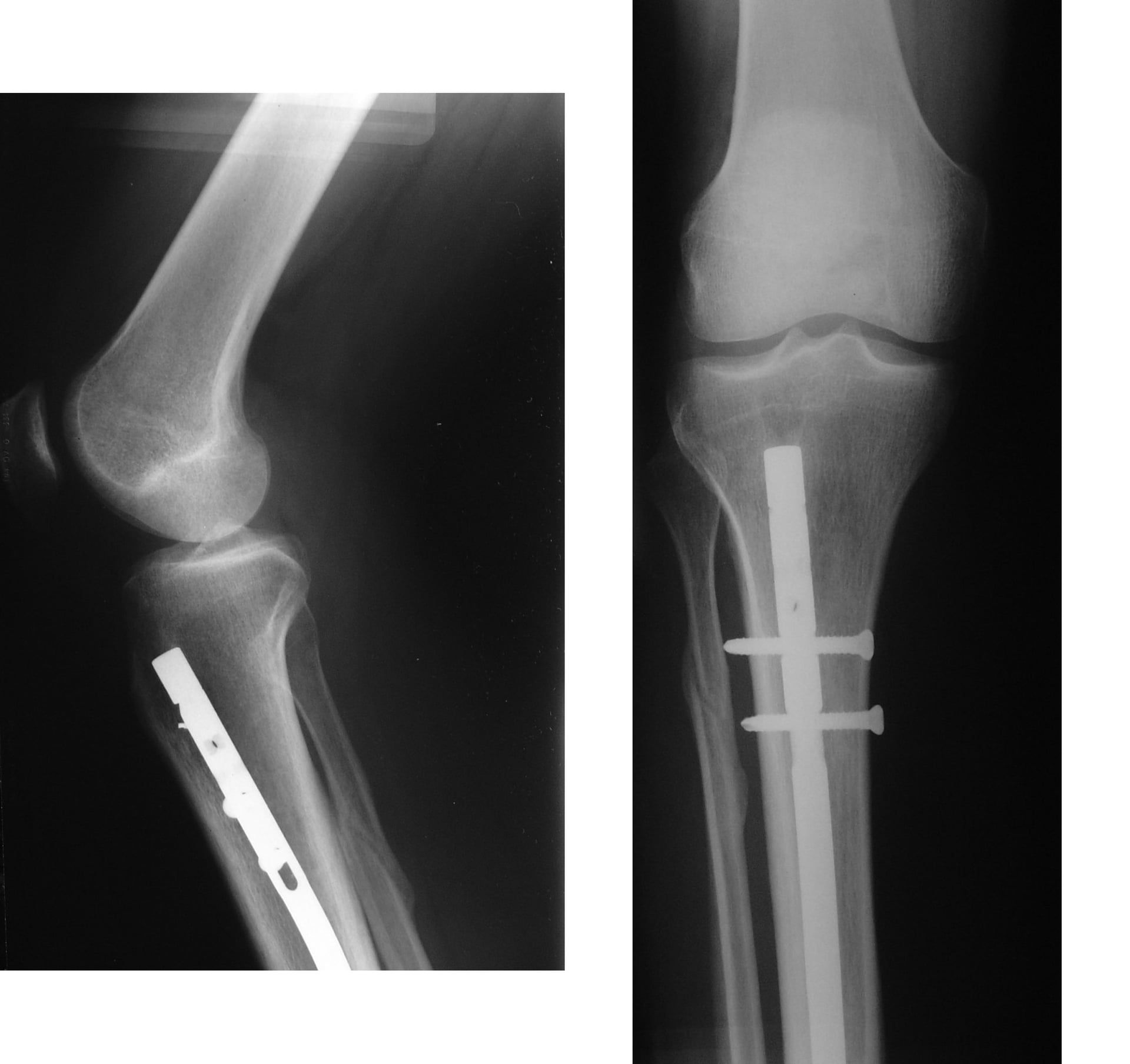
An incorrect start point is the single commonest technical cause of malreduction. Too medial drives the nail into valgus; too lateral risks the lateral plateau and varus; too posterior or too low produces apex-anterior angulation and a posterior nail track. Confirm the guide wire medial to the lateral spine on AP and on the anterior down-slope on lateral before opening the cortex, and avoid over-penetrating posteriorly toward the popliteal vessels with the wire or opening reamer.
Thermal necrosis and a ring sequestrum follow aggressive reaming of a tight canal, blunt reamers, or reaming with the tourniquet up. Use sharp sequential reamers with gentle advancing pressure, irrigate, deflate the tourniquet before reaming, and ream only about 1 to 1.5 mm over the chosen nail diameter.
The superficial peroneal nerve is at risk with lateral-to-medial distal screws. Make a real incision, open onto bone, protect the soft tissues with a sleeve, and reconfirm a true perfect circle before drilling to avoid a "near-far" miss of the locking hole. A single distal lock in a distal-third fracture gives inadequate rotational and angular control — use at least two, ideally multiplanar.
Before scrubbing I decide the approach from the fracture level. For a proximal-third or segmental fracture I choose the suprapatellar semi-extended approach because the relaxed extensor mechanism and easy lateral imaging help me avoid apex-anterior and valgus deformity. I template the nail off the good leg and plan exactly where my blocking screws will sit.
In the proximal third the wide metaphysis gives the nail little endosteal contact, so the fragment is controlled by soft-tissue forces, not the implant. The flexed-knee infrapatellar position and patellar-tendon pull extend the proximal fragment into procurvatum while the pes anserinus and medial structures drive valgus. The semi-extended suprapatellar position relaxes these forces, an anterior-enough start point counters procurvatum, and blocking screws on the concave side redirect and lock the nail.
Aftercare & Complications
Post-operative protocol | Phase | Timing | Weight-bearing & immobilisation | Therapy & monitoring | |-------|--------|----------------------------------|----------------------| | Early | Day 0 to 14 | Serial neurovascular and compartment checks; elevate the limb | Stable transverse or short-oblique patterns — weight-bearing as tolerated; comminuted, segmental or metaphyseal — protected partial weight-bearing. Multimodal analgesia (escalating need is a red flag for compartment syndrome); thromboprophylaxis | | Rehabilitation | 2 to 6 weeks | Advance weight-bearing as callus appears | Knee and ankle range of motion; serial AP and lateral radiographs to confirm union | | Return to function | 6 weeks to 6 months | Full weight-bearing once united | Strengthening and proprioception; light or desk work within a few weeks, manual work and sport typically once united (around 3 to 6 months) | | Hardware | Beyond union | Nail removal only if symptomatic, commonly beyond a year | Counsel that removal may not relieve anterior knee pain | Special situations - Open fractures. Give antibiotics early (ideally within an hour) and tetanus prophylaxis, and thoroughly debride devitalised tissue before definitive fixation. Nailing is acceptable for many open shafts after debridement — reamed and unreamed nailing show no significant difference in open fractures (SPRINT). Plan combined orthoplastic "fix-and-flap" cover for high-grade (IIIB) injuries, and use temporary damage-control external fixation first in the unstable polytrauma patient or a grossly contaminated wound, converting to a nail once the patient and soft tissues allow.
- Proximal third. Deforms into apex-anterior and valgus — use the semi-extended or suprapatellar approach, a correct anterior-enough start point, and blocking screws on the concave side; confirm alignment carefully on the lateral view, where procurvatum is easily missed.
- Distal third. Limited distal segment for fixation — use at least two, ideally multiplanar, distal interlocks, blocking screws, and consider fibular plating to restore length and resist valgus; plating is a reasonable alternative when nail control is inadequate. Complications
- Incidence
- Common (commonly around 47% with infrapatellar nailing)
- Recognition
- Pain on kneeling and at the front of the knee, often persisting long-term; the leading late complaint
- Prevention and management
- Prevention: consider the suprapatellar approach; avoid a proud proximal nail; correct entry technique. Management: physiotherapy and analgesia; nail removal may help but does NOT reliably resolve pain — counsel accordingly
- Incidence
- Higher in metaphyseal patterns (proximal-third apex-anterior or valgus is most classic)
- Recognition
- Angulation greater than 5 degrees, malrotation greater than 10 degrees, or shortening on post-op imaging or clinical assessment
- Prevention and management
- Prevention: correct entry point, semi-extended or suprapatellar position, blocking screws on the concave side, multiplanar distal locks, fibular fixation. Management: revision with blocking screws or re-nailing; corrective osteotomy if established and symptomatic
- Incidence
- Tibia is the commonest site; proximal third is highest risk
- Recognition
- Pain out of proportion, pain on passive stretch, tense compartments, paraesthesia (late); a clinical diagnosis
- Prevention and management
- Prevention: vigilance before and after nailing; do not rely on nailing to decompress. Management: urgent four-compartment fasciotomy; do not delay for pressure monitoring if the clinical picture is clear
- Incidence
- Higher in open and high-energy fractures
- Recognition
- Persistent pain and motion at the fracture, lack of bridging callus beyond expected time on serial radiographs
- Prevention and management
- Prevention: preserve soft tissues, optimal stability, reamed nailing in closed fractures, manage open fractures well. Management: dynamisation, exchange (reamed) nailing, bone grafting; address infection if present
- Incidence
- Higher in open fractures; deep infection uncommon in closed
- Recognition
- Pain, swelling, erythema, discharge, raised inflammatory markers; may present as infected nonunion
- Prevention and management
- Prevention: early antibiotics (within an hour) and thorough debridement in open fractures, sterile technique. Management: debridement, antibiotics, possible implant retention until union then removal, or staged revision for established infection
- Incidence
- Uncommon
- Recognition
- Broken locking screw or nail, loss of fixation and recurrence of deformity on radiographs (often at a long working length or nonunion)
- Prevention and management
- Prevention: appropriately sized nail, adequate interlocks, avoid excessive working length, achieve union promptly. Management: revise fixation or exchange nail, bone graft the nonunion that caused the cyclical loading failure
- Incidence
- Rare
- Recognition
- Cortical necrosis or ring sequestrum on later imaging; can predispose to nonunion or infection
- Prevention and management
- Prevention: sharp sequential reamers, gentle pressure, irrigation, tourniquet down during reaming, ream only modestly over nail size. Management: treat the resulting nonunion or sequestrum; debride and graft as needed
- Incidence
- Uncommon
- Recognition
- Superficial peroneal nerve numbness after distal locking; vascular injury from posterior over-penetration is rare but serious
- Prevention and management
- Prevention: open onto bone for distal locking and protect soft tissues; avoid posterior over-penetration near the popliteal vessels. Management: explore and repair nerve or vessel injury; document deficits early
Viva & Exam Focus
ENTRYENTRY — the tibial nail start point and set-up
TIBIATIBIA — assessing the tibial shaft fracture
The trap. An incorrect start point is the single commonest technical cause of malreduction. Too medial drives valgus; too lateral risks the lateral plateau and varus; too posterior causes apex-anterior angulation. The fix. AP target is just medial to the lateral tibial spine; lateral target is the anterior margin of the articular surface (the down-slope). Confirm both views on the image intensifier before reaming the entry.
Why. The wide proximal metaphysis gives the nail little canal contact, so the fragment is controlled by soft-tissue forces. The flexed-knee infrapatellar position and patellar-tendon pull produce apex-anterior (procurvatum) and valgus. The fix. Use the semi-extended suprapatellar approach, blocking screws on the concave side, and a correct anterior-enough start point.
Why critical. The tibia is the commonest site; the proximal third is the highest-risk region. Reaming and nailing do not relieve, and may raise, compartment pressure. The action. Serial clinical assessment before and after surgery — pain out of proportion and pain on passive stretch are the earliest signs. Maintain a low threshold for fasciotomy; do not be falsely reassured by definitive fixation.
Why. The distal metaphyseal flare offers poor nail-to-canal contact, allowing valgus or varus and translation with loss of rotational control. The fix. Use at least two distal interlocks (ideally multiplanar), blocking screws, and consider fibular plating to restore length and resist valgus.
Mechanism. Aggressive reaming in a tight canal, blunt reamers, or reaming with the tourniquet up generates heat that can cause cortical necrosis and a ring sequestrum. Prevention. Sharp sequential reamers, gentle pressure, irrigation, avoid a prolonged tourniquet during reaming, and ream only about 1 to 1.5 mm over the chosen nail diameter.
Principle. Most open tibial fractures are still amenable to nailing, but require early antibiotics, tetanus cover and thorough surgical debridement before definitive fixation. Evidence. Early antibiotics (within an hour) reduce infection; SPRINT found no significant difference between reamed and unreamed nailing in open fractures. Plan soft-tissue cover with plastics for high-grade injuries (fix-and-flap).
Clinical Decision Scenarios
Practise clinical reasoning and management decisions out loud
“A 32-year-old man sustains a closed proximal-third tibial shaft fracture in a motorcycle crash. You plan intramedullary nailing. How will you minimise the risk of malreduction, and what specific deformity are you guarding against?”
“Compare reamed and unreamed tibial nailing. What does the evidence say, and how does open versus closed status change your decision?”
“A patient develops increasing pain six hours after tibial nailing of a closed mid-shaft fracture, requiring escalating analgesia. The leg is splinted. What is your concern and how do you proceed?”
Indications and principles
- Indicated for most displaced or unstable diaphyseal tibial shaft fractures, closed and many open, segmental and polytrauma
- Harder with very proximal or distal metaphyseal patterns — the nail has poor canal contact there
- Contraindicated with active sepsis at the entry or canal and in skeletally immature patients (open physes)
- An IM nail is a LOAD-SHARING device along the mechanical axis; interlocks control length and rotation
- Working length is the unsupported nail across the fracture; a longer working length reduces stiffness
Entry point (the cardinal step)
- AP: just MEDIAL to the lateral tibial spine
- Lateral: at the ANTERIOR margin of the articular surface (the down-slope of the plateau)
- Too medial equals valgus; too lateral equals varus; too posterior or low equals apex-anterior
- Confirm BOTH fluoroscopic views before opening the cortex
- Alignment goals: less than 5 degrees angulation, less than 1 cm shortening, less than 10 degrees malrotation
Infrapatellar versus suprapatellar
- Infrapatellar: knee flexed; transtendinous or medial paratendinous; quick and familiar; more anterior knee pain; proximal-third deformity risk
- Suprapatellar: semi-extended; through the quadriceps tendon with a protective sleeve across the patellofemoral joint
- Suprapatellar advantages: relaxed extensor pull, easier lateral imaging, better for proximal, distal and segmental fractures
- Suprapatellar needs a healthy patellofemoral joint and meticulous cartilage protection
- The SUPER trial and related studies compared anterior knee pain between the approaches
Reamed versus unreamed (SPRINT)
- Reamed equals a larger and stiffer nail, larger locks, osteogenic debris, but disrupts the endosteal supply
- CLOSED fractures: reamed nailing reduced reoperation events (favoured)
- OPEN fractures: no significant difference between reamed and unreamed
- The periosteal supply (key in open fractures) is preserved by reaming
- Ream about 1 to 1.5 mm over nail size with sharp reamers, tourniquet down, to avoid thermal necrosis
Deformity tools
- Proximal-third classic deformity: apex-anterior (procurvatum) and valgus
- The semi-extended or suprapatellar position relaxes the deforming forces
- Blocking (Poller) screws on the CONCAVE side redirect and lock the nail path
- Distal-third: at least two, ideally multiplanar, distal interlocks for rotational and angular control
- Distal-third valgus instability: plate the fibula (often first) to restore length and resist valgus
Danger zones
- Incorrect entry point — the commonest technical cause of malreduction
- Compartment syndrome — the tibia is the commonest site; proximal third highest risk; nailing does not protect
- Superficial peroneal nerve at distal (lateral) interlocking — open onto bone and protect
- Popliteal vessels posteriorly — avoid over-penetration with the wire or reamer
- Patellofemoral cartilage in the suprapatellar approach — keep the protective sleeve seated
Complications
- Anterior knee pain: the commonest long-term complaint (around 47% infrapatellar); nail removal may not relieve it
- Malalignment: highest in metaphyseal patterns; use entry, position, blocking screws and fibular fixation
- Compartment syndrome: a clinical diagnosis; urgent four-compartment fasciotomy
- Nonunion or delayed union: higher in open or high-energy; treat with dynamisation, exchange nailing, grafting
- Infection: higher in open fractures — early antibiotics and debridement are protective
Open fractures
- Antibiotics early (ideally within an hour) — delay increases infection
- Tetanus prophylaxis and thorough debridement before definitive fixation
- Nailing is acceptable for many open shaft fractures after debridement
- Reamed and unreamed are equivalent in open fractures (SPRINT)
- Plan orthoplastic fix-and-flap cover for high-grade (IIIB) injuries; consider damage-control external fixation first if unstable
Background & Evidence
Epidemiology. The tibial shaft is the most frequently fractured long bone, classically in a bimodal distribution — high-energy fractures in young men (motor vehicle and sporting trauma) and low-energy fractures in older adults — with open fractures common because of the thin anteromedial soft-tissue envelope. The proximal third carries the highest risk of acute compartment syndrome. Surgical anatomy. The tibia is subcutaneous along its anteromedial border, which explains both the frequency of open fractures and the importance of careful soft-tissue handling. The proximal articular surface slopes posteriorly, so on the lateral image the anterior down-slope of the plateau marks the correct sagittal entry. The tibial tuberosity and patellar tendon are the infrapatellar landmarks; the quadriceps tendon and patella are the suprapatellar landmarks. Biomechanics of the intramedullary nail. An IM nail is a load-sharing device positioned along the mechanical axis — it shares load with the surrounding bone, unlike a plate which is largely load-bearing. Working length is the unsupported length of nail between the proximal and distal points of fixation across the fracture; a longer working length reduces bending and torsional stiffness, so minimising the comminution gap and using a well-fitting nail matter. A larger-diameter (reamed) nail has greater bending and torsional stiffness and allows larger interlocking screws, and the interlocking screws themselves control length and rotation, converting the nail into a stable construct in unstable patterns. Why proximal-third fractures deform. In the proximal third the wide metaphysis gives the nail little endosteal contact, so the fragment is controlled by soft tissues, not the implant. The classic deformity is apex-anterior (procurvatum) and valgus: the patellar tendon and extensor mechanism extend the proximal fragment (apex-anterior) when the knee is flexed for infrapatellar nailing, and the pes anserinus and medial structures contribute to valgus. The countermeasures are a sufficiently anterior start point, the semi-extended (suprapatellar) position, and blocking screws on the concave side. Neurovascular structures at risk. The superficial peroneal nerve is at risk with lateral-to-medial distal locking — use a careful open or free-hand perfect-circle technique and protect the soft tissues with a sleeve. The popliteal vessels lie posterior to the proximal tibia, so avoid over-penetration posteriorly with guide wires and reamers. The saphenous nerve and vein run along the medial leg and are at risk during medial approaches and distal medial locking. In the suprapatellar approach the patellofemoral cartilage is at risk, mitigated by a protective sleeve and case selection. The fibula in tibial fractures. In distal-third tibial fractures an associated fibular fracture allows the tibia to drift into valgus and shorten. Plating the fibula first restores length and resists valgus, improving the reduction before tibial nailing — particularly useful when distal nail control is limited. In mid-shaft fractures fibular fixation is usually unnecessary. Key evidence. The SPRINT trial (1319 patients across 29 centres) remains the landmark study: in closed tibial shaft fractures, reamed nailing reduced the rate of the primary composite outcome (reoperation events) compared with unreamed nailing, while in open fractures there was no significant difference between the two; it also advised not assessing reoperation events before six months. Katsoulis and colleagues established anterior knee pain as the leading long-term complaint after tibial nailing (commonly around 47% across pooled infrapatellar series), often only partially relieved by nail removal. Krettek and colleagues described the blocking (Poller) screw — placed on the concave side of the anticipated deformity to narrow the functional canal and steer the nail — for metaphyseal tibial fractures. Lack and colleagues showed that early antibiotic administration (within about an hour) reduces infection in open tibial fractures. The classic epidemiological description of tibial fracture patterns is Court-Brown and McBirnie, and the LEAP study informs limb salvage versus amputation decisions in severe open injuries.
References
Randomized trial of reamed and unreamed intramedullary nailing of tibial shaft fractures (SPRINT)
- 1319 adults with tibial shaft fractures randomised to reamed or unreamed intramedullary nailing across 29 centres
- In CLOSED fractures, reamed nailing reduced the rate of the primary composite outcome (reoperation events) compared with unreamed nailing
- In OPEN fractures there was no significant difference between reamed and unreamed nailing
- Re-operation events should not be assessed before 6 months as some early dynamisations or exchanges may be unnecessary
Suprapatellar versus infrapatellar tibial nailing and anterior knee pain
- Randomised comparison of suprapatellar (semi-extended) versus infrapatellar tibial nailing
- Anterior knee pain is the commonest long-term complaint after infrapatellar nailing; suprapatellar nailing aims to reduce this and eases reduction of proximal and distal patterns
- The suprapatellar approach traverses the patellofemoral joint, so cartilage protection with a dedicated sleeve and case selection (a healthy PFJ) are essential
Anterior knee pain after intramedullary nailing of the tibia
- Systematic review of anterior knee pain after tibial nailing
- Anterior knee pain was reported in a high proportion of patients (commonly around 47% across pooled series), making it the leading long-term complaint
- Pain is often only partially relieved by nail removal, so removal is not a guaranteed solution
The use of blocking (Poller) screws in nailing of proximal and distal tibial fractures
- Description of blocking (Poller) screws to narrow the functional medullary canal in the metaphysis and steer the nail
- The screw is placed on the CONCAVE side of the anticipated deformity to redirect the nail and resist recurrence of the malalignment
- Particularly effective for proximal-third (apex-anterior or valgus) and distal-third metaphyseal fractures where the nail otherwise has poor canal contact
Effect of timing of antibiotic administration on infection in open fractures
- Cohort of open tibial fractures examining antibiotic timing and debridement
- Delay in antibiotic administration beyond approximately 66 minutes was associated with a significantly increased infection rate
- Supports early (within one hour) antibiotics alongside timely debridement for open tibial fractures
The epidemiology of tibial fractures
- Classic epidemiological description of tibial shaft fracture patterns and demographics
- Established the bimodal age distribution and the high proportion of open fractures from the thin anteromedial soft-tissue envelope
Outcomes after reconstruction or amputation of leg-threatening injuries (LEAP)
- Prospective cohort of severe lower-limb trauma comparing reconstruction with amputation
- Outcomes at two years were similar between limb salvage and amputation for many patients, while functional outcomes depended heavily on soft-tissue and nerve injury
- Informs shared decision-making for limb salvage versus amputation in severe open tibial injuries