OrthoVellum
Knowledge Hub

Study

  • Topics
  • MCQs
  • ISAWE
  • Operative Surgery
  • Flashcards

Company

  • About Us
  • Editorial Policy
  • Contact
  • FAQ
  • Blog

Legal

  • Terms of Service
  • Privacy Policy
  • Cookie Policy
  • Medical Disclaimer
  • Copyright & DMCA
  • Refund Policy

Support

  • Help Center
  • Accessibility
  • Report an Issue
OrthoVellum

© 2026 OrthoVellum. For educational purposes only.

Not affiliated with the Royal Australasian College of Surgeons.

Blount Disease (Tibia Vara)

Back to Topics
Contents
0%

Blount Disease (Tibia Vara)

Comprehensive Orthopaedic exam guide to Blount disease including Langenskiöld classification, infantile vs adolescent forms, metaphyseal-diaphyseal angle measurement, bracing protocols, and surgical management strategies including proximal tibial osteotomy and guided growth techniques.

complete
Updated: 2024-12-18
High Yield Overview

BLOUNT DISEASE (TIBIA VARA)

Pathologic genu varum | Medial physeal growth disturbance | Infantile vs adolescent forms

Under 4 yearsInfantile onset
Over 8 yearsAdolescent onset
11 degreesCritical MDA (Drennan)
16 degreesNormal MDA threshold
3:1Male to female ratio

LANGENSKIÖLD CLASSIFICATION (Stages I-VI)

Stage I
PatternIrregular metaphysis, mild medial beaking
TreatmentObservation or bracing
Stage II
PatternDefined metaphyseal defect, medial depression
TreatmentBracing (KAFO)
Stage III
PatternMetaphyseal fragmentation, stepped plateau
TreatmentConsider surgery if under 4 years
Stage IV
PatternMedial metaphyseal overgrowth, physeal slope
TreatmentOsteotomy indicated
Stage V
PatternMedial epiphyseal separation, double metaphysis
TreatmentComplex osteotomy
Stage VI
PatternMedial physeal bar, permanent deformity
TreatmentBar resection + osteotomy

Critical Must-Knows

  • Differentiation from physiologic genu varum: Physiologic resolves by age 2-3 years, bilateral, MDA under 11 degrees, no radiographic metaphyseal changes
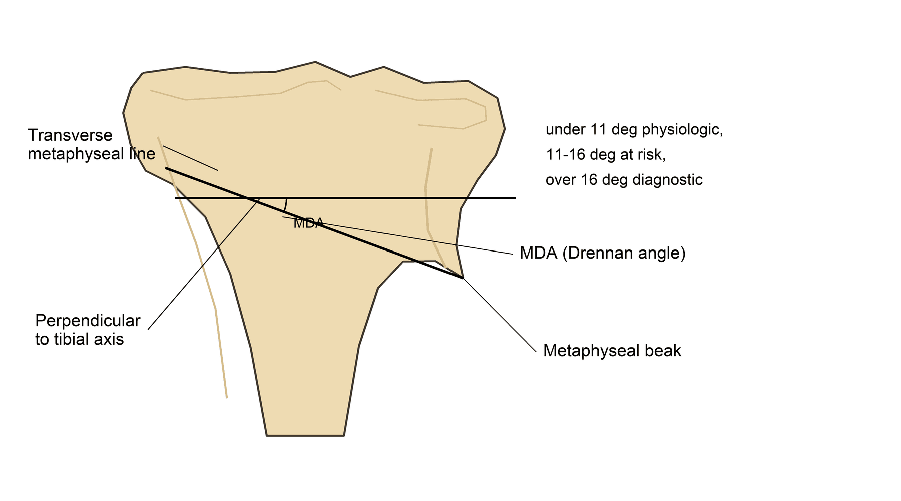
  • Critical imaging measurement: Metaphyseal-diaphyseal angle (Drennan) measured on AP radiograph - over 11 degrees predicts progression, over 16 degrees diagnostic
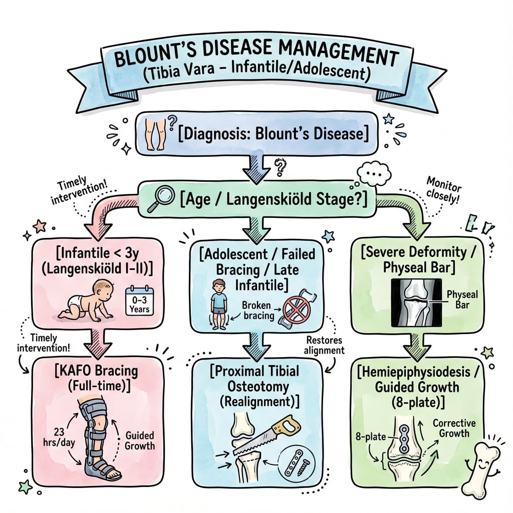
  • Two distinct forms: Infantile (under 4 years, often bilateral, better bracing response) vs adolescent (over 8 years, unilateral, obesity-related, poor bracing response)
  • Bracing window: Most effective for Stage I-II infantile disease in children aged 1-3 years, requires KAFO (knee-ankle-foot orthosis) worn 23 hours daily for 1-2 years
  • Surgical timing: Osteotomy preferred before age 4 years (better remodeling), after age 4 years less predictable, adolescent form requires osteotomy at presentation

Examiner's Pearls

  • "
    Langenskiöld Stage IV or higher = surgery required
  • "
    MDA over 16 degrees = pathologic, will not resolve spontaneously
  • "
    Beware combined femoral and tibial deformity in severe cases
  • "
    Physeal bar develops in neglected cases - Stage VI disease

Exam Warning

The Physiologic Mimic

Physiologic Genu Varum: Bilateral, resolves by age 2-3. MDA less than 11°. No metaphyseal changes.

Critical Differentiators

Rickets: Widened physis, biochemical abnormalities. FFCD: Unilateral, abrupt angulation. Dysplasia: Multiple joints, short stature.

Examiner's Key Tool

Drennan's Angle (MDA): The single most important measurement. Langenskiöld Staging: Determines treatment.

At a Glance

The OnePagerSummary above provides a comprehensive at-a-glance overview of Blount disease including the Langenskiöld classification, key statistics, must-know concepts, and exam pearls.

Mnemonic

MDA - Key Angles for Blount Disease

M
Measure MDA
Metaphyseal-diaphyseal angle on standing AP radiograph
D
Diagnostic threshold
MDA over 16 degrees = diagnostic of pathologic Blount disease
A
At-risk zone
MDA 11-16 degrees = at risk for progression, needs monitoring

Memory Hook:MDA guides everything - under 11° is physiologic, 11-16° is concerning, over 16° is diagnostic. Always measure the MDA first!

Overview and Epidemiology

Overview and Epidemiology

Blount disease (tibia vara) is a pathologic growth disturbance of the medial aspect of the proximal tibial physis, resulting in progressive genu varum deformity. It represents a failure of normal endochondral ossification at the medial metaphysis.

Two distinct clinical entities:

  1. Infantile form (most common):

    • Onset before age 4 years
    • Often bilateral (60-80%)
    • More common in early walkers
    • African and Hispanic descent (3-4x higher)
    • Better response to conservative management
  2. Adolescent form:

    • Onset after age 8-10 years
    • Usually unilateral (80%)
    • Strong association with obesity
    • Less predictable, worse prognosis
    • Requires surgical management

Australian epidemiology:

  • Rare in Caucasian Australian population
  • Higher prevalence in Aboriginal and Torres Strait Islander children
  • Increasing incidence correlating with childhood obesity epidemic
  • Male predominance (3:1 ratio)

Risk factors:

  • Early walking (before 10 months)
  • Obesity (especially adolescent form)
  • African or Hispanic descent
  • Vitamin D deficiency (controversial)
  • Mechanical stress on immature physis

The condition is progressive if untreated, leading to permanent deformity, lateral thrust gait, lateral compartment osteoarthritis, and functional disability.

Infantile Form Features

  • Onset: 1-3 years
  • Often bilateral (60-80%)
  • Responds to bracing if early
  • Better remodeling potential
  • Associated with early walking
  • Langenskiöld stages I-III at presentation

Adolescent Form Features

  • Onset: 8+ years
  • Usually unilateral (80%)
  • Poor response to bracing
  • Limited remodeling capacity
  • Strong obesity association
  • Requires surgical management

Key Differentiators

  • MDA over 11° = predictive of progression
  • MDA over 16° = diagnostic of Blount disease
  • Metaphyseal beaking on radiograph
  • Progressive deformity after age 2 years
  • Unilateral involvement suggests pathologic

Examination Red Flags

  • Lateral thrust gait (dynamic varus thrust)
  • Internal tibial torsion (common association)
  • Leg length discrepancy (medial overgrowth)
  • Ligamentous laxity (LCL stretching)
  • Fixed varus deformity (advanced disease)
Mnemonic

INFANT - Infantile Blount Disease Features

I
Infancy onset
Presents before age 4 years, typically 1-3 years
N
Normal at birth
Physiologic varus at birth becomes pathologic if does not correct
F
Frequently bilateral
60-80% of infantile cases are bilateral
A
African descent
Higher prevalence in African and Hispanic populations
N
Night bracing ineffective
Must be full-time (23 hrs/day) bracing to work
T
Three years critical
Age under 3 years for best bracing outcomes

Memory Hook:INFANT Blount - remember these key features of the infantile form to distinguish from physiologic bowing and guide appropriate bracing trial.

Anatomy and Pathophysiology

Normal Proximal Tibial Growth

Physis anatomy:

  • Proximal tibial physis contributes 55-60% of tibial growth
  • Medial and lateral physeal growth should be symmetric
  • Growth rate approximately 6mm per year (3mm medial, 3mm lateral)
  • Peak growth velocity in infancy and early adolescence

Normal lower limb alignment evolution:

  • Birth: 10-15 degrees varus (physiologic)
  • Age 12-18 months: Neutral mechanical axis
  • Age 2-4 years: 10-12 degrees valgus (physiologic)
  • Age 6-7 years: 5-7 degrees valgus (adult alignment)

Mechanical axis:

  • Line from femoral head center to ankle center
  • Normally passes through medial tibial spine (or just lateral)
  • In Blount disease, passes medial to knee (medial compartment overload)

Pathophysiology of Blount Disease

Hueter-Volkmann principle:

  • Increased compression → decreased growth
  • Decreased compression → increased growth
  • Medial physeal compression from mechanical overload → growth suppression
  • Creates vicious cycle: varus → increased medial stress → more varus

Histologic findings:

  • Disorganized physeal architecture (loss of columnar arrangement)
  • Fibrocartilaginous tissue replacing normal physeal cartilage
  • Thickened periosteum on medial aspect
  • Metaphyseal cartilage islands (failure of resorption)
  • Progressive changes from Stage I to Stage VI

Mechanical factors:

  • Early walking increases medial compartment stress
  • Obesity dramatically increases joint reaction forces
  • Internal tibial torsion increases frontal plane moment arm
  • Creates asymmetric loading of medial physis

Progressive deformity components:

  1. Varus angulation (primary deformity)
  2. Internal tibial torsion (60-80% of cases)
  3. Procurvatum (anterior bowing) in advanced cases
  4. Medial physeal bar formation (Stage VI)
  5. Leg length discrepancy (lateral overgrowth)
Mnemonic

BLOUNT - Key Pathophysiology Features

B
Bowing
Progressive varus deformity from medial physeal suppression
L
Loading
Asymmetric loading per Hueter-Volkmann principle causes growth inhibition
O
Obesity
Major risk factor, especially for adolescent form
U
Unilateral
More common in adolescent form (80%), infantile often bilateral
N
Necrosis
Not true necrosis but fibrocartilaginous replacement of physis
T
Torsion
Internal tibial torsion associated in 60-80% of cases

Memory Hook:Remember BLOUNT when you see a child with progressive genu varum - think about the Loading forces causing Bowing in Obese children.

Classification

Classification Systems

The Langenskiöld classification (1952, revised 1964) describes six progressive stages of infantile Blount disease based on radiographic appearance of the proximal medial tibial metaphysis and epiphysis. This is the gold standard classification used worldwide.

Critical for surgical decision-making: Stages I-III may respond to bracing, Stages IV-VI require surgery.

Irregularity and Mild Beaking
Defined Metaphyseal Defect
Metaphyseal Fragmentation
Medial Metaphyseal Overgrowth
Medial Epiphyseal Separation
Physeal Bar and Permanent Deformity
Langenskiöld classification stages I-VI for Blount disease
Click to expand
Langenskiöld classification of infantile Blount disease (tibia vara) showing six progressive stages of medial proximal tibial pathology: Stage I - mild metaphyseal beaking; Stage II - defined metaphyseal defect with epiphyseal wedging; Stage III - metaphyseal fragmentation; Stage IV - marked metaphyseal overgrowth with 'beak' beneath epiphysis; Stage V - medial epiphyseal separation with double contour; Stage VI - physeal bar formation with fusion. Stages I-III may respond to bracing, while stages IV-VI require surgical intervention.Credit: Khanfour AA et al., J Orthop Surg Res (PMC3490732) - CC BY 4.0

Exam Warning

Stage I vs II

Beak vs Defect: Stage II has a clear metaphyseal depression/defect.

Stage III vs IV

The Beak: Stage IV shows metaphyseal overgrowth projecting beneath epiphysis.

Stage V vs VI

The Bridge: Stage VI shows a medial physeal bar (bony bridge).

Management Rule

Treatment: Stages I-III = Brace (if less than 3y). Stages IV-VI = Surgery.

Drennan's Metaphyseal-Diaphyseal Angle (MDA):

  • Measured on AP radiograph
  • Line tangent to medial and lateral metaphyseal beaks
  • Line along longitudinal axis of tibial shaft
  • Normal: under 11 degrees
  • 11-16 degrees: Predictive of progression to Blount disease
  • Over 16 degrees: Diagnostic of pathologic tibia vara
  • Most important single measurement for differentiating physiologic from pathologic bowing

Tibiofemoral angle (TFA):

  • Angle between femoral and tibial mechanical axes
  • Normal at age 2-3 years: 0-5 degrees valgus
  • Blount disease: progressive varus (10-30 degrees or more)
  • Measured on standing long-leg radiograph

Levine and Drennan classification (alternative):

  • Group 1: Normal or physiologic (MDA under 11 degrees)
  • Group 2: Moderate changes (MDA 11-16 degrees, monitor closely)
  • Group 3: Severe changes (MDA over 16 degrees, Blount disease confirmed)

Schoenecker modification for adolescent Blount:

  • Describes angular deformity, physeal status, and associated leg length discrepancy
  • Less commonly used than Langenskiöld for infantile form

These classification systems guide treatment decisions based on disease severity and progression.

Mnemonic

MEDIAL - Langenskiöld Progressive Stages

M
Mild beaking
Stage I - Irregular metaphysis with mild medial beaking
E
Evident defect
Stage II - Defined medial metaphyseal defect/depression
D
Disrupted fragments
Stage III - Fragmentation and stepped appearance
I
Increased overgrowth
Stage IV - Medial metaphyseal overgrowth (beak)
A
Apart epiphysis
Stage V - Medial epiphysis appears separated
L
Locked/bar
Stage VI - Medial physeal bar with fusion

Memory Hook:The MEDIAL progression tracks increasing severity - from Mild beaking to Locked physeal bar. Stage IV is when surgery becomes mandatory.

Clinical Presentation

Clinical Presentation and History

Presenting complaint:

  • Parents notice bowed legs that are not improving or are worsening
  • Child has abnormal gait (waddling, lateral thrust)
  • Knee or leg pain in older children (uncommon in infantile form)
  • Concerns about shoe wear (lateral sole wear)

Age at presentation:

  • Infantile form: Typically 18 months to 3 years (after walking established)
  • Adolescent form: 8-15 years (often coincides with growth spurt)

Key history questions:

  • Age of first walking (under 10 months = risk factor)
  • Birth weight and current weight (obesity assessment)
  • Unilateral vs bilateral deformity
  • Progressive or static deformity
  • Family history of bowed legs or metabolic bone disease
  • Dietary history (vitamin D, calcium intake)
  • Previous bracing or treatment

Associated symptoms:

  • Pain uncommon in infantile form
  • Adolescent form may have knee pain (medial compartment overload)
  • Difficulty running or sports participation
  • Cosmetic concerns

Red flags for alternative diagnosis:

  • Short stature (consider skeletal dysplasia)
  • Other joint involvement (consider skeletal dysplasia)
  • Pathologic fractures (consider rickets or metabolic disease)
  • Developmental delay (consider genetic syndrome)

Physical Examination

Gait assessment (most important):

  • Lateral thrust gait: Dynamic varus thrust during stance phase
  • Indicates functional medial compartment insufficiency
  • Predictor of progression
  • Waddling gait if bilateral
  • Reduced cadence and stride length

Standing alignment:

  • Intercondylar distance: Measure between medial femoral condyles with ankles together
  • Normal under 6 cm in young children
  • Over 6-8 cm suggests pathologic bowing
  • Asymmetry if unilateral (compare knee heights)

Varus stress test:

  • Apply varus force to knee in extension
  • Increased medial gapping suggests LCL laxity (chronic lateral stretch)
  • Fixed varus = bony deformity (advanced disease)

Rotational profile:

  • Internal tibial torsion: Thigh-foot angle (normally 10 degrees external)
  • Present in 60-80% of Blount disease cases
  • Femoral version: Check internal rotation in prone position
  • May have combined femoral anteversion

Leg length measurement:

  • True leg length: ASIS to medial malleolus
  • Apparent leg length: Umbilicus to medial malleolus
  • Leg length discrepancy common (affected side may be shorter or longer)

Knee examination:

  • Range of motion: Usually full, unless severe deformity
  • Ligament stability: LCL may be attenuated
  • Tenderness: Medial metaphysis (uncommon)

Neurologic examination:

  • Document distal neurovascular status
  • Exclude cerebral palsy or neuromuscular disorder

General examination:

  • BMI calculation and obesity assessment
  • Height and weight percentiles
  • Signs of rickets (widened wrists, costochondral beading)
  • Signs of skeletal dysplasia
Mnemonic

THRUST - Key Clinical Examination Findings

T
Thrust gait
Lateral thrust during stance phase - pathognomonic sign
H
Height disparity
Leg length discrepancy and asymmetric knee heights
R
Rotational profile
Internal tibial torsion in 60-80% of cases
U
Unilateral vs bilateral
Unilateral more common in adolescent, bilateral in infantile
S
Stability testing
LCL laxity from chronic lateral stretch
T
True leg length
Measure ASIS to medial malleolus for discrepancy

Memory Hook:Watch for the lateral THRUST gait - it's the most important dynamic clinical sign that predicts progression and need for intervention.

Infantile vs Adolescent Blount Disease

Investigations

Investigations - Radiographic Imaging

Standing anteroposterior (AP) radiograph:

  • Gold standard for diagnosis and staging
  • Must be standing (weight-bearing) for accurate alignment assessment
  • Include both knees for comparison
  • Key measurements:
    • Metaphyseal-diaphyseal angle (MDA) - Drennan angle
    • Tibiofemoral angle (TFA)
    • Intercondylar distance
  • Langenskiöld staging based on medial metaphyseal changes
  • Look for: Medial beaking, metaphyseal defect, physis widening, epiphyseal wedging

Standing long-leg radiograph (full-length):

  • Mechanical axis alignment - line from femoral head to ankle
  • Should be obtained when planning surgery
  • Identifies femoral vs tibial contribution to deformity
  • Measures true leg length discrepancy
  • Mechanical axis deviation (MAD): Distance from knee center to mechanical axis

Lateral radiograph:

  • Assess for procurvatum deformity (anterior bowing)
  • Evaluate tibial slope
  • Less critical for diagnosis but useful for surgical planning

Oblique views:

  • Occasionally helpful for evaluating physeal bar in Stage VI
  • Not routinely required

Follow-up imaging schedule:

  • Initial diagnosis: Standing AP both knees
  • If under 3 years with Stage I-II and bracing: Repeat every 3-4 months
  • If observation: Repeat every 6 months until skeletal maturity
  • Post-operative: Immediate, 6 weeks, 3 months, then every 6 months

Advanced Imaging

CT scan:

  • Indications:
    • Suspected physeal bar (Stage VI)
    • Pre-operative planning for complex deformity
    • Assessment of three-dimensional deformity
  • Can create 3D reconstructions
  • Quantify physeal bar size and location
  • Radiation concern in young children

MRI:

  • Indications:
    • Evaluate physeal bar without radiation (preferred in children)
    • Assess cartilaginous structures not visible on radiograph
    • Differentiate Blount from focal fibrocartilaginous dysplasia
  • T2-weighted sequences best for physis evaluation
  • Can show physeal cartilage irregularities in early disease

Ultrasound:

  • Limited role
  • Can assess medial physeal cartilage in infants
  • Not routinely used in clinical practice

Nuclear medicine:

  • Not indicated

Laboratory Investigations

Routine bloods (to exclude metabolic causes):

  • Serum calcium: Normal in Blount, low in rickets
  • Serum phosphate: Normal in Blount, low in rickets
  • Alkaline phosphatase: Normal in Blount, elevated in rickets
  • 25-OH vitamin D: Rule out vitamin D deficiency rickets
  • Parathyroid hormone (PTH): Elevated in rickets if secondary hyperparathyroidism

Indications for laboratory testing:

  • Atypical presentation (younger than 18 months, short stature)
  • Radiographic features suggestive of metabolic disease (widened physis, metaphyseal fraying throughout skeleton)
  • Family history of metabolic bone disease
  • Dietary concerns (inadequate vitamin D, calcium)

Not routinely required if classic Blount disease presentation and radiographic findings.

Mnemonic

IMAGES - Essential Radiographic Assessment

I
Intercondylar distance
Measure between medial femoral condyles with ankles together
M
MDA (Drennan angle)
Metaphyseal-diaphyseal angle - over 11 degrees predicts progression
A
Axis (mechanical)
Long-leg radiograph shows mechanical axis deviation
G
Growth plate changes
Langenskiöld staging based on medial physis/metaphysis appearance
E
Epiphyseal wedging
Medial epiphyseal compression in advanced stages
S
Standing films
Weight-bearing radiographs mandatory for accurate assessment

Memory Hook:Get your IMAGES right - standing AP radiographs with MDA measurement and Langenskiöld staging are essential for diagnosis and management decisions.

Drennan Angle (MDA) Measurement

Technique:

  • Draw line tangent to medial and lateral metaphyseal beaks
  • Draw line along tibial shaft longitudinal axis
  • Measure angle between these lines

Interpretation:

  • Under 11° = physiologic, likely to resolve
  • 11-16° = at risk, close monitoring
  • Over 16° = pathologic Blount disease

Mechanical Axis Measurement

Technique:

  • Long-leg standing radiograph
  • Line from femoral head center to ankle center
  • Measure deviation from knee center (MAD)

Normal:

  • Passes through medial tibial spine or 0-10mm lateral

Blount disease:

  • Passes medial to knee (medial compartment overload)

When to Get Advanced Imaging

MRI indications:

  • Suspected physeal bar (Stage V-VI)
  • Pre-operative planning for bar resection
  • Unclear diagnosis (vs FFCD)

CT indications:

  • Confirm physeal bar anatomy
  • 3D deformity assessment
  • Complex surgical planning

Laboratory Testing Indications

Order labs if:

  • Age under 18 months at presentation
  • Short stature or failure to thrive
  • Radiographic metaphyseal changes throughout skeleton
  • Widened physis globally (not just medial)
  • Family history of metabolic bone disease
  • Dietary vitamin D/calcium deficiency

Differential Diagnosis

Key Differentials

The examiner will expect you to differentiate Blount disease from other causes of genu varum in children.

Blount Disease vs Key Differentials

Mnemonic

DIFFER - Differential Diagnosis Approach

D
Developmental (physiologic)
Most common - resolves by age 2-3, MDA under 11 degrees
I
Infantile Blount
MDA over 16 degrees, Langenskiöld changes, age 1-4 years
F
Fibrocartilaginous dysplasia
FFCD - unilateral, abrupt angulation, cortical defect
F
Foodborne deficiency
Rickets - widened physis globally, biochemical abnormalities
E
Endocrine/metabolic
Hypophosphatasia, renal osteodystrophy - check labs
R
Residual deformity
Post-trauma or infection physeal injury

Memory Hook:Think DIFFER when faced with genu varum - systematically rule out physiologic bowing, rickets, and FFCD before confirming Blount disease.

Exam Pearl

Q: How do you differentiate physiologic genu varum from infantile Blount disease?

A: Key discriminators:

  1. Age: Physiologic improves after age 2, Blount worsens
  2. MDA (Drennan angle): Under 11 degrees = physiologic, over 16 degrees = Blount
  3. Metaphyseal changes: Absent in physiologic, beaking/depression in Blount
  4. Progression: Physiologic resolves spontaneously, Blount progresses
  5. Langenskiöld staging: Specific radiographic stages only in Blount disease

The MDA over 16 degrees is the most reliable single predictor of Blount disease.

Management Algorithm

Management Algorithm

📊 Management Algorithm
blount disease management algorithm
Click to expand
Management algorithm for blount diseaseCredit: OrthoVellum

Treatment decision tree for Blount disease:

Step 1 - Age Assessment:

  • Under 2 years with MDA under 11 degrees → Observe (likely physiologic)
  • Under 3 years with MDA 11-16 degrees, Stage I-II → Bracing trial
  • Age 3-4 years → Consider bracing if compliant, but lower success
  • Over 4 years → Surgery (bracing ineffective)
  • Adolescent (over 8 years) → Surgery required

Step 2 - Langenskiöld Stage:

  • Stage I-II → Bracing may succeed if young (under 3 years)
  • Stage III → Borderline, may attempt bracing if very young
  • Stage IV or higher → Surgery mandatory

Step 3 - Response Assessment:

  • Bracing response positive (MDA decreasing) → Continue 12-24 months
  • No response by 6-12 months → Convert to surgery
  • Progression despite bracing → Proceed to surgery

Step 4 - Surgical Selection:

  • Young child (under 5 years), moderate deformity → Osteotomy ± external fixator
  • Child 5-10 years with growth remaining → Consider guided growth (8-plate)
  • Adolescent or severe deformity → Proximal tibial valgus osteotomy

This stepwise approach ensures appropriate treatment selection based on age, stage, and response to initial management.

Age GroupLangenskiöld StageMDATreatment
Under 2 yearsAnyUnder 11°Observe
1-3 yearsI-II11-16°Bracing trial
3-4 yearsI-IIIAnyBracing with caution
Over 4 yearsAnyAnySurgery
Over 8 yearsAnyAnySurgery

Key Points:

  • Age under 3 years is critical for bracing success
  • Stage IV or higher = surgery regardless of age
  • MDA over 16° = diagnostic of Blount disease
  • Overcorrect to 5-10° valgus to prevent recurrence

Use this quick reference table as a guide for initial treatment planning.

Management

Observation

Indications:

  • Physiologic genu varum in children under 2 years with MDA under 11 degrees
  • Close monitoring of borderline cases (MDA 11-16 degrees)
  • After skeletal maturity if minimal functional impairment

Follow-up protocol:

  • Clinical and radiographic assessment every 6 months
  • Standing AP radiographs to measure MDA and tibiofemoral angle
  • Monitor for progression (increasing MDA, advancing Langenskiöld stage)
  • Reassess obesity and mechanical factors

Parental counseling:

  • Natural history of physiologic bowing (resolves by age 2-3)
  • Warning signs of progression
  • Importance of weight management
  • When to return (worsening deformity, gait changes)

Bracing (KAFO)

Indications (all criteria must be met):

  • Infantile Blount disease (onset before age 4 years)
  • Age under 3 years (preferably 1-3 years)
  • Langenskiöld Stage I or II (occasionally Stage III if very young)
  • MDA 11-16 degrees (some authorities include up to 20 degrees)
  • Compliant family (must wear 23 hours daily)

Contraindications:

  • Age over 3-4 years (poor compliance and efficacy)
  • Langenskiöld Stage IV or higher
  • Adolescent Blount disease (ineffective)
  • Fixed deformity (bony block to correction)

Orthosis type:

  • KAFO (knee-ankle-foot orthosis) - gold standard
  • Valgus producing force at knee
  • Extends from upper thigh to ankle
  • Allows hinged knee motion or locked in slight valgus
  • Custom-molded for optimal fit

Protocol:

  • Wear 23 hours per day (remove for bathing only)
  • Duration: 12-24 months typically
  • Night-time only bracing is ineffective (must be full-time)
  • Serial clinical and radiographic monitoring every 3-4 months

Success criteria:

  • Improvement in MDA (decrease by 5+ degrees)
  • Improvement in tibiofemoral angle (less varus)
  • No progression of Langenskiöld stage
  • Goal: MDA under 11 degrees and mechanical axis correction

Failure indicators (proceed to surgery):

  • Progression of MDA despite bracing after 6-12 months
  • Advancement of Langenskiöld stage
  • Age reaching 4 years without improvement
  • Non-compliance with bracing

Expected success rate:

  • Stage I-II, age under 3 years: 60-80% success
  • Stage III: 20-40% success
  • Stage IV or older children: Essentially 0% success

Bracing Pitfalls

  • Night-time only bracing does NOT work - must be 23 hours daily
  • Age over 3-4 years = very low success rate, delays definitive surgery
  • Stage IV or higher = bracing will fail, proceed directly to surgery
  • Non-compliance = most common reason for failure; assess family ability to adhere
  • Do not persevere with failed bracing - recognize failure early and convert to surgical plan
Mnemonic

BRACING - Criteria for Conservative Management

B
Below 3-4 years
Age under 3 years preferred, efficacy drops dramatically after age 4
R
Radiographic Stage I-II
Langenskiöld Stage I or II (occasionally early Stage III)
A
Adequate compliance
Family must commit to 23 hours daily wear for 1-2 years
C
Correct orthosis type
KAFO (knee-ankle-foot orthosis) producing valgus force
I
Infantile form
Bracing only for infantile Blount, NOT effective for adolescent form
N
Not fixed deformity
Must have flexible deformity, not fixed bony deformity
G
Goals defined
Target MDA under 11 degrees, monitor every 3-4 months for response

Memory Hook:Remember BRACING criteria - all must be met for a trial of conservative management. If any criteria not met, proceed directly to surgical planning.

Exam Pearl

Q: A 2.5-year-old presents with bilateral genu varum, MDA 14 degrees, Langenskiöld Stage II. What are your management options?

A: This child meets criteria for bracing trial:

  • Age appropriate (under 3 years)
  • Stage II disease
  • MDA in range (11-16 degrees)

I would recommend full-time KAFO bracing (23 hours daily) for 12-24 months with serial radiographs every 3-4 months to assess response. Success rate approximately 60-80%. If no improvement by 6-12 months or progression occurs, would proceed to proximal tibial valgus osteotomy, ideally before age 4 years for optimal remodeling potential.

Must counsel family on importance of compliance and early recognition of failure.

III
Ferriter and Shapiro (1987)
📚 Effectiveness of Bracing in Infantile Blount Disease
Key Findings:
  • 78% success rate with bracing in Stage I-II disease under age 3 years
  • 0% success rate in children over age 4 years at brace initiation
  • Average bracing duration 14 months for successful treatment
  • MDA improvement of 10+ degrees in responders vs 2 degrees in non-responders
Clinical Implication: Established the critical importance of age under 3-4 years for bracing success. Defined that night-time only bracing is ineffective and full-time wear (23 hours daily) is required. This study remains the benchmark for patient selection for conservative management.

Indications for Surgery

Absolute indications:

  • Langenskiöld Stage IV or higher (medial metaphyseal overgrowth)
  • Failed bracing after 6-12 months in appropriate candidate
  • Age over 4 years at presentation (bracing unlikely to succeed)
  • Adolescent Blount disease (all cases require surgery)
  • Progressive deformity with functional impairment (lateral thrust gait)

Relative indications:

  • Stage III disease in child approaching age 4 years
  • MDA over 20 degrees even if young age
  • Severe deformity with mechanical axis far medial
  • Unilateral disease (less likely to respond to bracing)

Goals of surgery:

  • Correct varus deformity (restore mechanical axis)
  • Address internal tibial torsion if present
  • Correct procurvatum if present
  • Restore symmetric physeal growth (in growing children)
  • Prevent recurrence

Surgical Options

Proximal Tibial Valgus Osteotomy

Most common procedure for Blount disease with established deformity.

Indications:

  • Stage III-V disease
  • Significant angular deformity (varus over 15-20 degrees)
  • Age over 4 years or failed bracing
  • Can be performed at any age

Technique:

  • Proximal tibial metaphyseal osteotomy (distal to physis)
  • Opening wedge (medial), closing wedge (lateral), or dome osteotomy
  • Opening wedge: Advantages include gradual correction, addresses leg length; disadvantages include need for bone graft, lateral peroneal nerve at risk
  • Closing wedge: Advantages include inherent stability; disadvantages include shortening, lateral structures at risk
  • Dome osteotomy: Allows multiplanar correction, can adjust rotation and procurvatum
  • Internal fixation with plate and screws or external fixator
  • Correct to 5-10 degrees valgus (overcorrect to account for recurrence risk)

Fibular osteotomy:

  • Often required if closing wedge or large correction
  • Prevents tethering by fibula
  • Can be mid-shaft or proximal (safer for peroneal nerve)

Address rotation:

  • Internal tibial torsion corrected simultaneously
  • Typically 10-20 degrees external rotation (de-rotation)

Recurrence risk:

  • High if performed before age 4 years (30-50% recurrence)
  • Lower after age 8 years (under 10% recurrence)
  • Related to remaining physeal growth potential
  • Overcorrection to 5-10 degrees valgus helps prevent recurrence

Proximal tibial osteotomy remains the gold standard surgical treatment for established Blount disease.

Guided Growth (Tension Band Plating / 8-Plate)

Indications:

  • Early disease (Stage II-III)
  • Significant remaining growth (at least 2-3 years)
  • Age typically 5-10 years
  • Moderate deformity (varus 10-20 degrees)
  • Open physis with potential for remodeling

Technique:

  • 8-plate or similar tension band plate placed on lateral proximal tibia (and distal femur if femoral contribution)
  • Hueter-Volkmann principle: Compression inhibits growth laterally, medial side grows faster, creates valgus correction
  • Minimally invasive, preserves physis
  • Gradual correction over 12-24 months
  • Remove plate once correction achieved

Advantages:

  • Minimally invasive
  • Gradual, physiologic correction
  • No bone graft required
  • Easy to reverse if overcorrection

Disadvantages:

  • Slow correction (6-12+ months)
  • Requires growth remaining
  • Risk of rebound after removal
  • May require prolonged bracing after removal
  • Less predictable than osteotomy

Success rates:

  • Approximately 60-80% in carefully selected patients
  • Best in younger children with moderate deformity
  • Less effective in adolescent Blount or severe deformity

Guided growth is best suited for early-stage disease in patients with significant growth remaining.

Guided growth treatment for bilateral Blount disease with long leg X-rays
Click to expand
Three-panel long leg standing radiographs demonstrating guided growth for bilateral Blount disease in a 12-year-old male: Pre-operative images show bilateral varus deformity with mechanical axis deviation. Lateral hemiepiphysiodesis (tension band plating) was performed at both distal femur and proximal tibia bilaterally. Follow-up radiographs show progressive correction of the mechanical axis toward neutral alignment. Red lines indicate mechanical axis measurements at each time point.Credit: Kadhim M et al., J Child Orthop (PMC4940243) - CC BY 4.0

Lateral Epiphysiodesis (Hemiepiphysiodesis)

Indications:

  • Similar to guided growth
  • Permanent growth arrest of lateral physis
  • Used when nearing skeletal maturity (less growth remaining)

Technique:

  • Lateral proximal tibial physeal screws or bone bridge creation
  • Permanently stops lateral growth
  • Medial physis continues growing, creating valgus correction
  • Permanent procedure - cannot reverse

Advantages:

  • Simple, quick procedure
  • Permanent correction

Disadvantages:

  • Cannot reverse if overcorrection
  • Requires at least 1-2 years growth remaining
  • Risk of overcorrection

Physeal Bar Resection

Indications:

  • Langenskiöld Stage VI with documented physeal bar
  • Bar involves less than 50% of physis
  • Significant remaining growth (at least 2 years to skeletal maturity)

Technique:

  • MRI or CT to define bar anatomy and size
  • Surgical excision of bony bar
  • Interposition material (fat, Cranioplast, bone wax) to prevent reformation
  • Often combined with osteotomy to correct existing deformity
  • Lateral epiphysiodesis may be required to balance growth

Success rates:

  • Highly variable depending on bar size and location
  • Small peripheral bars (under 25% physis): 70-80% success
  • Large central bars (over 50% physis): Poor results, consider alternative

Gradual Correction with External Fixator (Ilizarov)

Indications:

  • Severe, complex deformity (multiplanar)
  • Very young children (under 4 years) where recurrence risk high with acute correction
  • Recurrent deformity after previous surgery
  • Limb length discrepancy requiring simultaneous lengthening
  • Stiff deformity requiring gradual correction

Technique:

  • Circular external fixator (Ilizarov, Taylor Spatial Frame, or similar)
  • Proximal tibial corticotomy
  • Gradual correction over weeks to months (1mm per day typically)
  • Can correct angular deformity, rotation, translation, and length simultaneously
  • Remove frame once consolidation complete (3-6 months typically)

Advantages:

  • Can correct complex, multiplanar deformity
  • Gradual correction reduces neurovascular risk
  • Can lengthen simultaneously if needed
  • Allows adjustment during treatment

Disadvantages:

  • Pin site infections common
  • Patient/family burden (frame care)
  • Prolonged treatment (3-6 months in frame)
  • Pin site scars
  • More expensive

Acute Tibial Lengthening with Deformity Correction

Indications:

  • Significant leg length discrepancy (over 3-4 cm) AND angular deformity
  • Typically in adolescent or older children near skeletal maturity

Technique:

  • Intramedullary lengthening nail (e.g., PRECICE) OR external fixator
  • Acute angular correction with osteotomy
  • Gradual lengthening over weeks

Advantages:

  • Addresses both length and angular deformity
  • Intramedullary nail avoids external fixator burden

Disadvantages:

  • Complex surgery
  • Risk of neurovascular compromise
  • Prolonged consolidation time
  • Expensive

These advanced procedures are reserved for complex cases or when standard osteotomy is insufficient.

Surgical Timing and Age Considerations

Under 4 years:

  • Optimal for acute osteotomy if bracing failed
  • Excellent remodeling potential
  • High recurrence risk (30-50%) - must counsel family
  • Overcorrect to 10 degrees valgus to account for recurrence
  • May require repeat surgery in adolescence
  • Consider gradual correction with external fixator to reduce recurrence

Age 4-8 years:

  • Osteotomy remains preferred
  • Moderate recurrence risk (20-30%)
  • Consider guided growth if significant growth remaining and moderate deformity
  • Good remodeling potential

Age 8-12 years:

  • Osteotomy or guided growth depending on deformity severity and growth remaining
  • Lower recurrence risk (under 20%)
  • Adolescent form typically presents in this age group

Adolescent (12+ years):

  • Acute osteotomy preferred (limited growth remaining)
  • Very low recurrence risk (under 10%)
  • Limited remodeling potential - must achieve accurate correction
  • May have physeal bar (Stage VI) requiring bar resection
  • Consider adult deformity principles
Mnemonic

SURGERY - Surgical Decision-Making Algorithm

S
Stage (Langenskiöld)
Stage IV or higher = osteotomy mandatory
U
Under 4 years
High recurrence risk, consider gradual correction or accept need for revision
R
Remaining growth
Significant growth (2+ years) = consider guided growth; limited = osteotomy
G
Growth plate status
Open physis = physeal-preserving options; bar = resection vs fusion
E
External fixator
For severe/complex deformity, young age, or combined length discrepancy
R
Rotational deformity
Don't forget to address internal tibial torsion (60-80% of cases)
Y
Young age best
Surgery before age 4 years has best remodeling but highest recurrence risk

Memory Hook:Think SURGERY when planning operative management - consider stage, age, growth remaining, and complexity. Osteotomy is the workhorse procedure, guided growth for selected early cases.

Surgical Options Comparison

Surgical Pitfalls - Peroneal Nerve Injury

The common peroneal nerve is at significant risk during proximal tibial osteotomy:

  • Nerve courses around fibular neck - proximity to surgical site
  • Risk highest with lateral closing wedge osteotomy
  • Risk increased with significant lengthening or stretch
  • Risk increased with fibular osteotomy (must be done safely)

Prevention strategies:

  • Know the anatomy - nerve is posterior to biceps femoris tendon, anterior to lateral gastrocnemius
  • Avoid excessive lateral dissection
  • Perform fibular osteotomy mid-shaft (safer) or protect nerve if proximal
  • Avoid over-lengthening (keep under 10-15mm acute lengthening)
  • Consider external fixator for gradual correction if high risk

If nerve injury occurs: Typically neurapraxia (stretching) that recovers over 3-6 months. Document baseline neuro exam pre-op and immediate post-op.

Exam Pearl

Q: A 6-year-old with infantile Blount disease had a proximal tibial valgus osteotomy at age 3.5 years with good initial correction. Now at age 6, the varus has recurred with MDA 18 degrees and Langenskiöld Stage IV. What is your management?

A: This is recurrent deformity after early osteotomy - a well-recognized complication in this age group (recurrence rate 30-50% if surgery under age 4).

Management options:

  1. Repeat proximal tibial valgus osteotomy - most common approach

    • Correct to 10 degrees valgus (overcorrect more than initial surgery)
    • Consider lateral hemiepiphysiodesis to prevent further recurrence
    • Remodeling potential still good at age 6
  2. Guided growth (8-plate) - alternative if moderate deformity

    • Requires at least 2-3 years growth remaining
    • Slower but minimally invasive
    • May need to remove plate and convert to osteotomy if inadequate response
  3. External fixator with gradual correction - for complex or severe recurrence

I would recommend repeat osteotomy with overcorrection to 10 degrees valgus, with consideration for lateral epiphysiodesis to reduce further recurrence risk. Counsel family that recurrence is a recognized complication of early surgery and multiple procedures may be needed until skeletal maturity.

III
Schoenecker et al (1999)
📚 Proximal Tibial Osteotomy for Infantile Blount Disease: Recurrence and Outcomes
Key Findings:
  • Recurrence rate 47% in children operated under age 4 years
  • Recurrence rate 16% in children operated age 4-8 years
  • Overcorrection to 5-10 degrees valgus reduces recurrence risk
  • Internal tibial torsion should be corrected simultaneously (present in 75% of cases)
  • Average time to recurrence was 2.8 years post-operatively
Clinical Implication: Landmark study establishing the high recurrence rate with early osteotomy and the importance of overcorrection. Guides surgical timing decisions and family counseling regarding realistic expectations for multiple procedures in young children.

IV
Stevens (2007)
📚 Guided Growth for Blount Disease: Systematic Review
Key Findings:
  • Guided growth (hemiepiphysiodesis or tension band plating) successful in 60-80% of selected cases
  • Best results in younger children (under 10 years) with moderate deformity (10-20 degrees varus)
  • Average correction rate 5-10 degrees per year
  • Rebound deformity occurred in 15-20% after plate removal
  • Failure rate higher in obese children and adolescent Blount disease
Clinical Implication: Established guided growth as a viable alternative to acute osteotomy in carefully selected patients. Patient selection is critical - need adequate growth remaining and moderate (not severe) deformity. Not appropriate for severe or late-stage disease.

Complications - Blount Surgery

Early Complications (under 6 weeks):

Peroneal nerve palsy:

  • Most feared complication of proximal tibial osteotomy
  • Incidence 1-5% depending on technique
  • Usually neurapraxia (stretching injury) that recovers over 3-6 months
  • Risk factors: Lateral closing wedge, excessive acute lengthening, fibular osteotomy
  • Prevention: Careful surgical technique, protect nerve, limit acute lengthening
  • Management: Document immediately post-op, foot-drop brace (AFO), monitor for recovery

Compartment syndrome:

  • Rare but devastating if missed
  • Risk increased with excessive soft tissue dissection or acute lengthening
  • High index of suspicion in immediate post-operative period
  • Management: Immediate fasciotomy if suspected

Late Complications (6 weeks+):

Recurrence of deformity:

  • Most common complication especially if operated under age 4 years
  • Incidence 30-50% if surgery before age 4, 15-20% if age 4-8, under 10% if after age 8
  • Prevention: Overcorrect to 10 degrees valgus, consider lateral epiphysiodesis
  • Management: Monitor annually, repeat osteotomy if significant recurrence

Physeal bar formation:

  • Occurs if physis violated during surgery or from progressive disease
  • Results in growth arrest and progressive angular deformity
  • Prevention: Avoid crossing physis with hardware

Immediate Post-Operative Management

Recovery room:

  • Neurovascular assessment (especially peroneal nerve function)
  • Document dorsiflexion, sensation (first web space), pulses
  • Pain control (multimodal analgesia)
  • Ice and elevation

Immobilization:

  • Long leg cast or hinged knee brace for 6 weeks typically
  • Weight-bearing status varies by surgeon and construct stability
  • Non-weight-bearing for 2-4 weeks if opening wedge with graft
  • Partial weight-bearing (50%) at 2-4 weeks
  • Full weight-bearing at 6-8 weeks once union confirmed

Radiographs:

  • Immediate post-operative (in recovery room or day 1)
  • Confirm alignment correction (target 5-10 degrees valgus)
  • Assess hardware position
  • Confirm no intra-operative fracture

Discharge:

  • Typically 1-3 days for osteotomy
  • Same day or overnight for guided growth (8-plate)
  • Longer for external fixator (pin site care education)

Follow-Up Protocol

Week 2:

  • Wound check
  • Remove sutures if non-absorbable
  • Cast intact, check for pressure areas

Week 6:

  • Clinical assessment
  • Radiographs: AP and lateral to assess healing
  • Remove cast if radiographic union evident (usually 6-8 weeks in children)
  • Begin gentle range of motion exercises
  • Progress weight-bearing if not already full

Month 3:

  • Radiographs to confirm union
  • Assess alignment (mechanical axis if standing long-leg available)
  • Full activities as tolerated if union confirmed
  • Return to sports typically 3-4 months post-op

Month 6, 12, then annually until skeletal maturity:

  • Monitor for recurrence (especially if surgery performed young)
  • Standing AP radiographs including MDA measurement
  • Assess for physeal bar development (if physis violated)
  • Monitor leg length discrepancy

Guided growth specific:

  • Radiographs every 3-4 months to monitor correction rate
  • Remove plate once correction achieved (typically 12-24 months)
  • Risk of rebound - may need bracing after removal
  • Monitor for 6-12 months after removal for rebound

External fixator specific:

  • Weekly adjustments per protocol (gradual correction)
  • Pin site care (daily cleaning, dressing changes)
  • Monitor for pin site infection (most common complication)
  • Frame removal once consolidation complete (3-6 months)

Rehabilitation

Phase 1: Protection (0-6 weeks):

  • Immobilization (cast or brace)
  • Non-weight-bearing or protected weight-bearing
  • Ankle pumps and toe exercises to prevent stiffness
  • Hip and core strengthening (non-impacted by immobilization)

Phase 2: Early Mobilization (6-12 weeks):

  • Remove immobilization once union confirmed
  • Gentle active and passive range of motion (knee, ankle)
  • Progress weight-bearing to full as tolerated
  • Quadriceps strengthening (straight leg raises, quad sets)
  • Pool therapy if available (low impact, buoyancy assisted)

Phase 3: Strengthening (3-6 months):

  • Progressive resistance exercises (quadriceps, hamstrings, hip abductors)
  • Proprioception and balance training
  • Gait re-education (correct any lateral thrust pattern)
  • Gradual return to functional activities

Phase 4: Return to Activity (6+ months):

  • Sport-specific training
  • Agility drills
  • Full return to sports once full strength and no pain
  • Monitor for recurrence with annual radiographs

Post-Operative Complications to Monitor

Early (under 6 weeks):

  • Peroneal nerve palsy (most feared) - document neuro exam immediately post-op
  • Compartment syndrome (rare but devastating) - monitor for pain out of proportion, pain with passive stretch
  • Loss of fixation - ensure construct stability
  • Wound infection or dehiscence

Late (6 weeks+):

  • Nonunion (rare in children, more common if opening wedge without graft)
  • Malunion (under-correction or over-correction)
  • Recurrence (30-50% if operated under age 4 years)
  • Physeal bar if physis violated during surgery
  • Leg length discrepancy from overcorrection or lateral physeal arrest

External fixator specific:

  • Pin site infection (30-50% of cases) - usually minor, treated with antibiotics and local care
  • Premature consolidation (too fast correction rate)
  • Loss of correction after frame removal
Mnemonic

FOLLOW - Post-Operative Monitoring Essentials

F
Function (nerve)
Peroneal nerve function - document dorsiflexion immediately post-op
O
Osteotomy union
Radiographic union typically 6-8 weeks, confirm before mobilization
L
Long-term recurrence
Annual radiographs until skeletal maturity, especially if operated young
L
Limb length
Monitor for leg length discrepancy from asymmetric physeal growth
O
Over-correction check
Confirm 5-10 degrees valgus achieved, assess mechanical axis
W
Weight-bearing status
Progress NWB → PWB → FWB over 6-8 weeks based on construct and union

Memory Hook:FOLLOW your post-operative patients closely - early nerve assessment, union confirmation, and long-term recurrence monitoring are essential for optimal outcomes.

Complications

Early Complications

Peroneal nerve palsy:

  • Most feared complication of proximal tibial osteotomy
  • Incidence 1-5% depending on technique
  • Usually neurapraxia (stretching injury) that recovers over 3-6 months
  • Risk factors: Lateral closing wedge, excessive acute lengthening, fibular osteotomy
  • Prevention: Careful surgical technique, protect nerve, limit acute lengthening
  • Management: Document immediately post-op, foot-drop brace (AFO), monitor for recovery, EMG at 6 weeks if no recovery

Compartment syndrome:

  • Rare but devastating if missed
  • Risk increased with excessive soft tissue dissection or acute lengthening
  • Presents with pain out of proportion, pain with passive stretch, tense compartments
  • High index of suspicion in immediate post-operative period
  • Management: Immediate fasciotomy if suspected - do NOT delay

Vascular injury:

  • Very rare
  • Anterior tibial artery at risk with proximal fibular osteotomy
  • Popliteal vessels at risk if posterior dissection
  • Prevention: Know anatomy, minimize dissection
  • Management: Vascular surgery consultation if suspected

Wound complications:

  • Infection, dehiscence, hematoma
  • More common with external fixator (pin sites)
  • Increased risk in obese patients
  • Prevention: Meticulous technique, minimize soft tissue stripping, prophylactic antibiotics
  • Management: Antibiotics for infection, debridement if necessary

Hardware issues:

  • Screw or plate breakage (rare in acute phase)
  • Prominent hardware causing irritation
  • Loss of fixation if inadequate construct

Late Complications

Recurrence of deformity:

  • Most common complication especially if operated under age 4 years
  • Incidence 30-50% if surgery before age 4, 15-20% if age 4-8, under 10% if after age 8
  • Due to ongoing asymmetric physeal growth
  • Prevention: Overcorrect to 10 degrees valgus, consider lateral epiphysiodesis, delay surgery if possible
  • Management: Monitor annually, repeat osteotomy if significant recurrence

Physeal bar formation:

  • Occurs if physis violated during surgery or from progressive disease (Stage VI)
  • Results in growth arrest and progressive angular deformity
  • Prevention: Avoid crossing physis with hardware, meticulous surgical technique
  • Management: Physeal bar resection if under 50% and growth remaining, otherwise osteotomy or accept deformity

Leg length discrepancy:

  • Can result from lateral physeal arrest (intentional hemiepiphysiodesis) or asymmetric growth
  • Also from acute lengthening if opening wedge osteotomy
  • Prevention: Measure carefully, correct to neutral if symmetric growth expected
  • Management: Shoe lift if mild (under 2 cm), contralateral epiphysiodesis if moderate (2-4 cm), lengthening if severe (over 4 cm)

Malunion:

  • Under-correction (persistent varus)
  • Over-correction (excessive valgus)
  • Residual rotational deformity (internal tibial torsion)
  • Prevention: Accurate pre-operative planning, intra-operative alignment verification
  • Management: Revision osteotomy if symptomatic or significant (over 10 degrees from target)

Nonunion:

  • Rare in children (excellent healing potential)
  • More common if opening wedge without bone graft
  • Risk factors: Inadequate fixation, infection, smoking (adolescents)
  • Management: Revision surgery with bone graft and stable fixation

Osteoarthritis:

  • Long-term complication if deformity not corrected
  • Medial compartment overload leads to early degenerative changes
  • Lateral compartment also at risk from chronic lateral thrust
  • Prevention: Early correction of deformity to restore mechanical axis
  • Management: Joint preservation strategies (osteotomy), eventual arthroplasty if severe

Complications of Conservative Management

Failed bracing:

  • Most common "complication" of conservative approach
  • Progression of deformity despite appropriate bracing
  • Delayed definitive treatment may worsen prognosis
  • Prevention: Early recognition of failure (3-6 months), prompt conversion to surgery

Skin complications from brace:

  • Pressure sores, skin breakdown
  • Particularly in obese patients
  • Prevention: Proper brace fitting, regular skin checks, hygiene
  • Management: Temporary removal for healing, refit brace

Psychosocial impact:

  • Cosmetic concerns from deformity
  • Social isolation from bracing (full-time wear)
  • Reduced activity participation
  • Management: Family support, realistic expectations counseling, transition to surgery if appropriate
Mnemonic

RECURS - Preventing Recurrence After Osteotomy

R
Right age (older better)
Surgery after age 4-8 years has lower recurrence rate
E
Excessive valgus target
Overcorrect to 5-10 degrees valgus to account for recurrence tendency
C
Consider epiphysiodesis
Lateral hemiepiphysiodesis may reduce recurrence risk in young children
U
Understand family expectations
Counsel that multiple procedures may be needed if operated young
R
Regular monitoring
Annual radiographs until skeletal maturity to detect recurrence early
S
Staged approach option
Consider guided growth or external fixator for gradual correction in high-risk patients

Memory Hook:Recurrence RECURS most commonly in young children - plan for it with overcorrection, monitoring, and realistic family counseling about potential need for revision surgery.

IV
Thompson et al (2015)
📚 Complications of Surgical Treatment for Blount Disease
Key Findings:
  • Overall complication rate 15-20% for proximal tibial osteotomy
  • Peroneal nerve palsy occurred in 2-4% of cases, 90% recovered fully by 6 months
  • Recurrence most common complication - 47% in children under age 4, 16% age 4-8, 8% over age 8
  • Nonunion rare (under 2%) due to excellent healing potential in children
  • Pin site infection in 30-40% of external fixator cases but usually minor
Clinical Implication: Established realistic complication rates for patient counseling. Emphasizes that recurrence is the major long-term concern and reinforces importance of surgical timing and overcorrection strategies. Peroneal nerve palsy risk must be discussed in consent process.

Outcomes and Prognosis

Outcomes and Prognosis

Infantile Blount Disease Prognosis:

  • Bracing success (Stage I-II, under age 3 years): 60-80% achieve correction
  • Surgical outcomes: 85-90% satisfactory alignment at maturity if operated appropriately
  • Recurrence rates:
    • Surgery under age 4 years: 30-50% recurrence
    • Surgery age 4-8 years: 15-20% recurrence
    • Surgery after age 8 years: under 10% recurrence
  • Long-term function: Excellent if treated early and appropriately
  • Osteoarthritis risk: Low if corrected before persistent lateral thrust develops

Adolescent Blount Disease Prognosis:

  • Bracing: Ineffective (0% success)
  • Surgical outcomes: 85-90% satisfactory short-term alignment
  • Recurrence: Lower than infantile (8-10%) due to less remaining growth
  • Long-term function: Good but higher rate of early osteoarthritis due to pre-existing overload
  • Obesity management: Critical for long-term success but difficult to achieve

Factors Affecting Prognosis:

  • Age at treatment: Younger = better bracing response, but higher surgical recurrence
  • Langenskiöld stage: Higher stage = worse natural history, more complex surgery
  • Obesity: Strong negative prognostic factor, increases recurrence risk
  • Compliance: Critical for both bracing and post-operative rehabilitation
  • Bilateral vs unilateral: Bilateral infantile often responds better to bracing

Evidence Base

Evidence Base

III
Levine and Drennan (1992)
📚 Natural History of Infantile Tibia Vara
Key Findings:
  • Physiologic genu varum resolves by age 2-3 years in 95% of children
  • MDA over 11 degrees at age 2 years predicts progression to Blount disease with 95% sensitivity
  • MDA over 16 degrees is diagnostic of pathologic tibia vara
Clinical Implication: Established the Drennan MDA as the gold standard measurement for differentiating physiologic from pathologic bowing.

III
Raney et al (1998)
📚 Bracing for Infantile Blount Disease: Long-term Follow-up
Key Findings:
  • Success rate: 78% Stage I, 65% Stage II, 20% Stage III
  • Best results under age 3 years with full-time (23 hours daily) wear
  • Night-time only bracing unsuccessful
Clinical Implication: Defined the success rates and patient selection criteria for bracing in Blount disease.

III
Schoenecker et al (1999)
📚 Proximal Tibial Osteotomy for Blount Disease
Key Findings:
  • Overall recurrence rate 28% with 7.5 years follow-up
  • Recurrence: 47% if under age 4, 16% age 4-8, 0% if over age 8
  • Overcorrection to 5-10 degrees valgus reduces recurrence from 43% to 12%
Clinical Implication: Established age-dependent recurrence rates and importance of overcorrection.

IV
Henderson et al (1992)
📚 Adolescent Blount Disease: Clinical Characteristics
Key Findings:
  • Adolescent form: unilateral (80%), strong obesity association (mean BMI 32)
  • Lower recurrence (8%) but worse long-term outcomes due to pre-existing overload
Clinical Implication: Defined adolescent Blount as a distinct clinical entity with different prognosis.

III
Guzman et al (2019)
📚 Guided Growth for Blount Disease: Systematic Review
Key Findings:
  • 68% success rate with guided growth in selected patients
  • Best results: Age under 10, moderate deformity (10-20 degrees), 2+ years growth remaining
  • 18% rebound deformity after plate removal
Clinical Implication: Provides evidence for guided growth as alternative to osteotomy in selected patients.

Exam Viva Scenarios

Practice these scenarios to excel in your viva examination

VIVA SCENARIOStandard

Scenario 1: Differentiating Physiologic from Pathologic Bowing

EXAMINER

"A 20-month-old child presents with bilateral genu varum. The parents are concerned that the bowing is worsening. The child walked at 11 months of age. The standing AP radiograph shows an MDA of 9 degrees bilaterally with no metaphyseal changes. How would you manage this child?"

EXCEPTIONAL ANSWER
This is a 20-month-old with bilateral genu varum and MDA of 9 degrees - at the borderline between physiologic and early Blount disease. I would take a systematic approach. **Assessment**: - The MDA is under 11 degrees which is in the normal/physiologic range - However, the child is nearing the age (2 years) when physiologic bowing should begin resolving - Early walking at 11 months is a risk factor for Blount disease - No metaphyseal changes on radiograph (no Langenskiöld staging) **Diagnosis**: Most likely **physiologic genu varum** but at risk for progression to infantile Blount disease given the borderline MDA and risk factors. **Management Plan**: 1. **Close observation** with clinical and radiographic follow-up 2. Repeat standing AP radiographs in **3-4 months** to assess for progression 3. Progression defined as: Increasing MDA (especially over 11 degrees), development of metaphyseal changes, worsening clinical deformity 4. If MDA remains stable or improves, continue observation every 6 months until age 2-3 years 5. If progression occurs (MDA increases to over 11 degrees or metaphyseal changes develop), initiate **bracing** with KAFO **Family counseling**: - Most children with physiologic bowing resolve by age 2-3 years - This child is at slightly increased risk due to early walking - Close monitoring essential to detect early progression - If Blount disease develops, bracing most effective if started before age 3 years I would arrange follow-up in 3-4 months with repeat radiographs and clinical assessment of intercondylar distance and gait pattern.
KEY POINTS TO SCORE
MDA under 11 degrees favors physiologic but close monitoring required
Early walking at 11 months is risk factor for Blount disease
Follow-up in 3-4 months essential to detect progression
Bracing indicated if MDA increases over 11 degrees or metaphyseal changes develop
Most physiologic bowing resolves by age 2-3 years without intervention
COMMON TRAPS
✗Do NOT brace this child yet - MDA is still in normal range and no definite Blount disease
✗Do NOT reassure and discharge - close monitoring required given borderline findings and risk factors
✗Do NOT order blood tests - no indication for metabolic workup with this presentation
✗Do NOT recommend surgery - far too early and diagnosis not confirmed
LIKELY FOLLOW-UPS
"Q: What if the MDA is 13 degrees at the 3-month follow-up? A: This represents progression to over 11 degrees which predicts Blount disease. Would initiate full-time KAFO bracing (23 hours daily) and monitor every 3-4 months for response."
"Q: The parents ask about vitamin D supplementation - would this help? A: While vitamin D deficiency has been suggested as a risk factor, there is no strong evidence that supplementation prevents or treats Blount disease. However, ensuring adequate vitamin D intake is reasonable for general bone health."
"Q: What metabolic causes can mimic this presentation? A: Rickets (widened physis throughout skeleton, biochemical abnormalities), skeletal dysplasia (short stature, multiple joint involvement), hypophosphatasia (very low alkaline phosphatase). None of these are suggested by the current presentation."
VIVA SCENARIOStandard

Scenario 2: Failed Bracing - Timing of Surgery

EXAMINER

"A 3.5-year-old child with bilateral infantile Blount disease has been compliant with full-time KAFO bracing for 18 months (started at age 2 years). Initial MDA was 14 degrees bilaterally, Langenskiöld Stage II. Current radiographs show MDA 17 degrees and progression to Stage III bilaterally. What is your recommendation?"

EXCEPTIONAL ANSWER
This is a 3.5-year-old with bilateral infantile Blount disease showing **progression despite appropriate bracing** - indicating failed conservative management. I would recommend proceeding to surgical intervention. **Assessment of Bracing Trial**: - Duration adequate (18 months) - Compliance confirmed (full-time wear) - Initial presentation appropriate for bracing (under 3 years, Stage II, MDA 14 degrees) - Clear progression: MDA increased from 14 to 17 degrees, Stage II to Stage III - **Conclusion**: Bracing has failed **Current Status**: - Age 3.5 years - approaching upper limit for effective bracing - Bilateral disease - Langenskiöld Stage III - may still have remodeling potential but surgery indicated given progression - MDA 17 degrees - diagnostic of Blount disease, will not resolve spontaneously **Surgical Plan**: I would recommend **bilateral proximal tibial valgus osteotomy**. **Technical considerations**: 1. **Staged vs simultaneous**: Prefer staged (right first, then left 3-6 months later) to allow mobilization and reduce anesthetic burden 2. **Technique**: Opening wedge, closing wedge, or dome osteotomy depending on deformity complexity 3. **Target correction**: 5-10 degrees valgus (overcorrection to account for recurrence risk) 4. **Rotation**: Assess and correct any internal tibial torsion simultaneously 5. **Fixation**: Internal fixation with plate and screws vs external fixator **Age considerations**: - Age 3.5 years is borderline - still at increased risk for recurrence (30-40%) given age under 4 years - However, bracing has failed and further delay not appropriate - Must counsel family about realistic recurrence risk **Post-operative plan**: - Immobilization 6 weeks, progressive weight-bearing - Annual monitoring until skeletal maturity for recurrence - Repeat osteotomy if significant recurrence develops **Family counseling**: - Bracing was appropriate trial but has not been successful - Surgery is now required to prevent progressive deformity - Recurrence risk 30-40% given young age, may require repeat surgery in future - Long-term prognosis good with appropriate surgical management
KEY POINTS TO SCORE
Progression despite appropriate bracing = failed conservative management
Stage III disease at age 3.5 years with progression requires surgery
Bilateral proximal tibial valgus osteotomy indicated - consider staged procedures
Overcorrect to 5-10 degrees valgus to reduce recurrence risk
Recurrence risk 30-40% given age under 4 years - counsel family
COMMON TRAPS
✗Do NOT continue bracing - clear progression after adequate trial indicates failure
✗Do NOT consider guided growth at this age with Stage III disease - unlikely to succeed
✗Do NOT under-correct to 'neutral' - must overcorrect to valgus given high recurrence risk
✗Do NOT forget to assess and correct rotational deformity (internal tibial torsion) during surgery
LIKELY FOLLOW-UPS
"Q: Would you consider external fixator instead of internal fixation for this child? A: External fixator is an option given young age and high recurrence risk. Advantages include gradual correction (less acute stress on neurovascular structures) and potential for ongoing adjustment. However, it requires prolonged frame wear (3-6 months), pin site care burden, and risk of pin site infections. I would discuss both options with family."
"Q: What if the parents want to try bracing for another 6 months? A: I would strongly counsel against this. The child has shown clear progression over 18 months of bracing, Stage III disease at age 3.5 years requires surgical intervention, and further delay risks worsening deformity and more complex surgery. Bracing at this point will not succeed and surgery is indicated."
"Q: How would you assess internal tibial torsion clinically? A: Measure thigh-foot angle with child prone, knees flexed 90 degrees. Normal is 10 degrees external rotation. If foot points inward (internal rotation), this indicates internal tibial torsion which should be corrected during osteotomy by external rotation of distal fragment."
VIVA SCENARIOChallenging

Scenario 3: Adolescent Blount Disease - Surgical Planning

EXAMINER

"A 13-year-old obese boy (BMI 35) presents with right knee pain and progressive varus deformity over the past year. Standing long-leg radiographs show MDA 22 degrees, Langenskiöld Stage IV, mechanical axis passes 4 cm medial to knee center, and 2 cm leg length discrepancy (right shorter). How would you manage this patient?"

EXCEPTIONAL ANSWER
This is adolescent Blount disease in a 13-year-old with significant deformity, pain, and leg length discrepancy. This requires surgical management. **Assessment**: - **Adolescent form** (onset after age 8 years) - **Unilateral** right-sided disease (typical for adolescent form) - **Severe deformity**: MDA 22 degrees, mechanical axis 4 cm medial (significant medial compartment overload) - **Langenskiöld Stage IV**: Medial metaphyseal overgrowth - surgery mandatory - **Symptoms**: Knee pain from compartment overload and lateral thrust - **Leg length discrepancy**: 2 cm (right shorter) - **Obesity**: BMI 35 - major risk factor and ongoing stress on correction **Non-operative management not appropriate**: - Bracing ineffective for adolescent Blount disease - Stage IV disease requires surgery - Significant symptoms present **Surgical Plan**: **Proximal tibial valgus osteotomy** is the primary procedure. **Pre-operative planning**: 1. **Standing long-leg radiographs** (already obtained) - confirm mechanical axis deviation 2. Assess **femoral vs tibial contribution** to deformity - if significant femoral varus, may need distal femoral osteotomy as well 3. Assess **internal tibial torsion** - common associated finding 4. Calculate **correction required**: Need to shift mechanical axis to lateral compartment or just lateral to midline (target 0-10mm lateral to knee center) 5. **CT or MRI** if concerned about physeal bar (Stage IV approaching Stage V) **Surgical technique**: 1. **Proximal tibial osteotomy**: Opening wedge (medial), closing wedge (lateral), or dome - My preference: **Dome osteotomy** allows multiplanar correction (frontal and sagittal) - Correct to **5-10 degrees valgus** to restore mechanical axis and overcorrect slightly 2. **Address leg length discrepancy**: - 2 cm discrepancy can be addressed with opening wedge osteotomy (adds length) - Alternatively, accept and use shoe lift if needed 3. **Correct rotational deformity** (internal tibial torsion) simultaneously if present 4. **Fibular osteotomy** if closing wedge or if fibula appears to tether 5. **Fixation**: Plate and screws, ensure rigid fixation given patient weight **Peroneal nerve protection**: - High priority given lateral approach and significant correction - Consider prophylactic decompression or very careful lateral dissection **Concurrent interventions**: - **Weight loss counseling**: Essential to reduce ongoing mechanical stress and improve surgical outcome - Referral to dietitian and pediatric endocrinology if available **Post-operative plan**: - Non-weight-bearing 4 weeks, progressive weight-bearing to full by 8 weeks - Long leg cast or hinged brace 6-8 weeks - Physical therapy for range of motion and strengthening - Monitor for recurrence (lower risk at this age but still possible) - Address obesity concurrently **Prognosis**: - Good with surgical correction and weight management - Recurrence risk lower than infantile form (under 10% at this age) - Knee pain should improve once mechanical axis corrected - Long-term risk of osteoarthritis if not corrected
KEY POINTS TO SCORE
Adolescent Blount disease requires surgery - bracing not effective
Proximal tibial valgus osteotomy is definitive treatment
Correct to 5-10 degrees valgus to restore mechanical axis
Address leg length discrepancy and rotational deformity simultaneously
Weight loss counseling essential for long-term success
COMMON TRAPS
✗Do NOT attempt bracing in adolescent Blount disease - ineffective
✗Do NOT under-correct - must restore mechanical axis and overcorrect slightly
✗Do NOT forget peroneal nerve is at risk with lateral dissection and significant correction
✗Do NOT ignore obesity as risk factor - must address concurrently with surgery
LIKELY FOLLOW-UPS
"Q: Would you consider guided growth (8-plate) for this patient? A: No. Guided growth is not appropriate for this patient because: (1) Severe deformity (MDA 22 degrees, mechanical axis 4 cm medial) requires acute correction, (2) Langenskiöld Stage IV disease, (3) Limited growth remaining at age 13 years (Tanner stage assessment needed but likely limited), (4) Obesity reduces effectiveness of guided growth. This patient needs acute osteotomy for definitive correction."
"Q: The family asks about external fixator vs internal fixation - what would you recommend? A: For this patient, I would recommend internal fixation with plate and screws. Advantages include: (1) Single-stage procedure, (2) No external hardware or pin site care, (3) Patient can shower once wounds heal, (4) Better cosmesis. External fixator would be considered if: (1) Severe complex multiplanar deformity, (2) Significant leg length discrepancy requiring concurrent lengthening, (3) Very young child with high recurrence risk needing gradual correction. This 13-year-old with good remaining bone quality is ideal for internal fixation."
"Q: What would you do if you found a physeal bar on MRI? A: If physeal bar present (Stage VI disease) and involves less than 50% of physis with at least 1-2 years growth remaining, would consider bar resection with interposition material (fat graft) to restore growth potential. However, at age 13 years with limited growth remaining, more likely to proceed with osteotomy alone to correct existing deformity and accept that growth will be limited. Lateral epiphysiodesis may be performed simultaneously to prevent further asymmetric growth if physis still open laterally."
VIVA SCENARIOChallenging

Scenario 4: Recurrence After Early Osteotomy

EXAMINER

"A 7-year-old child presents with recurrent varus deformity. She had bilateral proximal tibial valgus osteotomies at age 3.5 years for infantile Blount disease with good initial correction. Current radiographs show MDA 16 degrees bilaterally, Langenskiöld Stage IV bilaterally, and mechanical axis medial to knees. She has a lateral thrust gait. What would you do?"

EXCEPTIONAL ANSWER
This is **recurrent Blount disease** after early osteotomy - a well-recognized complication when surgery is performed under age 4 years. The recurrence rate is 30-50% in this age group. **Current Assessment**: - Age 7 years, 3.5 years post-initial surgery - Bilateral recurrence - MDA 16 degrees (diagnostic level) - Langenskiöld Stage IV (medial metaphyseal overgrowth - surgery mandatory) - Mechanical axis medial to knees (medial compartment overload) - Lateral thrust gait (functional instability, predictor of progression) **Diagnosis**: Bilateral recurrent infantile Blount disease requiring revision surgery. **Surgical Plan**: **Bilateral revision proximal tibial valgus osteotomy**. **Key technical considerations**: 1. **Staging**: Staged procedures preferred (right first, left 3-6 months later) given bilateral disease and prior surgery 2. **Previous hardware**: Will need to remove prior hardware if still present 3. **Scar tissue**: Expect more difficult dissection, careful neurovascular protection 4. **Target correction**: More aggressive overcorrection to **10 degrees valgus** given history of recurrence 5. **Address rotation**: Internal tibial torsion likely present or recurrent 6. **Consider adjuncts to prevent further recurrence**: - **Lateral hemiepiphysiodesis** (permanent arrest of lateral physis) - most effective - Alternative: Temporary tension band plating laterally after osteotomy consolidation **Age-appropriate considerations**: - Age 7 years: Still at risk for recurrence but lower than initial surgery age - Approximately 2-3 years growth remaining to skeletal maturity (physis closure at age 10-12 years typically for proximal tibia) - **Hemiepiphysiodesis** is reasonable option at this age given recurrence history **Technical approach**: 1. Remove prior hardware if present 2. Proximal tibial osteotomy (opening wedge preferred to add some length if needed) 3. Correct to **10 degrees valgus** (more than standard 5-10 degrees) 4. Correct any rotational deformity 5. Rigid internal fixation 6. Consider **lateral hemiepiphysiodesis** (screws across lateral physis) to prevent further recurrence **Post-operative management**: - Standard protocol: Immobilization 6 weeks, progressive weight-bearing - **Intensive monitoring**: Every 6 months until skeletal maturity given recurrence history - Brace after healing if family willing to reduce recurrence risk **Family counseling** (critical): - Recurrence after early osteotomy is well-recognized complication (30-50% incidence) - Initial surgery was appropriate given failed bracing at age 3.5 years - Current recurrence requires repeat surgery - Using more aggressive overcorrection and considering hemiepiphysiodesis to reduce further recurrence risk - May still need additional surgery if recurrence occurs again, but risk lower now at age 7 years - Realistic expectations: Multiple procedures may be needed until skeletal maturity **Long-term prognosis**: - Good functional outcome expected with appropriate surgical management - Must achieve correction before skeletal maturity to prevent premature osteoarthritis - Annual monitoring until full skeletal maturity
KEY POINTS TO SCORE
Recurrence rate 30-50% when initial surgery under age 4 years
Revision osteotomy required - more aggressive overcorrection to 10 degrees valgus
Consider lateral hemiepiphysiodesis to prevent further recurrence
Staged bilateral procedures preferred
Realistic family counseling about recurrence risk essential
COMMON TRAPS
✗Do NOT blame the initial surgery - it was appropriate given failed bracing, recurrence is expected complication
✗Do NOT under-correct again - need more aggressive overcorrection (10 degrees valgus) given recurrence history
✗Do NOT forget to consider hemiepiphysiodesis to prevent third recurrence
✗Do NOT forget this is bilateral - will need staged procedures for both sides
LIKELY FOLLOW-UPS
"Q: Could you have prevented this recurrence? A: Recurrence after osteotomy under age 4 years is a well-known complication with incidence 30-50%. Strategies to reduce risk include: (1) Delaying surgery until older if possible (but bracing had failed), (2) More aggressive overcorrection (10 degrees valgus instead of 5 degrees), (3) Lateral hemiepiphysiodesis at time of initial surgery (controversial, not standard practice), (4) Gradual correction with external fixator (may reduce recurrence in very young children). In this case, surgery at age 3.5 years was appropriate given failed bracing, and recurrence is an expected complication of treating young children with Blount disease."
"Q: What is your technique for lateral hemiepiphysiodesis? A: Two main options: (1) **Permanent epiphysiodesis** with transphyseal screws - place 2-3 screws across lateral proximal tibial physis, creating permanent bony bar to stop lateral growth while medial physis continues growing, correcting any residual or recurrent varus. (2) **Tension band plating (8-plate)** laterally - temporary implant that can be removed if overcorrection occurs. I prefer permanent epiphysiodesis in this recurrent case given high risk of further recurrence and limited growth remaining (2-3 years)."
"Q: What if the family declines surgery? A: I would strongly counsel that surgery is required. Current MDA 16 degrees and Stage IV disease will not improve spontaneously. Progressive deformity will result in: (1) Worsening lateral thrust gait and functional disability, (2) Knee pain from medial compartment overload, (3) Premature medial compartment osteoarthritis (may require joint replacement as young adult), (4) Progressive leg length discrepancy, (5) Reduced ability to participate in activities and sports. Risks of surgery are acceptable given the consequences of non-treatment. If family still declines, document thoroughly and arrange close follow-up."

MCQ Practice Points

MCQ Practice Points

The following topics are frequently tested in Orthopaedic exams related to Blount disease:

Exam Pearl

Q: What is the most important radiographic measurement for predicting progression of physiologic genu varum to Blount disease?

A: Drennan's metaphyseal-diaphyseal angle (MDA).

  • MDA under 11 degrees: Physiologic, likely to resolve
  • MDA 11-16 degrees: At risk for progression, close monitoring required
  • MDA over 16 degrees: Diagnostic of Blount disease, will not resolve spontaneously

MDA is measured on standing AP radiograph as the angle between a line tangent to the medial and lateral metaphyseal beaks and a line along the tibial shaft longitudinal axis.

Exam Pearl

Q: A 2-year-old child with bilateral genu varum has an MDA of 13 degrees and Langenskiöld Stage I changes. What is the most appropriate initial management?

A: Full-time KAFO (knee-ankle-foot orthosis) bracing for 12-24 months worn 23 hours daily.

This child meets criteria for bracing trial:

  • Age appropriate (under 3 years)
  • Stage I disease
  • MDA in predictive range (11-16 degrees)

Success rate approximately 60-80% for Stage I-II disease under age 3 years. Must monitor every 3-4 months for response. If no improvement by 6-12 months, convert to surgical plan.

Critical point: Night-time only bracing is ineffective - must be full-time (23 hours daily).

Exam Pearl

Q: What Langenskiöld stage represents the threshold beyond which bracing is ineffective and surgery is required?

A: Stage IV (medial metaphyseal overgrowth).

Stages I-III may respond to bracing if age appropriate and early disease. Once Stage IV changes are present (medial metaphyseal beak projecting beneath epiphysis), the deformity is too advanced for bracing and osteotomy is required.

Stage V and VI have even more severe changes (epiphyseal separation and physeal bar respectively) and absolutely require surgery.

Exam Pearl

Q: A 3.5-year-old child undergoes proximal tibial valgus osteotomy for bilateral Blount disease. What is the most common complication?

A: Recurrence of varus deformity.

Recurrence rates:

  • Under age 4 years: 30-50%
  • Age 4-8 years: 15-20%
  • Over age 8 years: Under 10%

Prevention strategies include overcorrection to 5-10 degrees valgus, lateral hemiepiphysiodesis, and intensive monitoring until skeletal maturity. Family counseling about recurrence risk is essential, especially when operating on young children.

Exam Pearl

Q: What nerve is most at risk during proximal tibial osteotomy for Blount disease?

A: Common peroneal nerve.

The nerve courses around the fibular neck in proximity to the surgical site. Risk highest with:

  • Lateral closing wedge osteotomy
  • Proximal fibular osteotomy
  • Excessive acute lengthening (over 10-15mm)
  • Significant lateral soft tissue dissection

Prevention: Know anatomy, careful surgical technique, protect nerve if identified, avoid excessive acute lengthening, consider mid-shaft fibular osteotomy if needed.

Most injuries are neurapraxia (stretching) that recover over 3-6 months. Document baseline neuro exam pre-op and immediately post-op.

Exam Pearl

Q: What is the target correction angle for proximal tibial valgus osteotomy in Blount disease?

A: 5-10 degrees valgus (overcorrection from neutral).

Rationale:

  • Compensates for recurrence tendency, especially in young children
  • Restores mechanical axis to neutral or slightly lateral to knee center
  • Reduces medial compartment stress

Under-correction to "neutral" (0 degrees) is inadequate and results in higher recurrence rate. More aggressive overcorrection (10 degrees) may be used in recurrent cases or very young children.

Exam Pearl

Q: What percentage of Blount disease cases have associated internal tibial torsion?

A: 60-80% of cases have associated internal tibial torsion.

Clinical assessment:

  • Measure thigh-foot angle with child prone, knees flexed 90 degrees
  • Normal is 10 degrees external rotation
  • Internal rotation indicates internal tibial torsion

Critical surgical point: Internal tibial torsion should be corrected simultaneously during proximal tibial osteotomy by externally rotating the distal fragment 10-20 degrees. Failure to address rotation results in persistent functional impairment despite angular correction.

Exam Pearl

Q: Which form of Blount disease (infantile vs adolescent) has better prognosis with surgical treatment?

A: Adolescent form has better prognosis regarding recurrence (under 10% recurrence rate vs 30-50% for infantile operated under age 4).

However, adolescent form has worse prognosis regarding overall outcome because:

  • Often presents late with severe deformity
  • Limited remodeling potential in adolescence
  • Strong association with obesity (ongoing mechanical stress)
  • Higher risk of early osteoarthritis due to chronic overload prior to correction

Infantile form treated early (age 1-3 years with bracing) has best overall prognosis. Infantile form treated surgically after age 4 years has good results with low recurrence.

Australian Context

Australian Context

Epidemiology in Australia:

Blount disease is relatively rare in the Australian population but shows specific patterns:

  • Lower overall prevalence compared to North American and African populations
  • Higher incidence in Aboriginal and Torres Strait Islander children
  • Increasing incidence correlating with childhood obesity epidemic
  • More common in tropical northern Australia where early walking is prevalent

Australian Treatment Considerations:

  • Bracing access: KAFO braces require specialist orthotist prescription
  • Monitoring: Follow-up through major pediatric orthopaedic centers
  • Surgical access: Proximal tibial osteotomy available at all tertiary pediatric centers
  • Regional considerations: Remote communities may have delayed presentation

Key Australian Guidelines:

  • Royal Australasian College of Surgeons (RACS) supports Ponseti-type principles for conservative management
  • Australian Orthopaedic Association guidelines align with international standards for surgical timing
  • NDIS may provide funding for bracing and therapy in eligible children

Culturally Appropriate Care:

  • Consider traditional practices and family involvement in Indigenous communities
  • Ensure appropriate interpreter services for CALD families
  • Weight management counseling should be culturally sensitive

Exam Day Cheat Sheet

Blount Disease - Orthopaedic Exam Essentials

High-Yield Exam Summary

Must-Know Classification

  • •**Langenskiöld Stages I-VI**: I (mild beaking) → II (metaphyseal defect) → III (fragmentation) → IV (metaphyseal overgrowth) → V (epiphyseal separation) → VI (physeal bar)
  • •**Stage IV = surgery mandatory threshold**
  • •**Drennan MDA**: Under 11° (physiologic), 11-16° (at risk), over 16° (diagnostic)
  • •**Two forms**: Infantile (under 4 years, bilateral, better bracing response) vs Adolescent (over 8 years, unilateral, obesity-related, poor bracing response)

Critical Differentials

  • •**Physiologic genu varum**: Resolves by age 2-3, MDA under 11°, bilateral, no metaphyseal changes
  • •**Rickets**: Widened physis throughout skeleton, frayed metaphysis, low Ca/PO4, elevated ALP
  • •**Focal fibrocartilaginous dysplasia**: Unilateral, abrupt angulation, cortical defect, self-limiting
  • •**Skeletal dysplasia**: Multiple joint involvement, short stature, family history

Bracing Protocol (MUST KNOW ALL CRITERIA)

  • •**Age under 3 years** (preferably 1-3 years) - critical factor
  • •**Langenskiöld Stage I-II** (occasionally early Stage III)
  • •**MDA 11-16 degrees** (some include up to 20 degrees)
  • •**KAFO (knee-ankle-foot orthosis) worn 23 hours daily** for 12-24 months
  • •Night-time only bracing = ineffective
  • •Success rate 60-80% for Stage I-II under age 3 years
  • •Monitor every 3-4 months, recognize failure by 6-12 months

Surgical Indications

  • •**Langenskiöld Stage IV or higher** (absolute indication)
  • •**Failed bracing** after 6-12 months appropriate trial
  • •**Age over 4 years at presentation** (bracing unlikely to succeed)
  • •**All adolescent Blount disease** (bracing ineffective)
  • •**Progressive deformity** with lateral thrust gait

Surgical Principles

  • •**Proximal tibial valgus osteotomy** = workhorse procedure
  • •**Target correction: 5-10 degrees valgus** (overcorrect to prevent recurrence)
  • •**Address internal tibial torsion** simultaneously (present in 60-80%)
  • •**Fibular osteotomy** often required (mid-shaft safer for peroneal nerve)
  • •**Recurrence rates**: Under age 4 = 30-50%, age 4-8 = 15-20%, over age 8 = under 10%

Complications (High Yield)

  • •**Peroneal nerve palsy** (2-4% incidence, usually recovers in 3-6 months)
  • •**Recurrence** (most common long-term complication, age-dependent)
  • •**Compartment syndrome** (rare but devastating - high index of suspicion)
  • •**Physeal bar formation** (Stage VI disease or from surgery crossing physis)
  • •**Malunion** (under-correction or over-correction)

Viva Scenario Approach

  • •**Always measure MDA** - under 11° vs 11-16° vs over 16° guides management
  • •**Stage the disease** using Langenskiöld - Stage IV is surgery threshold
  • •**Assess age** - under 3 years consider bracing, age 3-4 borderline, over 4 surgery
  • •**Rule out metabolic causes** if atypical (young age, short stature, bilateral symmetric)
  • •**Counsel about recurrence** if operating on young children (30-50% if under age 4)
  • •**Don't forget rotation** - internal tibial torsion in 60-80%, must correct simultaneously

Quick Exam Tips

  • •MDA over 16° = diagnostic of Blount disease (will NOT resolve)
  • •Stage IV = medial metaphyseal beak = surgery required
  • •Age under 3 + Stage I-II = try bracing (23 hrs/day, not night-only)
  • •Peroneal nerve at risk - ALWAYS mention in surgical consent/complications
  • •Overcorrect to 5-10° valgus to prevent recurrence
  • •Recurrence highest if surgery under age 4 years (30-50%)
  • •Adolescent form = unilateral, obesity, poor bracing response, needs surgery
  • •Internal tibial torsion present in 60-80% - correct during osteotomy

Summary

Blount disease (tibia vara) is a pathologic growth disturbance of the medial proximal tibial physis resulting in progressive genu varum deformity. Two distinct forms exist: infantile (onset under 4 years, often bilateral) and adolescent (onset after 8 years, obesity-related, usually unilateral).

Key diagnostic features:

  • Drennan's metaphyseal-diaphyseal angle (MDA): Over 16 degrees diagnostic
  • Langenskiöld classification (Stages I-VI): Stage IV (medial metaphyseal overgrowth) is threshold for surgery

Management:

  • Observation: Physiologic genu varum with MDA under 11 degrees
  • Bracing (KAFO): Age under 3 years, Stage I-II, MDA 11-16 degrees, 23 hours daily for 12-24 months (60-80% success)
  • Surgery: Stage IV or higher, failed bracing, age over 4 years, all adolescent Blount disease

Surgical options:

  • Proximal tibial valgus osteotomy (workhorse) - correct to 5-10 degrees valgus
  • Guided growth (8-plate) for selected early cases with growth remaining
  • External fixator for severe/complex deformity or very young children

Complications:

  • Recurrence (30-50% if surgery under age 4 years)
  • Peroneal nerve palsy (2-4% incidence)
  • Long-term osteoarthritis if uncorrected

Critical exam points:

  • Differentiate from physiologic genu varum using MDA and age
  • Stage IV = surgery mandatory
  • Overcorrect to 5-10 degrees valgus to prevent recurrence
  • Address internal tibial torsion (present in 60-80%)
  • Recurrence counseling essential when operating on young children

Blount disease requires early recognition, appropriate conservative management when indicated, and timely surgical intervention for progressive or advanced disease. Long-term monitoring until skeletal maturity is essential, particularly in children operated at young ages due to high recurrence risk.

Quick Stats
Reading Time215 min
Related Topics

Adolescent Idiopathic Scoliosis

Atlantoaxial Instability

Brachial Plexus Birth Palsy

Cavus Foot (Pediatric)