Non-Progressive UMN Lesion | GMFCS and Hip Surveillance
- Definition: A group of permanent disorders of the development of movement and posture, causing activity limitation, that are attributed to non-progressive disturbances that occurred in the developing fetal or infant brain.
- Hip Surveillance: ALL children with CP need hip surveillance. The frequency is determined by GMFCS level (Level V = Every 6 months).
- GMFCS: The most robust predictor of motor development and hip displacement risk.
- Spasticity vs Contracture: Spasticity is velocity-dependent tone (dynamic). Contracture is fixed shortening (static). Differentiate them using examination (R1/R2) or EUA.
- Lever Arm Dysfunction: Torsional deformities (femoral anteversion, tibial torsion) degrade the power generation of muscles. Must be corrected in SEMLS.
- “GMFCS Level is the single most important prognostic factor.
- “Hip dislocation is silent in CP! Hence surveillance.
- “Never lengthen the Achilles in a crouch gait (makes it worse).
- “Hemiplegic kids nearly always walk (Level I/II).
Pain free? hips can dislocate without pain initially. By the time they hurt, the head is destroyed. Screening is mandatory.
Surgery Risk. Dyskinesia (Dystonia/Chorea) responds POORLY to orthopaedic surgery. Rule out dystonia before cutting tendon/bone.
Single Event. Avoid "Birthday Syndrome" (surgery every year). Aim for Single Event Multi-Level Surgery (SEMLS) at age 8-10.
Do NOT lengthen T-Achilles. In crouch gait, the T-Achilles is often already long (over-lengthened). Lengthening it further causes calcaneal gait (disaster).
- Spasticity
- Velocity-dependent resistance
- Dystonia
- Involuntary muscle contractions/postures
- Spasticity
- Clasp-knife
- Dystonia
- Lead-pipe / Fluctuating
- Spasticity
- Persists (reduced)
- Dystonia
- Disappears
- Spasticity
- Responds well
- Dystonia
- Contraindicated / Unpredictable
Climb, Cane, Crutch, Car, CartGMFCS Levels
Hook:The 5 C's of GMFCS (Simplified).
1234Hemiplegia Patterns (Winters)
Hook:Winters Classification for Hemiplegia.
POSTERRisk Factors
Hook:POSTER child for CP.
Overview and Epidemiology
Cerebral Palsy (CP) is defined by the Rosenbaum (2005) consensus: "A group of permanent disorders of the development of movement and posture, causing activity limitation, that are attributed to non-progressive disturbances that occurred in the developing fetal or infant brain. The motor disorders are often accompanied by disturbances of sensation, perception, cognition, communication, and behavior, by epilepsy, and by secondary musculoskeletal problems."
- Incidence: 2-2.5 per 1000 live births (Stable despite obstetric advances, due to survival of extreme preterms).
- Risk Factors: Prematurity (strongest), Low Birth Weight, Multiple gestation, Infection (Chorioamnionitis).
- Asphyxia: Intrapartum asphyxia accounts for only 10% of cases.
- Magnesium Sulfate: Given to mothers in threatened very preterm labour for fetal neuroprotection. Pooled meta-analysis (Cochrane) shows roughly a one-third relative reduction in CP; individual trials were under-powered for CP alone (see Evidence Base).
- Cooling (Therapeutic Hypothermia): Standard of care for term infants with HIE. Reduces mortality and severe disability.
- Corticosteroids: Antenatal steroids for lung maturity also reduce IVH risk.
Pathophysiology and Mechanisms
- Periventricular Leukomalacia (PVL): Necrosis of white matter near lateral ventricles. Affects medial fibres of the corticospinal tract (legs affected more than arms). Classic cause of Spastic Diplegia in premature infants.
- Intraventricular Hemorrhage (IVH): Common in preterms.
- HIE (Hypoxic Ischemic Encephalopathy): Global injury. Often leads to Quadriplegia or Dyskinetic CP.
- Stroke (MCA Infarct): Cause of Hemiplegia.
The brain lesion is static, but the MSK issues are progressive.
- Primary: Loss of selective motor control, spasticity, balance loss.
- Secondary: Muscle contracture (myostatic contracture), lever arm dysfunction (torsion).
- Tertiary: Bony deformity (hip dislocation, scoliosis), joint degeneration.
Skeletal deformities reduce the efficiency of muscles.
- Femoral Anteversion: Intoeing. Glutes lose abduction power.
- Tibial Torsion: External. Foot pressure axis lateral.
- Pes Valgus: Midfoot break. Gastroc power lost (lever arm shortens).
Classification
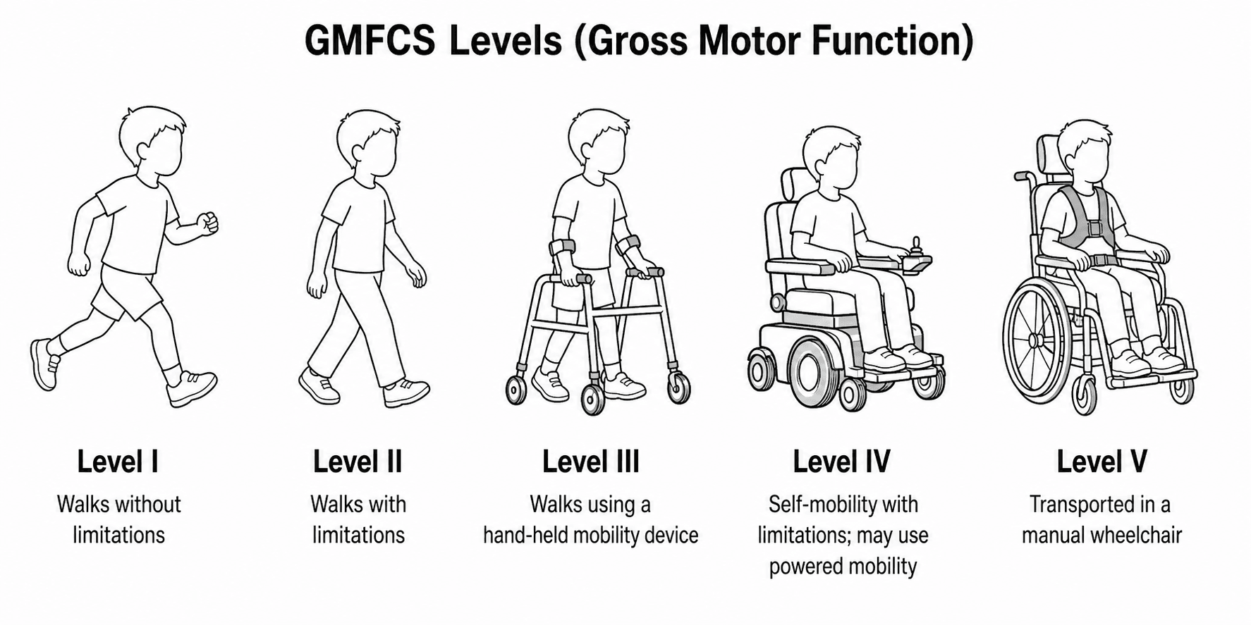
Gross Motor Function Classification System (GMFCS)
The gold standard for prognosis and communication. Based on self-initiated movement sitting/walking.
- Level I: Walks without limitations. Runs/Jumps.
- Level II: Walks with limitations (railings, uneven ground). No running.
- Level III: Walks with handheld mobility device (Walker/Crutches). Wheelchair for long distance.
- Level IV: Self-mobility with limitations (Powered chair). Can stand for transfers.
- Level V: Transported in manual wheelchair. Head control issues.
GMFCS is stable over time.
Companion Classifications: MACS, CFCS and EDACS
GMFCS describes gross motor function, but the examiner expects you to know that the modern CP "toolbox" classifies the whole child across four domains - all five-level, all stable over time.
- Domain
- Gross motor (sitting, walking, self-mobility)
- What level V means
- Transported in a manual wheelchair
- Domain
- How the hands handle objects in daily tasks
- What level V means
- Does not handle objects; severely limited
- Domain
- Everyday sending and receiving of communication
- What level V means
- Seldom effective even with familiar partners
- Domain
- Safety and efficiency of eating and drinking
- What level V means
- Unable to eat or drink safely; tube feeding considered
Each runs from level I (independent or effective) to level V (dependent), and a mini-MACS exists for the 1-to-4-year age group. They are complementary, not interchangeable - a hemiplegic child can be GMFCS I yet MACS II-III because the affected hand limits manual ability.
GMFCS is only the gross-motor axis. Pair it with MACS (hand/manual ability), CFCS (communication) and EDACS (eating and drinking) - each a stable, five-level (I independent to V dependent) system. They are independent: a child can be GMFCS I but MACS III, so always classify hand and communication function separately rather than inferring them from walking ability.
Clinical Assessment
- Birth history (Gestation, ICU stay).
- Milestones (Sit by 2? Walk by ?).
- Communication/Feeding status.
- Tone: Modified Ashworth Scale (0-4). Tardieu Scale (R1/R2).
- R1: Angle of first catch (velocity dependent).
- R2: Angle of Max passive range (static length).
- R2-R1: Dynamic component (Spasticity).
- Selective Motor Control (SMC): Ability to isolate joint movement.
- Rotational Profile: Anteversion, Tibial Torsion.
- Spine: Scoliosis check.
- Hips: Abduction range (Risk of dislocation if less than 45).
- Spine: Scoliosis check. Sitting balance. Pelvic obliquity.
This is the ability to isolate joint movement.
- Test: Ask patient to dorsiflex ankle without flexing hip/knee.
- Significance: Poor SMC predicts poor outcome from tendon transfers. If SMC is absent, transfer will not work "in phase" (but may act as a tenodesis).
- Sagittal Plane: Look for the "Gait Deviations".
- True Equinus: Hips extended, Knee extended, Ankle plantarflexed.
- Jump Gait: Hip flexed, Knee flexed, Ankle plantarflexed.
- Apparent Equinus: Hip flexed, Knee flexed, Ankle neutral (but looks equinus due to knee flexion).
- Crouch Gait: Hip flexed, Knee flexed (greater than 30 deg), Ankle dorsiflexed (Calcaneus).
- Coronal Plane:
- Scissoring: Adductor spasticity.
- Trendelenburg: Abductor weakness.
- Transverse Plane:
- Intoeing: Femoral anteversion vs Internal Tibial Torsion.
- Outtoeing: External Tibial Torsion (often iatrogenic or compensatory).
- Respiratory: High risk of aspiration (swallow dysfunction) and post-op pneumonia.
- Seizures: Ensure anticonvulsants are continued.
- Latex Allergy: Higher prevalence in CP/Spina Bifida.
- Positioning: Contractures make positioning on the table difficult. Pad purely bony prominences.
- Pain Assessment: FLACC scale for non-verbal children. High risk of under-treatment.
- Cerebral Palsy
- Birth / Infancy (Static)
- Hereditary Spastic Paraparesis
- Childhood / Adult (Progressive)
- Cerebral Palsy
- Rare
- Hereditary Spastic Paraparesis
- Common (Autosomal Dominant)
- Cerebral Palsy
- Abnormal (PVL/IVH)
- Hereditary Spastic Paraparesis
- Normal
- Cerebral Palsy
- Non-progressive (MSK worsens)
- Hereditary Spastic Paraparesis
- Neurology worsens
Investigations
1. Hip Surveillance (X-rays):
- Why?: To prevent dislocation. Dislocation leads to pain, scoliosis, and hygiene issues in GMFCS V.
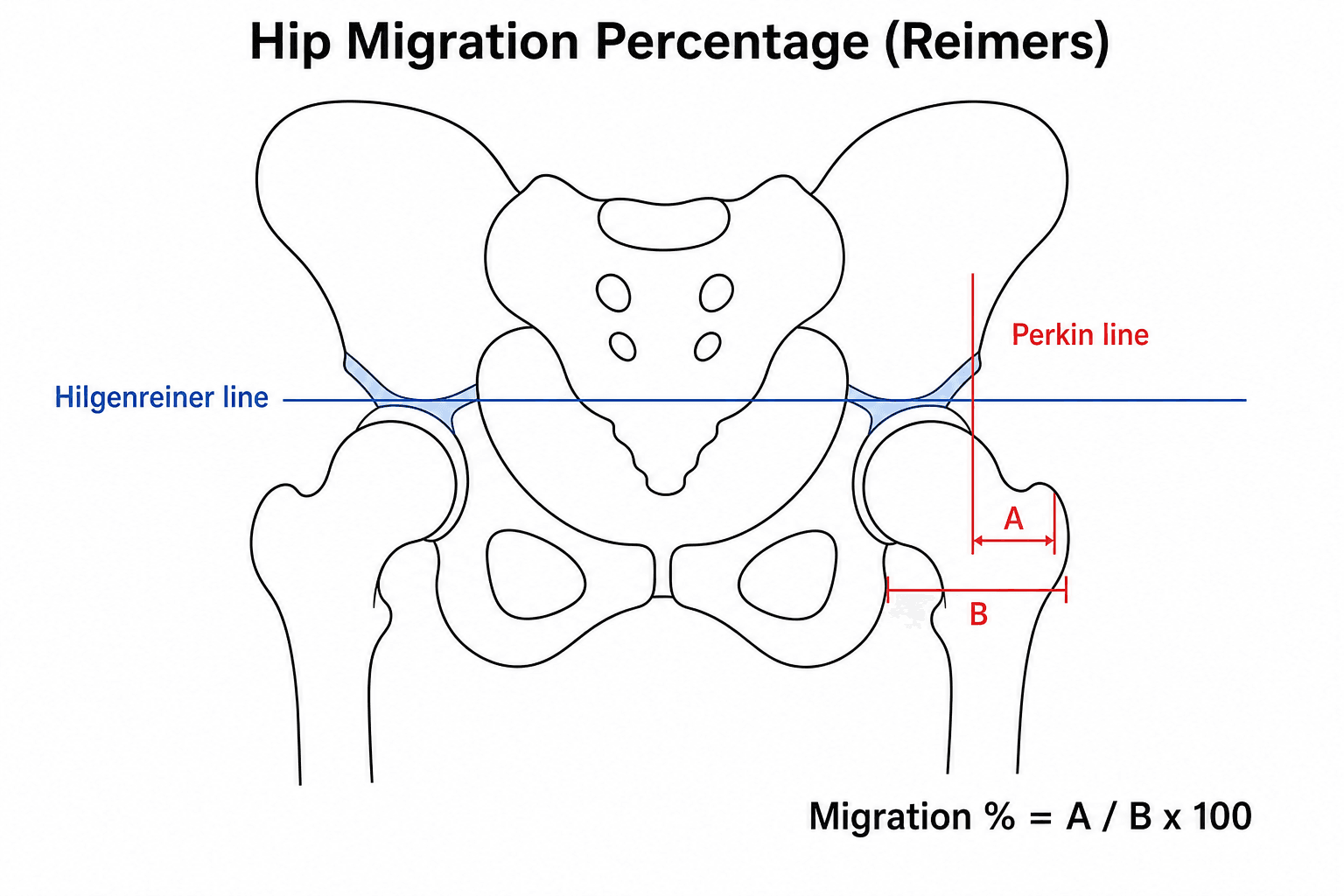
- Metric: Reimer's Migration Percentage (MP).
- Calculation: Percentage of the femoral head lateral to Perkins' line (Lateral edge of acetabulum).
- Normal: Less than 10% in normal children. Less than 30% acceptable in CP.
- Risk: Greater than 30% ("Hip at Risk"). Often "Silent" (Pain free).
- Dislocated: Greater than 100%. The head is completely lateral to the acetabulum.
- Gold Standard for surgical planning in walkers (GMFCS I-III).
- Kinematics: Joint angles.
- Kinetics: Forces/Moments (Joint powers).
- EMG: Muscle firing timing (rectus spasticity in swing?).
- Pedobarography: Foot pressure. 3. Gait Velocity and Oxygen Cost:
- Children with CP use 3-5x more energy to walk than peers.
- Oxygen Cost: measured in mL/kg/m.
- Goal of Surgery: Improve efficiency (Lower Oxygen cost).
- A single number representing gait pathology.
- 100: Normal.
- Every 10 points below 100: One standard deviation from normal.
- Typical CP: GDI 60-70.
- Post-SEMLS: Expect increase of 5-10 points (Clinically significant).
- GMFM (Gross Motor Function Measure): Even more detailed than GMFCS. Used to track change over time (e.g. pre/post SDR).
- GMFM-88: Validated for CP and Down Syndrome. Includes lying/rolling.
- GMFM-66: Rasch-scaled version. Only for CP.
- FMS (Functional Mobility Scale): Rates mobility at 3 distances (Home, School, Community).
- 5 meters: Home.
- 50 meters: School.
- 500 meters: Community.
- Rating: 1 (Crawler) to 6 (Independent on all surfaces).
- CP-CHILD: Caregiver-reported Quality of Life measure (Comfort, Positioning).
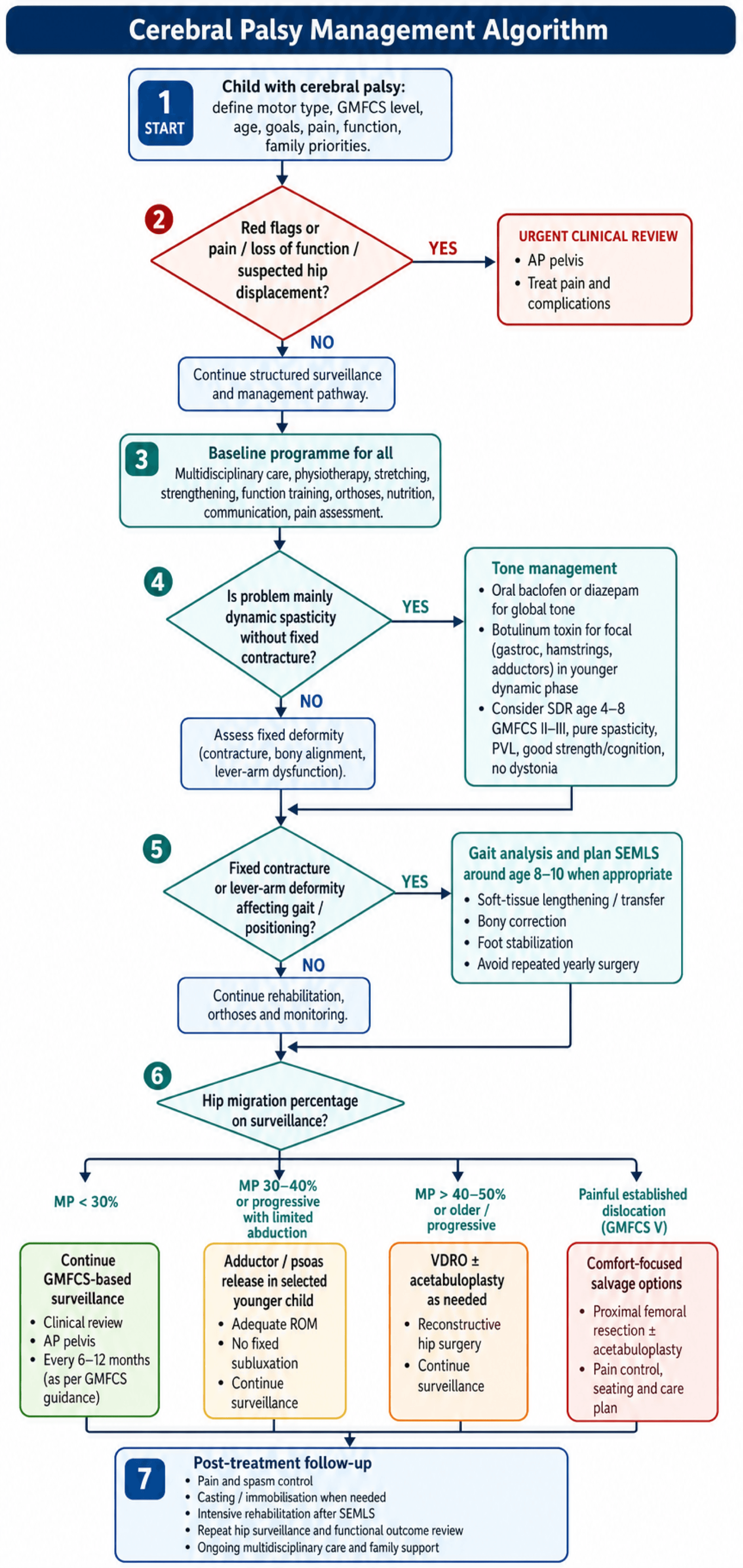
Management Algorithm
Multidisciplinary Management
- Physiotherapy: Stretch, Strengthen, Functional training.
- Orthotics:
- AFO (Ankle Foot Orthosis): Solid (for crouch/equinus) or Hinged (for simple drop foot).
- GRAFO: Ground Reaction AFO (Pre-tibial shell) for CROUCH gait (prevents tibial advancement).
- Tone Management:
- Oral: Baclofen, Diazepam (global effect).
- Botox: Focal spasticity. Good for young kids (dynamic phase). Target: Gastroc, Hamstrings, Adductors.
- Intrathecal Baclofen (ITB): For severe severe quadriplegia (GMFCS IV/V) with dystonia.
Botox is most effective in the "Dynamic Phase" (Age 2-6).
Choosing Between SDR and Intrathecal Baclofen
The topic describes SDR and intrathecal baclofen (ITB) separately; the examiner will make you choose between them for a given child. They suit opposite ends of the CP spectrum.
- SDR
- Permanent sectioning of a proportion of dorsal (sensory) rootlets under intra-operative EMG
- Intrathecal Baclofen (ITB)
- Implanted programmable pump delivering baclofen to the CSF
- SDR
- No - permanent and irreversible
- Intrathecal Baclofen (ITB)
- Yes - titratable and reversible (can be turned down or removed)
- SDR
- Spasticity only (NOT dystonia)
- Intrathecal Baclofen (ITB)
- Spasticity AND dystonia
- SDR
- Ambulant spastic diplegia, GMFCS II-III, age about 4-8, pure spasticity, good strength/selective control/cognition, PVL on MRI
- Intrathecal Baclofen (ITB)
- Severe generalised hypertonia, GMFCS IV-V, comfort/care/positioning goals, mixed spasticity-dystonia
- SDR
- One operation then intensive physiotherapy; risk of unmasking weakness, sensory change and later spinal deformity
- Intrathecal Baclofen (ITB)
- Lifelong refills and device upkeep; catheter/pump failure, infection, and life-threatening overdose or withdrawal
Oral agents (baclofen, diazepam) cover mild global tone and botulinum toxin covers focal, dynamic spasticity in the young child; SDR and ITB are the options when tone is severe enough to need a definitive or device-based solution.
SDR = permanent, spasticity-only, for the ambulant GMFCS II-III spastic diplegic with good strength and PVL (the spasticity is masking usable function). ITB = reversible and titratable, treats spasticity AND dystonia, for the severe GMFCS IV-V child where the goal is comfort, care and positioning. Dystonia steers you away from SDR; the need for adjustability and a whole-body effect steers you towards ITB - which, unlike SDR, carries the ongoing risks of pump failure, overdose and withdrawal.
Surgical Technique
Hip Reconstruction
Hip Surveillance Surgery
For Migration greater than 30-40% or progressive subluxation.
- Soft Tissue (Preventive): Adductor/Psoas Release.
- Indication: MP greater than 30% in young child (less than 4) with abduction less than 30 deg.
- Bony Reconstruction: VDRO + Acetabuloplasty (Dega/San Diego).
- Indication: MP greater than 40-50% in older child (greater than 4).
- VDRO: Shorten (release tension), Varus (better cover), Derotate (fix anteversion).
- Pelvis: Hinge osteotomy to cover anterior/lateral head.
- Dega Technique: Curvilinear cut above acetabulum (leaving posterior cortex intact). Lever down the roof. Bone graft wedge. Ideal for deficient anterior/lateral coverage.
- San Diego: Similar to Dega but extends to sciatic notch (more coverage).
- Salter: Less common in CP (re-directs whole acetabulum, but creates retroversion which is bad in CP).
- Salvage: Castle Procedure (Resection Interposition) or Proximal Femoral Replacement.
- Indication: Painful GMFCS V dislocated hip with destroyed head.
Total Hip Arthroplasty is generally contraindicated in GMFCS V due to high dislocation risk.


Complications
- Risk Factor
- TAL (Tendo-Achilles)
- Management
- Calcaneal gait (Crouch). Hard to fix.
- Risk Factor
- Young age (less than 6) at time of surgery
- Management
- Repeat surgery
- Risk Factor
- GMFCS V / Scoliosis
- Management
- Salvage surgery
- Risk Factor
- Osteopenia / Cast immobilization
- Management
- Gentle handling / Bisphosphonates
- Risk Factor
- Catheter kink / Infection / Overdose
- Management
- Emergency pump interrogation
Baclofen Pump Failure:
- Overdose: Coma, Respiratory depression, Hypotension. Support airway. Physostigmine (controversial).
- Withdrawal: Itchy, Agitated, Rigid, Seizures, Hyperthermia. Life threatening. Restore Baclofen (oral/intrathecal) immediately.
Postoperative Care
Pain management is critical, especially in Spastic CP where pain triggers spasm, which triggers more pain.
- Spasm Protocols: Benzodiazepines (Diazepam) + Gabapentin.
- Immobilization: Petrie Casts (Broomstick) or Spica for hips.
- Rehab: Intensive inpatient rehab for 6-12 weeks post-SEMLS.
Outcomes
- Walking: GMFCS I/II walk well. III walk with aids. IV/V do not walk.
- Employment: Competitive employment rates are low (GMFCS dependent).
- Pain: Over 50% of adults with CP report chronic musculoskeletal pain.
- Life Expectancy: Reduced in GMFCS V (respiratory issues). Near normal in I-III.
Guidelines, Registries & Global Practice
Global Epidemiology:
- Prevalence: ~2.0-2.5 per 1000 live births in high-income settings; higher in low- and middle-income countries (LMICs) where prevalence may exceed 3 per 1000, with a higher proportion of severe, GMFCS IV-V and post-neonatally acquired CP (infection, kernicterus, birth asphyxia).
- Trend: Prevalence has plateaued or fallen modestly in high-income countries despite improved survival of extreme preterms, attributed to antenatal magnesium sulfate, antenatal steroids, and therapeutic hypothermia.
- Distribution: Spastic subtype ~80%; spastic diplegia/hemiplegia predominate; bilateral GMFCS IV-V drive the orthopaedic burden (hip, spine).
- Hip Surveillance
- GMFCS-stratified schedule; 6-monthly films for GMFCS IV-V
- Tone & Surgery Emphasis
- Early preventive soft-tissue release; SEMLS for ambulant CP
- Hip Surveillance
- Care pathways endorse GMFCS-based screening
- Tone & Surgery Emphasis
- Multidisciplinary tone clinics; SDR in selected GMFCS II-III
- Hip Surveillance
- National CP Integrated Pathway (e.g. CPIPS) — GMFCS-based films
- Tone & Surgery Emphasis
- Surveillance-led prevention; NICE NG62 supports MDT spasticity care
- Hip Surveillance
- Founding surveillance registry; near-zero dislocation
- Tone & Surgery Emphasis
- Registry-driven prevention; benchmark for outcomes
- CPUP (Sweden/Norway/Denmark/Scotland): The reference hip and spine surveillance registry; demonstrated near-elimination of hip dislocation.
- SCPE (Surveillance of Cerebral Palsy in Europe): Harmonised CP definitions and population prevalence data across European centres.
- CPIPS / UK national CP integrated pathway and Australian CP Register: National-scale GMFCS-based surveillance and outcome tracking.
- High-resource: Routine GMFCS-based radiographic hip surveillance, 3D gait labs for SEMLS planning, intrathecal baclofen, SDR programmes, neonatal neuroprotection bundles.
- Limited-resource: Later presentation with established dislocation/contracture; emphasis on physiotherapy, low-cost orthoses, and salvage rather than preventive surgery; gait labs and SDR rarely available. Prevention of kernicterus and birth-asphyxia CP is a major public-health priority.
Controversies & Areas of Uncertainty
- SDR vs intrathecal baclofen vs SEMLS: For ambulant spastic diplegia there is no high-quality head-to-head RCT. Long-term cohorts (Tedroff) show SDR durably reduces tone but does not prevent later orthopaedic surgery or contracture — challenging the idea that early tone reduction alone changes natural history.
- Does SEMLS truly change long-term function? Gait-lab kinematics (GDI) improve, but functional/participation gains and durability are debated, and the evidence base lacks RCTs (McGinley).
- Optimal hip surveillance threshold for intervention: Migration percentage thresholds (30% vs 40% vs trend/velocity) and whether soft-tissue release alone suffices vary between programmes; reoperation rates remain high (Kiapekos).
- Magnesium sulfate magnitude of effect: Individual trials (including ACTOMgSO4/Crowther) were under-powered for CP as a sole endpoint; the neuroprotective recommendation rests on pooled meta-analysis, and optimal dosing/timing still varies.
- Single-event vs staged surgery: "Birthday syndrome" is condemned, yet very large single events carry higher perioperative and rehabilitation burden — the ideal bundling remains individualised.
- Botulinum toxin and muscle morphology: Concern that repeated BTX-A injections may cause long-term muscle atrophy/fibrosis in the developing muscle, tempering enthusiasm for high-frequency dosing.
Viva Scenarios
Clinical Decision Scenarios
Practise clinical reasoning and management decisions out loud
“Discuss your management approach.”
Clinical Decision Scenarios
Practise clinical reasoning and management decisions out loud
“What is your plan?”
MCQ Practice Points
Q: What is the most common physiologic type of CP? A: Spastic (Pyramidal) - approx 80%. Dyskinetic is 10-15%. Ataxic less than 5%.
Q: How often should a GMFCS Level V child have a hip X-ray? A: Every 6 months. (High risk of rapid displacement). Level I usually discharged at 5 years.
Q: What determines the difference between GMFCS II and III? A: Handheld Mobility Device. Level II walks without aids (may use rail). Level III needs crutches/walker.
Q: What determines the difference between GMFCS IV and V? A: Self-Mobility. Level IV can drive a powered chair or mobilize short distances. Level V has no means of independent mobility (must be pushed).
Q: What is the likelihood of a child with Hemiplegic CP walking? A: Nearly 100% (Usually GMFCS I or II). If a hemiplegic child is not walking, reconsider diagnosis.
Q: Periventricular Leukomalacia (PVL) is most strongly associated with which CP pattern? A: Spastic Diplegia. The medial fibers (legs) of the corticospinal tract are affected.
GMFCS Levels
- I: Walks / Runs
- II: Walks / Railing / Uneven issues
- III: Handheld Device (Walker)
- IV: Powered Mobility
- V: Pushed (Head control issues)
Hip Surveillance
- Gold Standard: Reimer's MP
- Normal: less than 10%
- Risk: greater than 30%
- Freq: GMFCS V = 6 monthly
Management
- Botox: Focal Dynamic Spasticity
- Baclofen: Global Spasticity
- SEMLS: Age 8-10, Bony + Soft Tissue
- SDR: Pure Spasticity, GMFCS II/III
Key Concepts
- Non-progressive brain lesion
- Progressive MSK deformity
- Lever Arm Dysfunction
- Avoid Birthday Syndrome
Evidence Base
GMFCS — The Classification Paper
- Delphi consensus among 48 experts produced the 5-level GMFCS.
- Interrater reliability (kappa) 0.75 for children aged 2-12 years; lower (0.55) under 2 years.
- Designed as a staging system analogous to medical grading scales.
Hip Surveillance Prevents Dislocation (CPUP)
- 20-year results of the Swedish CPUP surveillance programme.
- Hip dislocation fell from 8% in the historical control group to effectively 0% in surveilled cohorts (p less than 0.001).
- The only 2 study-group children who dislocated were too unwell for preventive surgery.
- Of 689 surveilled children, 13% required preventive surgery.
Primary Surgery for Hip Displacement
- Compared adductor-iliopsoas tenotomy (APT) vs femoral osteotomy (FO) as primary preventive surgery.
- At minimum 5 years, reoperation was 43% after APT and 39% after FO.
- Residual migration percentage over 50% in 2% (APT) vs 9% (FO).
- After soft-tissue release, higher preoperative migration percentage predicted failure.