'Dripping Candle Wax' Sclerosing Bone Dysplasia | Sclerotomal Distribution | MAP2K1 Mutation
- Dripping candle wax appearance - pathognomonic dense cortical hyperostosis flowing along bone cortex on radiographs
- Sclerotomal distribution - classically follows a single sclerotome (dermatomal equivalent for bone)
- MAP2K1 somatic mutation - identified in approximately 50% of cases, activates MAPK/ERK pathway
- Pain and stiffness - predominant symptoms; contractures from soft tissue involvement
- Surgery has high recurrence - limited surgical role; excision complicated by disease recurrence and joint stiffness
- “Describe the classic radiographic appearance: dense, irregular cortical hyperostosis resembling 'dripping candle wax' flowing down the bone
- “Know the differential diagnosis: osteopoikilosis (round lesions), osteopathia striata (linear striations), parosteal osteosarcoma, myositis ossificans
- “Understand management is primarily conservative: NSAIDs, physiotherapy, bisphosphonates trial; surgery reserved for severe contractures or deformity
- “Surgical complications include high recurrence, stiffness, and incomplete symptom relief
Overview and Epidemiology
Melorheostosis (Greek melos, limb; rhein, flow; osteon, bone) is a rare, sporadic sclerosing bone dysplasia of dense cortical hyperostosis, recognised on radiographs by its dripping candle wax appearance. Leri and Joanny first described it in 1922, and fewer than 500 cases have been reported, which makes it one of the rarest bone conditions. The 2018 discovery of somatic MAP2K1 mutations by Kang and colleagues transformed understanding of how it arises.
Who. The estimated incidence is 1 per 1,000,000. It can present at any age, but 50% present before age 20. The sexes are equally affected, although the Mayo Clinic cohort reported a 4:1 female predominance. It is sporadic: the mutation is a somatic mosaic one, not inherited.
Natural history. Onset is insidious, anywhere from childhood to adulthood, and the disease progresses slowly over decades. Pain is the most common symptom; stiffness and contractures also occur. It is benign but disabling, with no malignant transformation.
Where. Disease is usually unilateral and confined to one limb:
- Lower limb - 70% of cases
- Upper limb - 20%
- Axial skeleton - rare (10%), usually in combination with limb disease
Monostotic and polyostotic disease have been quoted as equally common, with polyostotic disease often in adjacent bones; in the Mayo Clinic cohort, two-thirds had polyostotic involvement.
Pathophysiology and Molecular Genetics
The mutation. Kang and colleagues identified somatic activating mutations in MAP2K1 (mitogen-activated protein kinase kinase 1), the gene encoding MEK1, a dual-specificity kinase, in melorheostosis lesions; the mutation is found in approximately 50% of cases. It is post-zygotic and present in affected tissue only, not blood, which confirms somatic mosaicism and explains both the sporadic, non-inherited nature of the disease and its mosaic distribution.
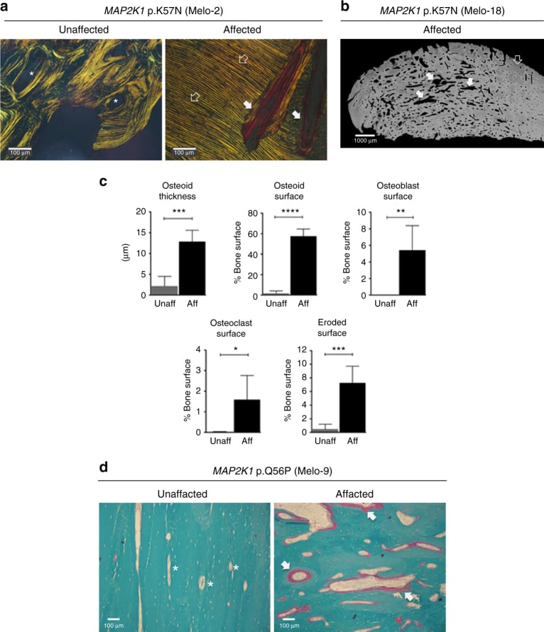
The pathway. The mutation drives constitutive ERK activation in osteoblasts through the RAS-RAF-MEK-ERK cascade, and the result is osteoblast hyperactivity and excessive bone formation. The mechanism is subtler than too much bone: the Kang study showed that the mutation increases osteoblast proliferation yet inhibits BMP2-mediated mineralisation and differentiation, producing excess unmineralised osteoid rather than simply more normal bone. The consequences reach beyond the osteoblast:
- Bone remodelling - uncoupled, favouring formation
- Soft tissues - fibroblast activation and fibrosis
- Vasculature - vascular malformations in some cases
- Skin - scleroderma-like changes over affected areas
Two genetic routes. The common, sporadic isolated disease is driven by the post-zygotic MAP2K1 (MEK1) mutation, and a minority carry related somatic RAS-MAPK changes (KRAS, SMAD3). The rarer familial route runs through germline loss-of-function LEMD3 (MAN1) mutations, which cause osteopoikilosis and Buschke-Ollendorff syndrome (osteopoikilosis plus connective-tissue naevi); melorheostosis can arise within these families as the osteopoikilosis-associated, or mixed sclerosing bone dysplasia, form. LEMD3 normally antagonises TGF-beta/BMP signalling.
Why the route matters. Isolated sporadic disease is a somatic MAP2K1 problem and carries no inheritance risk. Melorheostosis appearing alongside osteopoikilosis or connective-tissue naevi points to a germline LEMD3 disorder, which is autosomal dominant, and that distinction changes family counselling (the osteopoikilosis topic covers the LEMD3 detail).
The sclerotomal theory. The classic explanation for the distribution is that the disease affects bones, muscles, fascia and skin derived from a single embryonic segment. The sclerotome is described both as the somite derivative that forms a vertebra and its associated bones and as the territory of a single sensory nerve root, which is why the disease runs along one side of a bone in linear, segmental fashion, often confined to one limb, and stops at a segmental boundary rather than obeying vascular or muscular anatomy. The same segment explains why skin and soft-tissue changes overlie the affected bones in a dermatomal pattern.
Where the theory fails. A post-zygotic mutation affecting a single cell lineage fits the sclerotomal pattern. Some cases do not fit it, however, suggesting alternative mechanisms or earlier mutations affecting multiple lineages.
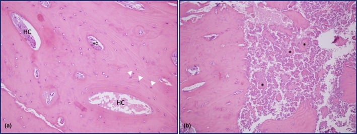
Histopathology. Affected bone is dense, mature, well-organised lamellar bone, not woven, with normal osteons (Haversian systems preserved), thickened trabeculae and layers of periosteal new bone on the cortical surface. Adjacent soft tissues show dense fibrosis. Two negatives matter for the differential:
- No inflammatory infiltrate - distinguishes it from chronic osteomyelitis
- No nuclear atypia - distinguishes it from malignancy
Clinical Presentation
The spectrum. Presentation is highly variable, from incidental discovery to severe disability, and many cases are discovered incidentally on imaging. Onset is insidious and the course slowly progressive over years. When symptoms bring the patient, they are these:
- Pain (50-80%) - dull and aching, often worse with activity
- Stiffness (60-80%) - joint contractures and reduced range of motion
- Deformity (40-60%) - limb length discrepancy or angular deformity
- Skin changes (20-40%) - scleroderma-like hardening over affected areas
- Limb swelling (30%) - from bone enlargement and soft-tissue involvement
Pain. Deep, aching bone pain over the affected segment, often worse with activity and sometimes present at night. It worsens slowly over years.
Contractures. They affect the joints adjacent to affected bones and arise from soft-tissue fibrosis and capsular involvement. Common sites are the knee, elbow, ankle and fingers. Severity ranges from mild limitation to a severe fixed contracture, and it gradually worsens over time.
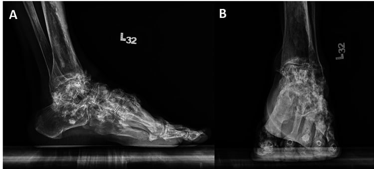
Look. The limb may be asymmetric, shorter or angulated. The skin may be thickened and indurated with a scleroderma-like appearance, bone enlargement is visible where the bone is superficial, and muscle wastes from disuse or direct involvement.
Feel. Hard, irregular bone masses lie along the cortical surface and are non-tender or mildly tender. The skin may feel indurated and bound down, with reduced soft-tissue mobility over affected areas.
Move. Adjacent joints are contracted, fixed flexion deformities are common, and stretching produces end-range pain.
By age. The presentation shifts with age:
- Childhood (under 10 years) - limb length discrepancy, angular deformity and delayed motor milestones; it may be mistaken for a developmental condition
- Adolescence and young adulthood - pain with increasing activity, stiffness affecting sport and activities, and cosmetic concerns; this is the peak time for diagnosis
- Adulthood - progressive pain and stiffness, occupational limitations and osteoarthritis in affected joints; the patient may have been symptomatic for decades before diagnosis
Across ages, examination should relate the visible deformity and contracture to the same segmental distribution seen on imaging.
Imaging and Diagnosis
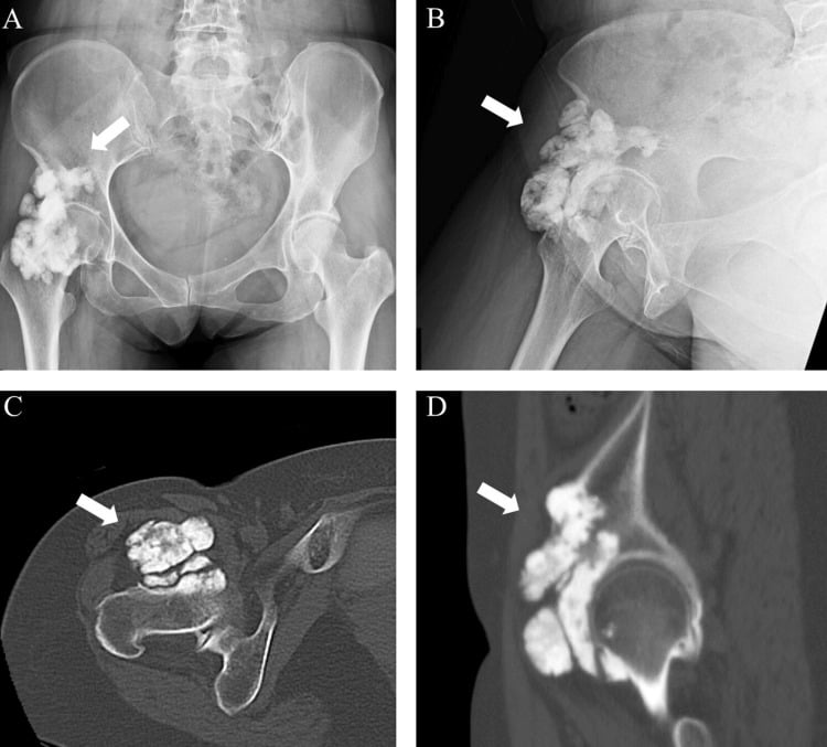
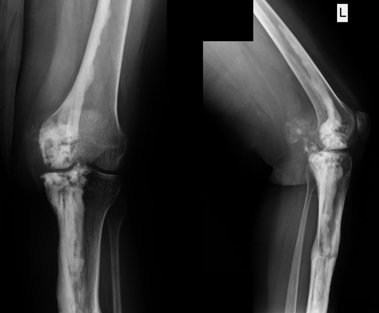
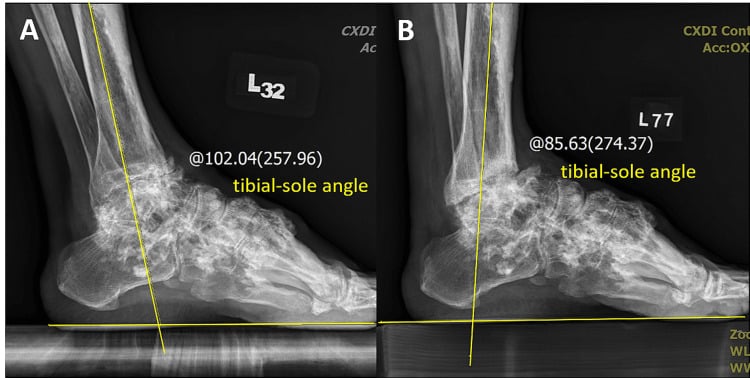
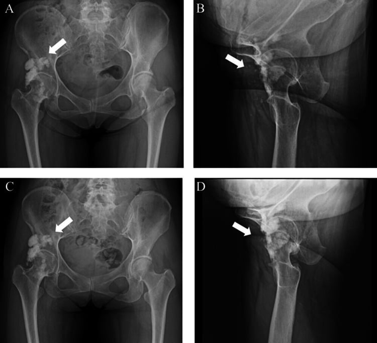
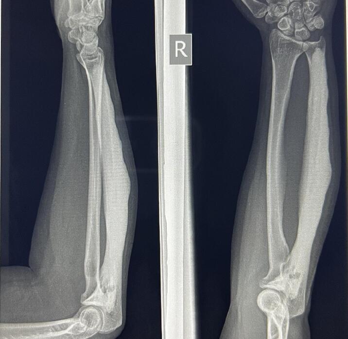
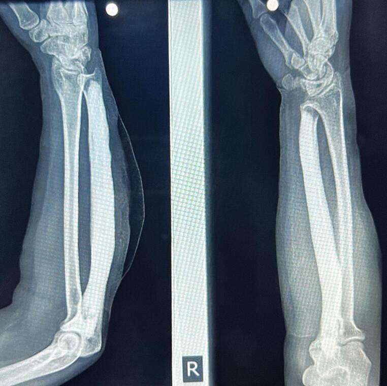
The radiograph makes the diagnosis. The dripping candle wax appearance is virtually pathognomonic: dense, irregular, flowing cortical hyperostosis along one side of the bone, like wax that has dripped down a candle and hardened. The sclerosis is eccentric, typically on one cortex, with an irregular wavy outer margin and an inner margin that may encroach on the medullary canal. It may cross joints to involve adjacent bones in the same sclerotome. There is no periosteal reaction, which distinguishes it from infection and tumour, and adjacent soft-tissue ossification may be seen. Radiographs serve for surveillance as well as initial diagnosis.
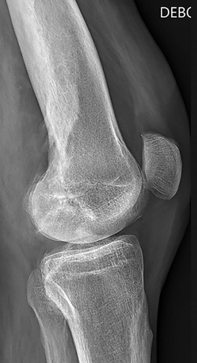
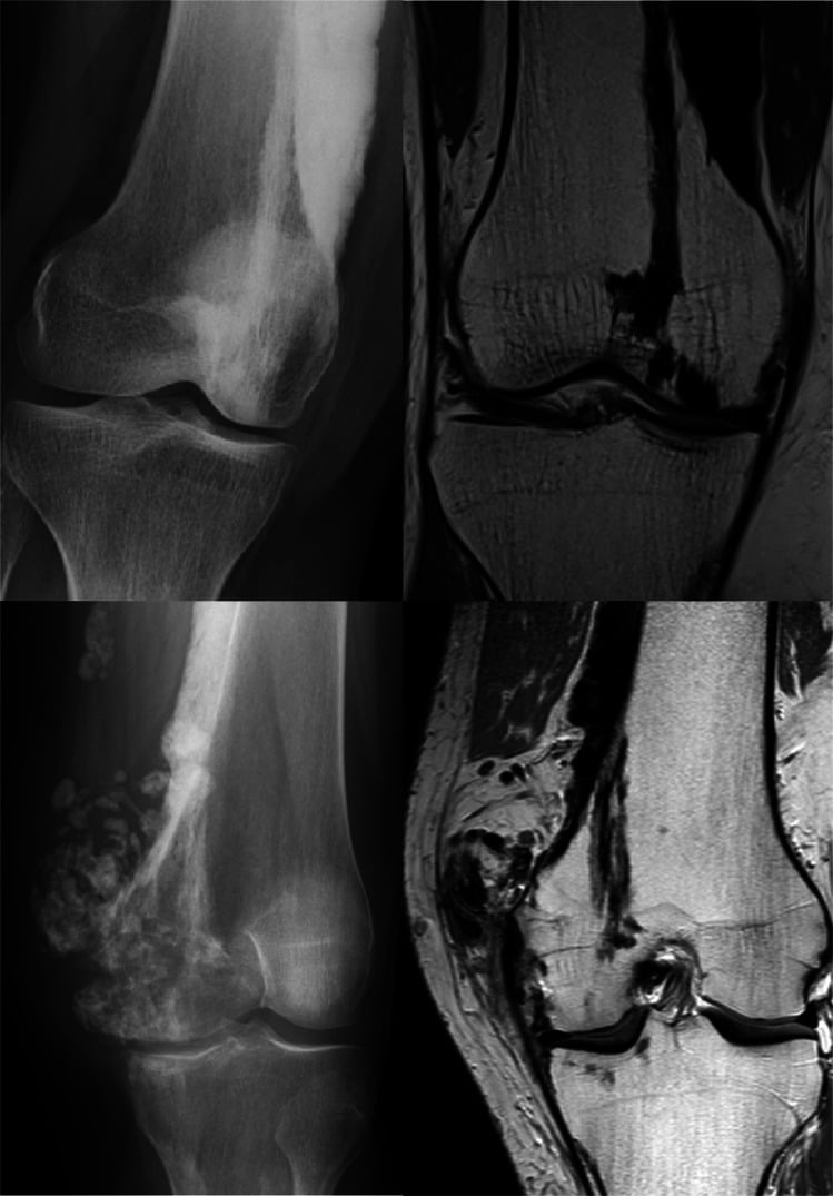
The patterns. The appearance is not always the classic one:
- Classic dripping candle wax - dense cortical hyperostosis flowing along one side of the cortex, as above
- Spotted, osteopoikilosis-like - focal sclerotic foci within the medullary cavity, sometimes coexisting with the classic pattern; termed the melorheostotic variant of mixed sclerosing bone dysplasia
- Linear striations, osteopathia striata-like - linear striations of dense bone, less common than the classic form and often an overlap syndrome rather than a distinct disease
- Soft-tissue ossification - ossification extending from bone into adjacent soft tissue, which can mimic myositis ossificans or parosteal osteosarcoma
Absence does not exclude. The classic pattern is described as the most common, but in Freyschmidt's series it was present in only 5 of 23 cases, so its absence does not exclude the diagnosis.
The dangerous mimic. Soft-tissue ossification is the pattern that gets a benign, non-neoplastic condition biopsied or resected as a sarcoma, so correlate deliberately. No antecedent trauma argues against myositis ossificans, and the long, painless, sclerotomal distribution argues against a surface osteosarcoma.
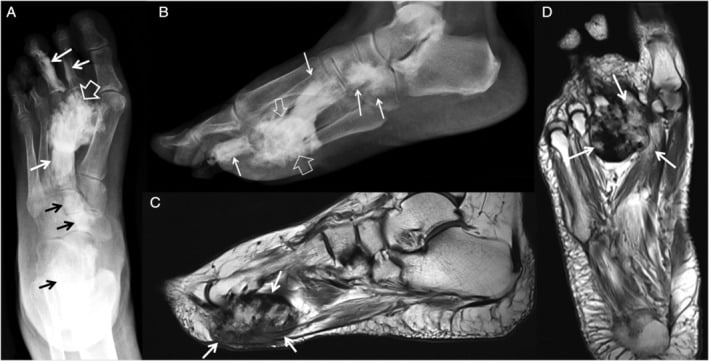
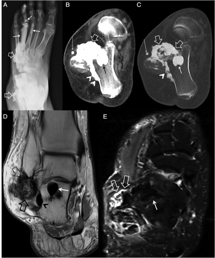
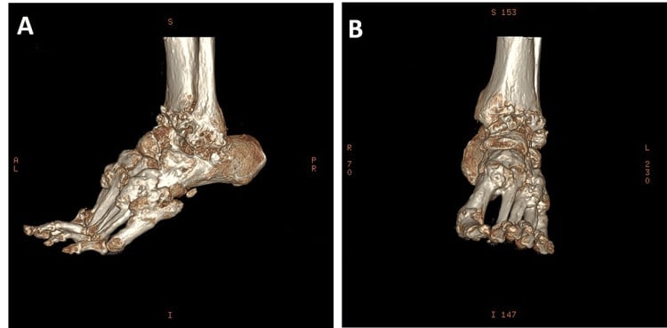
CT. CT delineates the sclerotic bone precisely: its cortical extent, extension into the medullary canal, intra-articular extension and osteoarthritis. It detects soft-tissue ossification better than radiographs, and 3D reconstruction helps plan surgery in complex cases.
MRI. MRI assesses the soft tissues and nerves. Soft-tissue fibrosis is low signal on T1 and T2; muscle shows atrophy, fibrosis or fatty replacement; the joint may show synovitis and capsular thickening; and marrow signal in sclerotic areas is usually normal. Neurovascular compression is important for surgical planning.
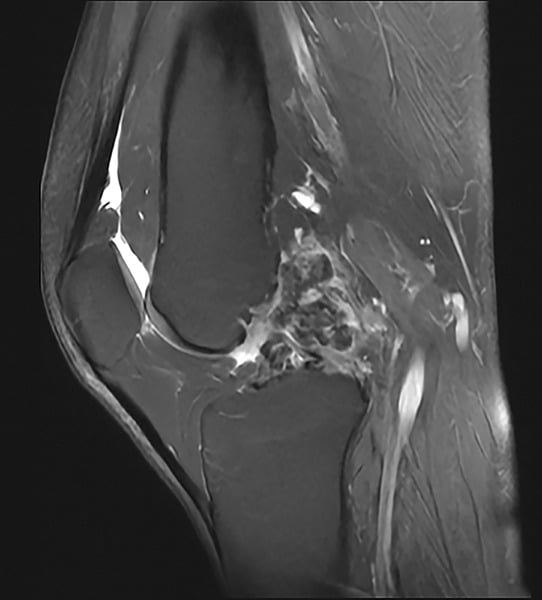
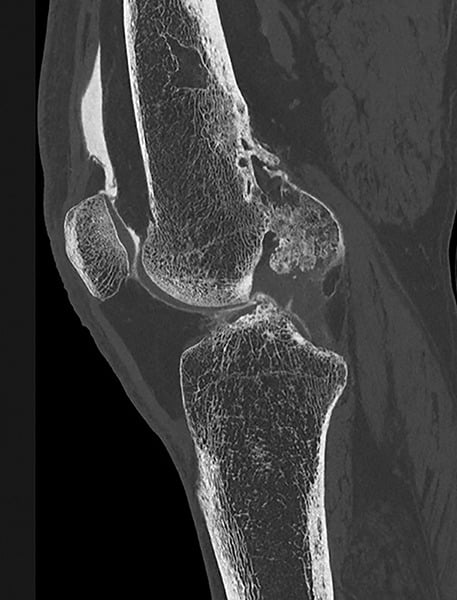
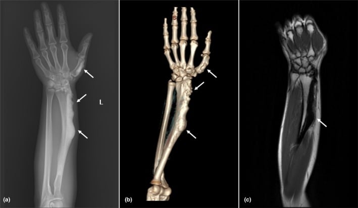
Bone scintigraphy. Uptake is increased in affected areas and may show involvement beyond the radiographic findings, and its intensity may correlate with disease activity. Specificity is limited, because it cannot distinguish melorheostosis from other sclerotic conditions. It maps metabolically active disease but does not replace pattern recognition on radiographs and cross-sectional imaging.
Differential Diagnosis
Pattern and symmetry separate the sclerosing dysplasias. Osteopoikilosis (spotted bones) consists of multiple small, round, well-defined sclerotic foci clustered near joints, completely different from the flowing, linear dripping wax of melorheostosis. Osteopoikilosis and osteopathia striata are bilateral and symmetric, where melorheostosis follows one sclerotome on one side.
- Radiographic Pattern
- Dripping candle wax
- Distribution
- Sclerotomal, unilateral
- Key Distinguishing Feature
- Flowing cortical hyperostosis along one cortex
- Radiographic Pattern
- Multiple round foci (spotted bones)
- Distribution
- Bilateral, symmetric
- Key Distinguishing Feature
- Round/oval sclerotic foci near joints
- Radiographic Pattern
- Linear striations
- Distribution
- Bilateral, symmetric
- Key Distinguishing Feature
- Longitudinal dense lines in metaphyses
- Radiographic Pattern
- Fusiform cortical thickening
- Distribution
- Bilateral, symmetric, diaphyseal
- Key Distinguishing Feature
- Camurati-Engelmann, inherited, systemic symptoms
Mixed sclerosing bone dysplasia. Some patients show features of more than one sclerosing dysplasia, melorheostosis with osteopoikilosis or osteopathia striata with osteopoikilosis. The overlap is not coincidence: these dysplasias share disturbances in the same bone-forming signalling pathways, which suggests a common pathogenetic mechanism.
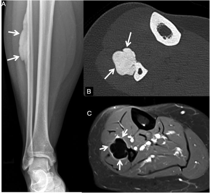
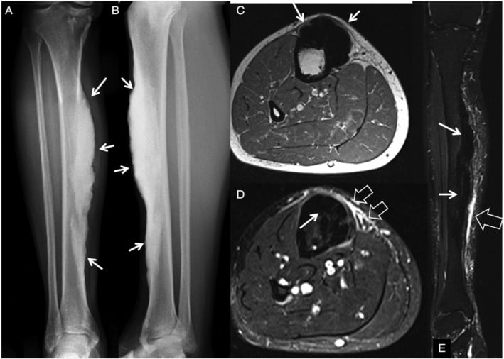
When to biopsy. The diagnosis is usually radiographic, and biopsy is kept for the cases that leave doubt:
- Diagnosis uncertain despite classic imaging
- Atypical features suggesting malignancy
- Rapid progression, which is unusual for melorheostosis
- A soft-tissue mass concerning for parosteal osteosarcoma
- A patient under 5 years, which is very rare for melorheostosis; consider other causes
Management
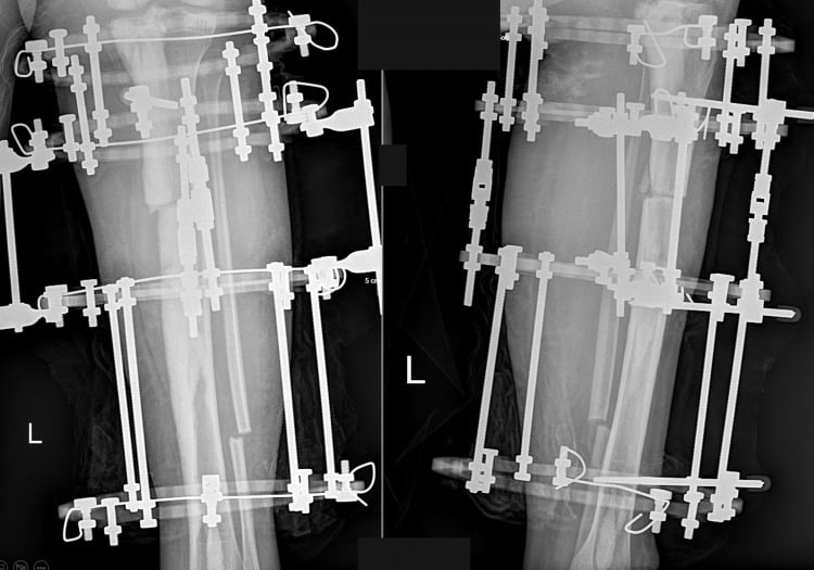
Conservative management is the mainstay. Most patients can be managed with analgesics, physiotherapy and supportive measures. Surgery has unpredictable results with high recurrence rates, and it is reserved for severe contractures or deformity causing functional impairment after conservative treatment has failed.
- Key Features
- Characteristic imaging, no symptoms
- Management
- Observation, reassurance, no treatment
- Exam Pearl
- Many cases are discovered incidentally on imaging
- Key Features
- Symptomatic but functional
- Management
- NSAIDs, physiotherapy, bisphosphonate trial
- Exam Pearl
- Conservative management is first-line for most cases
- Key Features
- Functional impairment, failed conservative Rx
- Management
- Surgical release, osteotomy, or excision
- Exam Pearl
- Warn patient about high recurrence and stiffness risk
Analgesia. NSAIDs are the first-line analgesics, but the response is variable and relief often partial:
- NSAIDs - first line
- Paracetamol - adjunct for mild pain
- Neuropathic agents - gabapentin or pregabalin if there is nerve involvement
- Opioids - reserved for severe, refractory pain
- Local measures - heat, ice, TENS
Physiotherapy. The goals are to maintain range of motion and prevent contracture progression. Regular stretching of the affected joints and strengthening to maintain muscle function are the core; hydrotherapy may help pain and mobility, and among orthotics, splints maintain position and AFOs manage drop foot.
Bisphosphonates. They inhibit osteoclast activity and reduce bone turnover. Melorheostosis is primarily an osteoblastic condition and their effect on bone formation is limited, and the excess osteoid of the Kang work is the reason merely restraining resorption is only partly helpful. Even so, they may reduce pain in a subset of patients, so a trial is reasonable for the symptomatic patient:
- Alendronate 70mg weekly, or
- Zoledronic acid 5mg IV annually
- Trial for 6-12 months to assess response
Evidence for bisphosphonate efficacy in melorheostosis is limited to case reports and small case series. Response is variable, with some patients experiencing significant pain relief and others showing no benefit. A therapeutic trial is reasonable but patients should be counselled about uncertain efficacy.
MEK inhibitors. Because the activating mutation makes MEK1 signal constitutively to ERK, a MEK inhibitor (for example trametinib or selumetinib) blocks the mutated kinase directly. That makes it the rational pathway-directed therapy, from a drug class already established in other RAS-MAPK RASopathies (selumetinib is approved for NF1 plexiform neurofibromas). It remains investigational: use in melorheostosis is off-label and unproven, so it is not standard care and conservative management stays first-line.
Other drugs. Nifedipine, a calcium channel blocker, has been reported to reduce pain in isolated cases. Denosumab, a RANKL inhibitor, has a theoretical benefit and case reports only.
Guidelines, Registries & Global Practice
Melorheostosis is too rare for any society (AAOS, BOA, EFORT, AO, IOF) to issue a dedicated guideline, and it has no disease registry. Evidence is confined to case reports and a few retrospective single-centre cohorts (Mayo Clinic n=24; Freyschmidt n=23). The most authoritative reference dataset is the NIH/NIAMS natural-history programme that defined the somatic MAP2K1 basis. The examiner's expectation is therefore that you cite the level of evidence honestly and default to multidisciplinary, predominantly non-operative care.
Global Epidemiology
- Incidence: approximately 1 per 1,000,000 — among the rarest sclerosing bone dysplasias; no geographic clustering reported
- Distribution: lower-limb predominant (about two-thirds), polyostotic more common than monostotic in cohort series; female predominance (up to 4:1) in the Mayo cohort, though earlier series report an equal sex ratio
- Age: any age; most present as young adults, with diagnostic delay of years to decades being typical worldwide
Reference Sources Instead of Guidelines
- Type
- Natural-history + translational
- Contribution
- Established somatic MAP2K1 (and minority RAS-MAPK) genetic basis; phenotype-genotype correlation
- Type
- Retrospective
- Contribution
- Symptom frequencies, interdisciplinary care model
- Type
- Translational
- Contribution
- Rationale for targeted therapy under investigation; no approved indication
Practice Variation by Resource Setting
- Diagnosis
- MRI + CT, lesional genetic testing (MAP2K1), referral to bone-dysplasia/sarcoma MDT; biopsy only if malignancy suspected
- Management
- Analgesia, physiotherapy, selective bisphosphonate trial; complex deformity/contracture surgery at specialist centres; trial access to MEK inhibitors
- Diagnosis
- Diagnosis on plain radiographs alone (dripping candle-wax pattern); genetic testing usually unavailable
- Management
- Conservative analgesia and physiotherapy; surgery reserved for disabling deformity, with realistic counselling about recurrence
Pragmatic Global Principles
- Diagnosis is usually radiographic; reserve biopsy for cases where parosteal osteosarcoma or another malignancy cannot be excluded
- Manage at, or in consultation with, a centre experienced in sclerosing bone dysplasias or musculoskeletal oncology
- Bisphosphonate and MEK-inhibitor use is off-label/investigational everywhere — counsel on uncertain efficacy and document informed consent
- Intensive, sustained physiotherapy is the single most important determinant of a durable result after any contracture surgery, irrespective of health system
Viva Practice Scenarios
Practise clinical reasoning and management decisions out loud
“A 28-year-old woman presents with a 5-year history of progressive right knee stiffness and pain. Radiographs show dense cortical hyperostosis along the medial femur and tibia with a 'dripping candle wax' appearance. How would you assess and manage this patient?”
“You are shown a radiograph demonstrating dense cortical sclerosis along one cortex of the femur. What is your differential diagnosis and how would you distinguish between these conditions?”
“A patient with melorheostosis affecting the knee has a 45-degree fixed flexion contracture despite 12 months of conservative management. They are requesting surgery. How would you counsel them?”
Definition and Key Facts
Molecular Pathogenesis
Clinical Presentation
Radiographic Features
Differential Diagnosis
Management
Exam Pearls
Evidence Base
Somatic activating MAP2K1 mutations cause melorheostosis
- Whole-exome sequencing identified somatic mosaic MAP2K1 mutations in affected (but not unaffected) bone of all 8 unrelated patients studied
- Activating mutations (Q56P, K57E, K57N) cluster tightly in the MEK1 negative regulatory domain, raising p-ERK1/2
- Mosaicism was also detected in skin overlying bone lesions in 4 of 5 patients tested
- Mutations increase osteoblast proliferation yet INHIBIT BMP2-mediated mineralization/differentiation, explaining the excess unmineralized osteoid
- Implicates MEK1 inhibition as a candidate therapeutic avenue
Melorheostosis: a review of 23 cases
- Series of 23 consecutive cases; equal gender ratio, mean age 34 years
- Five distinct radiographic patterns: osteoma-like (n=7), classic candle-wax (n=5), osteopathia striata-like (n=6), mixed (n=4), myositis ossificans-like (n=1)
- NON-classic patterns prevailed — the 'classic' dripping candle-wax was present in only 5/23, so absence does not exclude the diagnosis
- Lower extremity involvement predominated (16/23)
- Author proposes somatic mosaicism (not embryonic sensory-nerve infection) to explain the sporadic, asymmetric segmental pattern
Melorheostosis and its treatment with intravenous zoledronic acid
- Middle-aged man with painful tibial melorheostosis (confirmed on imaging and biopsy)
- Early symptom control after a single infusion of IV zoledronic acid
- Prolonged symptom relief accompanied by sustained suppression of the bone-resorption marker beta-CrossLaps
- Supports a trial of IV bisphosphonate for pain, monitored with resorption markers
- Single case report — efficacy remains anecdotal, no controlled data
Melorheostosis: a retrospective clinical analysis of 24 patients at the Mayo Clinic
- Largest single-institution cohort: 24 patients (1972-2010); female:male ratio 4:1, mean age at presentation 36.5 years
- Lower extremity most commonly affected (66.6%), then upper extremity (33.3%), spine (16.6%)
- Two-thirds had multiple-bone (polyostotic) involvement; one-third monostotic
- Pain was the dominant presenting symptom (83.3%), followed by deformity (54.1%) and limited movement (45.8%); numbness/weakness reflect nerve entrapment
- Supports an interdisciplinary, predominantly non-operative pathway with surgery reserved for selected cases
Melorheostosis and osteopoikilosis: a review of clinical features and pathogenesis
- Most melorheostosis arises from somatic MAP2K1 mutations; a minority involve related RAS-MAPK genes (e.g. KRAS)
- MAP2K1-mutant cases are more likely to show the classic dripping candle-wax appearance
- Osteopoikilosis (germline LEMD3/MAN1, perturbed TGF-beta/BMP signalling) can co-occur, supporting a shared pathway
- Four distinct radiographic patterns are recognised with substantial overlap with mixed sclerosing bone dysplasia
- Bone regrowth after surgery is uncommon, but recurrent soft-tissue contractures are the major surgical problem; RASopathy-directed therapies are a future avenue
A multi-omics approach expands the mutational spectrum of MAP2K1-related melorheostosis
- Confirmed the previously reported K57N and K57E negative-regulatory-domain variants in affected tissue of sporadic patients
- Identified a novel catalytic-domain variant (C121Ser) that also constitutively activates ERK — broadening the MAP2K1 mutational spectrum beyond the regulatory hotspot
- Transcriptomic (RNA-seq) analysis showed upregulation of proliferative pathways in affected tissue
- One patient with classic radiographic features had no detectable variant in MAP2K1, SMAD3, LEMD3 or KRAS, indicating locus heterogeneity
- Reaffirms MAP2K1 as the major cause of melorheostosis
References
- Kang H, Jha S, Deng Z, et al. Somatic activating mutations in MAP2K1 cause melorheostosis. Nature Communications. 2018;9(1):1390. PMID: 29643386.
- Freyschmidt J. Melorheostosis: a review of 23 cases. European Radiology. 2001;11(3):474-479. PMID: 11288855.
- Hollick RJ, Black A, Reid D. Melorheostosis and its treatment with intravenous zoledronic acid. BMJ Case Reports. 2010;2010:bcr0420091757. PMID: 22479293.
- Smith GC, Pingree MJ, Freeman LA, et al. Melorheostosis: a retrospective clinical analysis of 24 patients at the Mayo Clinic. PM&R. 2017;9(3):283-288. PMID: 27485676.
- Wordsworth P, Chan M. Melorheostosis and osteopoikilosis: a review of clinical features and pathogenesis. Calcified Tissue International. 2019;104(5):530-543. PMID: 30989250.
- De Ridder R, Boudin E, Zillikens MC, et al. A multi-omics approach expands the mutational spectrum of MAP2K1-related melorheostosis. Bone. 2020;137:115406. PMID: 32387835.
- Hellemans J, Preobrazhenska O, Willaert A, et al. Loss-of-function mutations in LEMD3 result in osteopoikilosis, Buschke-Ollendorff syndrome and melorheostosis. Nature Genetics. 2004;36(11):1213-1218. PMID: 15489854.