Tibial Tubercle Apophysitis | Adolescent Athletes | Activity Modification | Self-Limiting
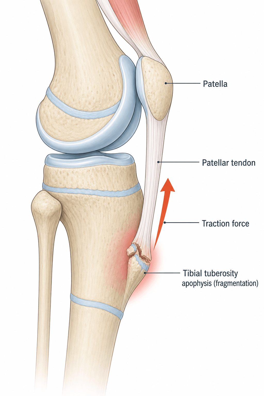
- Traction apophysitis: Repetitive stress on tibial tubercle from patellar tendon during growth spurt
- Self-limiting condition: Resolves when physis closes (typically 1-2 years), may leave painless bump
- Clinical diagnosis: Point tenderness over tibial tubercle, worsened by resisted knee extension
- Treatment is conservative: Activity modification, ice, stretching - rarely any surgery needed
- X-rays not routine: Reserve for atypical features or to rule out other pathology
- “Most common cause of anterior knee pain in athletic adolescents
- “Bilateral in 20-30% but usually asymmetric
- “Persistent bump over tibial tubercle after resolution is normal - not a complication
- “Rarely, a loose ossicle may require excision if symptomatic in adulthood
Traction apophysitis during growth spurt. Repetitive contraction of quadriceps pulls on tibial tubercle via patellar tendon. Peak age 10-15 years (earlier in girls due to earlier growth spurt). More common in jumping/running sports (basketball, soccer, gymnastics).
Point tenderness over tibial tubercle is pathognomonic. Palpable bump (enlarged tubercle). Pain worsened by running, jumping, kneeling, squatting, stairs. Reproduced by resisted knee extension and passive flexion. No effusion, no instability.
X-rays NOT required for diagnosis - clinical diagnosis. If obtained, may show soft tissue swelling, fragmentation, or irregular ossification of tubercle. Reserve X-rays for: atypical presentation, ruling out tumor/infection, failed treatment, suspected avulsion fracture.
Most cases managed in primary care. Refer if: symptoms persist beyond skeletal maturity, complete avulsion fracture suspected, concern for other diagnoses (tumor, infection), severe symptoms not responding to conservative care after 6 months.
- Key Features
- 10-15y athletes, activity-related pain, bump
- Tenderness Location
- Tibial tubercle (anterior, distal to patella)
- Management
- Activity modification, stretching, ice
- Key Features
- Similar age/mechanism, pain at inferior pole
- Tenderness Location
- Interior pole of patella
- Management
- Same conservative treatment as OSD
- Key Features
- Anterior knee pain, worse sitting/stairs
- Tenderness Location
- Diffuse peripatellar, retropatellar
- Management
- VMO strengthening, patellar taping
- Key Features
- Older athletes (16+), jumping sports
- Tenderness Location
- Inferior pole patella to tendon insertion
- Management
- Eccentric exercises, load management
- Key Features
- Acute traumatic event, swelling, unable to extend
- Tenderness Location
- Tibial tubercle with deformity
- Management
- URGENT - surgical fixation usually needed
SCHLATTERSCHLATTER - Differential Diagnosis
Hook:Think through SCHLATTER differentials - especially rule out SCFE with hip exam in any pediatric knee pain.
Overview and Epidemiology
Osgood-Schlatter Disease (OSD) is a common traction apophysitis affecting the tibial tubercle in active adolescents. It is characterized by localized pain, swelling, and tenderness at the insertion of the patellar tendon on the tibial tubercle.
Epidemiology:
- Most common cause of knee pain in athletic adolescents
- Peak incidence: males 12-15 years, females 10-13 years (earlier due to earlier puberty)
- Male to female ratio approximately 3:1 (historical), but increasing in females with sports participation
- 20-30% bilateral (often asymmetric in severity)
- Most common in running, jumping, kicking sports (soccer, basketball, gymnastics, volleyball)
The tibial tubercle apophysis (secondary ossification center) is biomechanically weaker than mature bone or the patellar tendon. During the adolescent growth spurt, rapid bone growth increases muscle-tendon tension while the apophysis has not yet fused. Repetitive traction from the powerful quadriceps causes microtrauma, inflammation, and sometimes fragmentation of the apophysis.
Etiology and Risk Factors:
- Age: Skeletal immaturity with open tibial tubercle physis
- Sex: Male more common (puberty timing, sports participation)
- Rapid growth: Growth spurt increases tension
- Quadriceps tightness: Tight quads increase traction force
- Hamstring tightness: Adds load to extensor mechanism
- High-impact sports: Running, jumping, kicking
- Training volume: Intense practice schedules during growth
- Poor conditioning: Sudden increase in activity level
- Hard playing surfaces: Increase impact loading
- Inadequate rest periods: No recovery time between training
Natural History:
- Most cases resolve spontaneously when the tibial tubercle apophysis fuses (typically 1-2 years)
- Residual bony prominence (bump) persists in most patients - this is NORMAL, not a complication
- 5-10% have persistent symptoms into adulthood, usually from ossicle within tendon
- Rare complications: tibial tubercle avulsion fracture, persistent ossicle requiring excision
A common viva extension is "name other traction apophysites" - OSD is the prototype of a family of overuse injuries at a tendon/muscle insertion onto a secondary ossification centre (apophysis) during the growth spurt, all sharing the same biology and conservative management:
- Osgood-Schlatter - tibial tubercle (patellar tendon insertion).
- Sinding-Larsen-Johansson - inferior pole of the patella (patellar tendon origin) - same knee, one level higher.
- Sever disease - calcaneal apophysis (Achilles insertion) - the commonest cause of paediatric heel pain.
- Iselin disease - base of the 5th metatarsal (peroneus brevis insertion).
- Medial epicondyle apophysitis ("Little League elbow") - medial humeral epicondyle (common flexor-pronator/UCL traction).
- Others: iliac crest / ASIS / AIIS apophysitis (pelvic avulsion spectrum), and the olecranon apophysis in throwers/gymnasts.
The unifying principles: a growing athlete, activity-related pain at the named bony insertion, a clinical diagnosis (imaging only for atypical features/avulsion), and self-limiting with load management, stretching and time until the apophysis fuses.
Exam point: when asked, frame OSD as a traction apophysitis and name the parallel sites (SLJ, Sever, Iselin, Little League elbow) with the shared "growing athlete + insertional pain + conservative + self-limiting" rule.
Pathophysiology and Mechanisms
Tibial Tubercle Anatomy
The tibial tubercle is the bony prominence on the anterior proximal tibia where the patellar tendon inserts. It originates from a secondary ossification center (apophysis) that appears around age 8-12.
- Age (years)
- 0-8
- Description
- Entirely cartilage, no ossification
- Clinical Relevance
- Rarely symptomatic at this age
- Age (years)
- 8-12
- Description
- Secondary ossification center appears
- Clinical Relevance
- Beginning of vulnerability period
- Age (years)
- 12-14
- Description
- Ossification extends proximally from apophysis
- Clinical Relevance
- Peak vulnerability - highest OSD incidence
- Age (years)
- 14-18
- Description
- Apophysis fuses to tibial epiphysis
- Clinical Relevance
- OSD symptoms resolve with fusion
The cartilaginous apophysis is the weak link. During the epiphyseal stage, the apophysis may avulse as a whole with acute trauma (jumping sports, landing). Unlike OSD, avulsion is an ACUTE injury with sudden pain, inability to extend knee, and visible/palpable deformity. Treat as a fracture - usually requires surgical fixation.
Extensor Mechanism Biomechanics
Quadriceps muscle generates force that is transmitted through:
- Quadriceps tendon → inserts on patella
- Patella → acts as sesamoid/pulley
- Patellar tendon → inserts on tibial tubercle
In growing adolescents, the tibial tubercle apophysis experiences HIGH tensile stress at the tendon-bone junction.
- Fulcrum effect: Patella increases quadriceps moment arm 30-50%
- Peak stress at insertion: Tendon-bone junction concentrates force
- Weak link: Cartilaginous apophysis weaker than tendon or bone
- Growth-related tension: Rapid femoral growth increases quad tightness
Increased Q-angle (greater than 15 degrees in males, greater than 18 degrees in females) may increase lateral patellar tracking and contribute to patellofemoral symptoms. However, the relationship between Q-angle and OSD specifically is less clear. Q-angle is more relevant to patellofemoral pain syndrome.
Classification Systems
Clinical Severity Grading
- Symptoms
- Pain only after activity, no swelling at rest
- Impact on Activity
- Can complete training sessions
- Treatment Approach
- Ice after activity, stretching, continue sports
- Symptoms
- Pain during AND after activity, mild swelling
- Impact on Activity
- Performance affected, some activity limitation
- Treatment Approach
- Modify activity level, patellar strap, formal physio
- Symptoms
- Pain at rest, limits daily activities, walking painful
- Impact on Activity
- Unable to participate in sport
- Treatment Approach
- Rest from sport, possible short immobilization, physio
Mild OSD may improve in 6-8 weeks with basic activity modification. Moderate OSD typically requires 3-6 months of modified activity. Severe OSD may need 6-12 months, possibly with periods of complete rest. All grades typically resolve when the physis closes, but symptom duration is proportional to severity.
Clinical Assessment
History:
- Age and sex: Peak 10-15 years, more common in males
- Sports participation: Type, frequency, intensity
- Training changes: Recent increase in volume or intensity?
- Pain characteristics: Location, timing, aggravating factors
- Bilateral symptoms: 20-30% bilateral
- Previous injury: Rule out acute trauma
- Night pain or rest pain: Consider tumor, infection
- Acute traumatic onset: Avulsion fracture
- Knee effusion: Unusual for OSD - suggests other pathology
- Systemic symptoms: Fever, weight loss - infection, malignancy
- Very young child: Under age 8 - OSD rare, investigate
- Hip symptoms: SCFE, Perthes - examine hip in all knee pain
Physical Examination:
Systematic Examination
- Visible bump over tibial tubercle (enlarged, prominent)
- Compare bilateral - may be asymmetric if bilateral OSD
- Look for effusion (unusual for OSD - suggests other diagnosis)
- Assess overall limb alignment, muscle bulk
- Point tenderness over tibial tubercle - pathognomonic
- Palpate entire patellar tendon (rule out tendinopathy)
- Palpate inferior pole of patella (Sinding-Larsen-Johansson)
- Palpate medial and lateral joint lines (meniscal pathology)
- Palpate patella and peripatellar tissues
- Knee ROM: Usually full, may have slight flexion discomfort
- Passive flexion: May be painful at end range (compresses tubercle)
- Quadriceps and hamstring flexibility: Often reduced
- Hip ROM: Must examine hip to rule out referred pain (SCFE)
- Resisted knee extension: Reproduces pain over tibial tubercle
- Active knee extension: May be painful against resistance
- Patellar mobility: Normal in OSD (unlike patellofemoral syndrome)
- Ligament stability: Should be normal
- Meniscal tests: Should be negative
SCFE and Perthes disease commonly present as knee pain in children due to referred pain along the obturator nerve. In ANY child presenting with knee pain, especially if obesity or limited hip ROM is present, you MUST examine the hip. Missing SCFE can lead to avascular necrosis and hip destruction.
OSGOODOSGOOD - Clinical Features
Hook:OSGOOD disease has all the Good features - obvious on exam, self-limiting, no surgery needed.
Investigations
Osgood-Schlatter is a CLINICAL diagnosis. Imaging is NOT required in typical cases. Key clinical features (point tenderness over tibial tubercle in athletic adolescent during growth spurt) are sufficient for diagnosis. Reserve imaging for atypical presentations or to exclude other pathology.
When to Order Imaging:
- Acute traumatic event (rule out avulsion fracture)
- Atypical age (too young or too old for OSD)
- Night pain or rest pain (rule out tumor)
- Effusion (unusual for OSD)
- Failure to improve with 3-6 months conservative treatment
- Suspected loose ossicle in symptomatic adult
- Soft tissue swelling anterior to tibial tubercle
- Fragmentation of tubercle ossification center
- Irregular ossification of apophysis
- Separate ossicle in patellar tendon (may persist)
- Prominent tibial tubercle post-fusion
Other Imaging:
- Indication
- First-line if imaging needed
- Findings
- Soft tissue swelling, fragmentation, ossicle
- Clinical Use
- Confirm diagnosis, rule out fracture
- Indication
- Assess patellar tendon, soft tissues
- Findings
- Tendon thickening, fragmentation, bursa
- Clinical Use
- Useful if tendinopathy suspected
- Indication
- Rule out tumor, stress fracture, infection
- Findings
- Edema at tubercle, soft tissue changes
- Clinical Use
- Rarely needed - reserve for red flags

Management Algorithm
Non-Operative Management (First-Line for ALL Cases)
95%+ of OSD resolves with conservative treatment alone.
Conservative Treatment Protocol
- Do NOT stop all activity - reduce intensity and volume
- Avoid painful activities (deep squats, jumping, kneeling)
- Continue sport at reduced level if tolerable
- Cross-train with low-impact activities (swimming, cycling)
- Ice after activity (15-20 minutes) for symptom relief
- Quadriceps stretching: Reduces traction force on tubercle
- Hamstring stretching: Decreases quadriceps demand
- Hip flexor stretching: Improves mechanics
- Hold stretches 30 seconds, 3-4 times daily
- Dynamic warm-up before activity
- Eccentric quadriceps: Once acute pain settles
- Core stability: Reduce load on knee during activity
- Hip strengthening: Improve biomechanics
- Progress gradually as symptoms allow
- Patellar strap/brace: Reduces traction on tubercle
- Knee pad: Protects tubercle when kneeling
- NSAIDs: Short-term for acute flares (not long-term)
- Ice/heat: Symptom relief
Prolonged complete rest is NOT recommended. It weakens muscles, detrains the athlete, and delays return to sport. Modify activity to a tolerable level rather than stopping entirely. The exception is severe cases with rest pain, which may need a short period of immobilization.
TENDERTENDER - Treatment Approach
Hook:Be TENDER with treatment - this is self-limiting, don't over-treat.
Surgical Technique
Ossicle Excision (Adults Only)
Surgery for Osgood-Schlatter disease is extremely rare and reserved for symptomatic ossicle in adults (post-skeletal maturity) or cosmetic removal of prominent tibial tubercle. Surgery is NOT indicated for active OSD in adolescents.
- Skeletal maturity (closed tibial tubercle physis)
- Persistent focal pain over ossicle
- Failed conservative management (6+ months)
- Imaging confirms symptomatic ossicle within tendon
- Longitudinal or transverse incision over tibial tubercle
- Identify ossicle within patellar tendon substance
- Excise ossicle, debride tendon edges
- Repair tendon if needed
- Some surgeons also smooth prominent tibial tubercle
This is a straightforward procedure with excellent outcomes in appropriately selected patients.
Complications
- Incidence
- Very common (50%+)
- Risk Factors
- Part of natural history
- Management
- Reassurance - cosmetic only, does not affect function
- Incidence
- Rare (less than 1%)
- Risk Factors
- Acute trauma in severe OSD
- Management
- Surgical ORIF if displaced, cast if non-displaced
- Incidence
- 5-10%
- Risk Factors
- Large initial ossicle, non-compliance
- Management
- Ossicle excision if failed conservative treatment
- Incidence
- Uncommon
- Risk Factors
- Return to sport too early, ongoing overuse
- Management
- Eccentric loading program, activity modification
- Incidence
- 10-20%
- Risk Factors
- Prominent tubercle persists
- Management
- Knee pads, reassurance, rarely surgical reduction
Tibial tubercle avulsion fracture is DIFFERENT from OSD. Avulsion is acute, traumatic, with sudden pain and inability to extend knee. It requires urgent orthopaedic referral. OSD is chronic, overuse, with gradual onset and preserved extension. Do not confuse these conditions.
The vivas above ask "what is genu recurvatum and why does it occur?" - here is the examinable answer, which is a paediatric physeal problem distinct from any adult cause:
- What it is: a knee that hyperextends (a recurvatum deformity) - the tibia sits in extension beyond neutral.
- Why it occurs in this context: the tibial tubercle apophysis is the anterior extension of the proximal tibial physis. An injury to that anterior physis - from a tubercle avulsion fracture, from its surgical fixation across the physis in a young child, or (rarely) from severe apophyseal disruption - can cause a localised anterior growth arrest. The posterior physis keeps growing while the front does not, so the proximal tibia tilts into recurvatum (an asymmetric physeal arrest deformity).
- Who is at risk: the skeletally immature child with substantial growth remaining - which is exactly why hardware is ideally kept out of/parallel to the physis and why these children need growth monitoring after tubercle injury or surgery.
- Why OSD itself rarely causes it: ordinary OSD does not arrest the physis (Krause's natural-history series found NO premature epiphyseal arrest) - recurvatum is a complication of the avulsion/iatrogenic end of the spectrum, not of uncomplicated apophysitis.
Exam point: genu recurvatum after a tibial tubercle injury = anterior proximal-tibial physeal arrest with continued posterior growth in a skeletally immature child - prevent it by respecting the physis and monitoring growth; uncomplicated OSD does not cause it.
Postoperative Care and Rehabilitation
Rehabilitation for Ossicle Excision (Adults)
Post-Ossicle Excision Protocol
- Weight-bearing as tolerated
- ROM exercises as comfort allows
- Ice, elevation for swelling
- Gentle quadriceps sets
- Full ROM expected by 6 weeks
- Progressive strengthening
- Bike, swimming for cardio
- Avoid deep squats, jumping
- Sport-specific training
- Plyometrics progression
- Jogging, running progression
- Full return based on strength testing
- Typically 3-4 months for full competition
- Ongoing maintenance stretching
Rehabilitation for Conservative OSD (Adolescents)
This is the main patient population - structured rehab program while continuing modified activity:
- Daily stretching: Quadriceps, hamstrings, hip flexors (30 sec × 3)
- Eccentric strengthening: When acute pain settles
- Core stability exercises
- Sport modification: Continue at reduced level
- Ice after activity
- Gradual progression as symptoms allow
Outcomes
- Excellent prognosis - most cases resolve completely with skeletal maturity
- Symptoms typically improve within 1-2 years (when tubercle physis fuses)
- Persistent tibial tubercle prominence is common but NOT a functional problem
- Most athletes return to full sport without long-term issues
- 5-10% have some adult symptoms, usually mild or related to ossicle
- Severe initial presentation
- Delay in activity modification
- Ongoing intense sports participation without modification
- Bilateral involvement
Parents often worry about the residual bump over the tibial tubercle. Reassure them this is part of the normal healing process - the prominence represents bone that formed during the inflammatory phase and is now incorporated into the mature tubercle. It is cosmetic only and does not affect function or sports performance.
Guidelines, Registries & Global Practice
Global Epidemiology:
- OSD is the most common cause of activity-related anterior knee pain in adolescents worldwide. In youth elite male football the clinical point prevalence is around 17%, yet roughly 80% of affected players have no time-loss (Schultz et al, Phys Ther Sport 2022, PMID 35305497).
- Peak age tracks the pubertal growth spurt: boys approximately 12-15 years, girls approximately 8-13 years (earlier maturation). The historical male predominance has narrowed substantially as female sports participation has risen.
- Bilateral involvement occurs in 20-30% of cases, frequently asymmetric. A history of another apophysitis (Sever disease) is a strong associated factor (OR 16.8 in elite footballers, PMID 35305497).
- Adolescent knee pain (including OSD) is not uniformly trivial: in an individual-participant-data meta-analysis, 51% still reported knee pain at 12 months, with female sex and bilateral pain predicting a poorer course (Holden, Rathleff et al, Pain 2021, PMID 33449504).
Side-by-side Guidance and Authoritative Sources:
- Position on Imaging
- Clinical diagnosis; radiographs only for atypical features or to exclude other pathology
- First-line Treatment
- Activity modification, relative rest, stretching, ice, analgesia as needed
- Surgery
- Reserved for skeletally mature patients with a symptomatic ossicle after failed conservative care
- Position on Imaging
- No routine imaging; X-ray only if red flags or diagnostic doubt
- First-line Treatment
- Reassurance, self-care, activity modification, simple analgesia, physiotherapy
- Surgery
- Specialist referral only for persistent or atypical symptoms
- Position on Imaging
- Clinical diagnosis; image to exclude avulsion, tumour or infection if red flags
- First-line Treatment
- Education, load management, eccentric and flexibility programme
- Surgery
- Rarely indicated; ossicle excision in mature symptomatic patients
- Position on Imaging
- Ultrasound increasingly used to confirm tubercle changes; X-ray for red flags
- First-line Treatment
- Conservative load management; injection therapy investigational only
- Surgery
- Arthroscopic or open ossicle excision after physeal closure
No society publishes a formal high-level (Grade A) treatment guideline for OSD because randomised evidence is sparse. Recommendations are consensus/expert-opinion level, converging on the same message: clinical diagnosis, conservative load-managed treatment, and surgery only for the skeletally mature refractory ossicle. Injection therapies (including prolotherapy) remain investigational with low-quality evidence (Sanderson and Bryant, J Foot Ankle Res 2015, PMID 26500703).
- There is no dedicated OSD registry; population-level signal comes from claims and cohort datasets. A large US commercial-claims analysis of adolescent sports injuries found female adolescents more likely than males to undergo surgery for OSD (adjusted OR 1.8, 95% CI 1.38-2.39), although operative rates overall remained low and stable (Bonazza et al, Arthrosc Sports Med Rehabil 2019, PMID 32266341).
- Arthroplasty-style implant registries (e.g. AOANJRR, NJR, AJRR) are not relevant to OSD as it is non-arthroplasty paediatric pathology.
- Across health systems, OSD is managed predominantly in primary care, general practice, and sports-medicine/physiotherapy services, with orthopaedic referral reserved for suspected avulsion fracture or the refractory skeletally mature patient.
- Common sporting contexts worldwide include soccer, basketball, netball and similar youth sports. Access to physiotherapy and sports medicine services varies by health system but follows the same conservative principles globally.
- Practice variation centres on adjuncts (patellar straps, injections) and the threshold for imaging rather than on the core conservative principle, which is globally consistent.
MCQ Practice Points
Q: An adolescent presents with knee pain after jumping. How do you distinguish OSD from avulsion fracture? A: OSD has GRADUAL onset, pain during/after activity, ability to extend knee, and no visible deformity. Avulsion has ACUTE onset after trauma, inability to extend knee, visible/palpable deformity, and severe pain. X-ray shows displaced fragment in avulsion vs fragmentation in OSD.
Q: Which of the following is an indication for X-ray in suspected OSD? A: Night pain (rule out tumor), acute traumatic onset (rule out avulsion), failure to improve after 6 months conservative treatment, atypical age (less than 8 or after skeletal maturity). Routine OSD does NOT require imaging.
Q: What is the recommended activity level for adolescent with OSD? A: Activity MODIFICATION, not complete rest. Continue sport at reduced level if tolerable. Avoid painful activities (deep squats, kneeling, jumping). Cross-train with low-impact activities. Complete rest weakens muscles and delays return.
Q: What happens to the bump after OSD resolves? A: The tibial tubercle prominence typically persists as a painless, cosmetic bump in 50-75% of patients. This represents ossification that occurred during the healing process and is now incorporated into the mature tubercle. It is NOT a complication and does not affect function.
Q: A 12-year-old with knee pain has point tenderness at the INFERIOR POLE of patella. What is the diagnosis? A: Sinding-Larsen-Johansson syndrome - traction apophysitis at the inferior pole of patella (where central patellar tendon originates). Same mechanism as OSD but at the proximal end of the patellar tendon. Treatment is identical - activity modification, stretching, ice.
Medicolegal Considerations
- Clear history of gradual onset and activity-related symptoms
- Documentation of point tenderness specifically over tibial tubercle
- Hip examination performed (to exclude SCFE/Perthes)
- Discussion of self-limiting nature and expected timeline
- Activity modification advice given (not complete rest)
- Risk of persistent symptoms
- Hardware removal may be needed
- Scar
- Stiffness
SCFE commonly presents as knee pain in obese adolescent males. Missing SCFE diagnosis causes significant morbidity from AVN. Document hip examination in ALL adolescents presenting with knee pain. This is a frequent source of litigation in pediatric orthopaedics.
Clinical Decision Scenarios
Practise clinical reasoning and management decisions out loud
“A 13-year-old boy who plays soccer presents with 3 months of anterior knee pain. The pain is worse after training and he has noticed a bump at the front of his knee.”
“An 11-year-old female gymnast presents with bilateral anterior knee pain. She trains 20 hours per week and has competition in 6 weeks. Parents want a solution so she can compete.”
“A 14-year-old basketball player jumps for a rebound and lands with sudden severe knee pain. He cannot extend his knee and you notice a visible deformity over the tibial tubercle.”
Diagnosis
- Clinical diagnosis - imaging not required
- Point tenderness over tibial tubercle
- Pain with resisted knee extension
- 10-15 years, athletic, growth spurt
Treatment
- Activity MODIFICATION not complete rest
- Stretching: quads, hamstrings, hip flexors
- Ice after activity (15-20 min)
- Patellar strap during sport
- NSAIDs short-term for flares only
Imaging Indications
- Acute traumatic onset (avulsion)
- Night pain (tumor)
- Failure after 6 months conservative
- Atypical age or presentation
Red Flags
- Acute trauma with inability to extend = AVULSION
- Night/rest pain = tumor, infection
- Knee effusion = not typical for OSD
- Hip symptoms = SCFE, Perthes
Prognosis
- 90%+ resolve with skeletal maturity
- Duration 1-2 years (until physis closes)
- Bump persists but is painless
- 5-10% adult symptoms (usually ossicle)
Evidence Base
According to PubMed, the evidence base below has been verified against the primary literature. Osgood-Schlatter disease (OSD) has few high-level trials; the strongest data concern its benign natural history and the outcomes of ossicle excision in the small refractory subgroup.
Natural History of Osgood-Schlatter Disease
- Retrospective review of the natural history of 69 knees in 50 patients with untreated OSD
- 76% reported no limitation of activity at follow-up, but 60% still could not kneel without discomfort
- Two groups emerged: those with radiological fragmentation (often left a separated ossicle or abnormally ossified tuberosity) and those with soft-tissue swelling only (asymptomatic at review)
- Low incidence of patellar instability or anterior knee pain, and NO case of premature proximal tibial epiphyseal arrest
Osgood Schlatter Syndrome - Contemporary Review
- Critical review establishing OSD as a traction apophysitis of the tibial tubercle from repetitive strain on the secondary ossification centre
- Approximately 90% of patients respond well to non-operative treatment (rest, ice, activity modification, rehabilitation exercises)
- Boys typically 12-15 years, girls 8-12 years; symptoms exacerbated by jumping sports and direct kneeling contact
- Surgical excision of the ossicle/free cartilage reserved for skeletally mature patients who remain symptomatic
Prognostic Factors for Adolescent Knee Pain (IPD Meta-analysis)
- Individual-participant-data meta-analysis of 1281 unique adolescents (10-19y) with non-traumatic knee pain across 13 prospective studies (including OSD)
- 51% still reported knee pain at 12 months - adolescent knee pain is NOT reliably trivial or short-lived
- Poorer prognosis predicted by higher baseline pain frequency, lower quality of life, female sex, and bilateral pain
- BMI, pain sensitivity, and knee strength were NOT associated with prognosis
Ossicle Resection with Tibial Tubercleplasty for Unresolved OSD
- Retrospective review of 16 knees in 15 patients undergoing ossicle excision plus tibial tubercleplasty after failed non-operative treatment
- 12 of 15 patients (75%) returned fully to pre-operative activities and sport; 2 partial, 1 did not return
- Mean post-operative Lysholm 76.5, mean IKDC 75, mean Tegner activity level 6.8
- Authors recommend deferring surgery until skeletal maturity and adding tubercleplasty when resecting the ossicle
Arthroscopic Ossicle Excision in Athletes with Unresolved OSD
- 11 competitive athletes (mean age 23y) with persistent tibial tubercle pain (mean 15.5 months) treated arthroscopically
- Mean Kujala score improved 82.9 to 98.5 and Lysholm 87.5 to 96.9 (both significant at p of 0.01) at mean 66-month follow-up
- Mean return to sport-specific training 6.7 weeks; all returned to the same competitive level
- Arthroscopic approach reported to speed recovery and avoid open patellar-tendon damage
Prevalence and Time-loss of OSD in Youth Elite Football
- Cross-sectional and nested case-control study of youth elite male footballers (cross-sectional n = 127)
- Clinical point prevalence of OSD was 17%, yet 80% had NO time-loss despite symptoms
- Previous Sever disease was strongly associated with OSD (OR 16.8, 95% CI 1.6-174.5)
- Growth velocity and ultrasonographic bone-maturity (Ehrenborg) stage were NOT associated in age-matched analysis
Prolotherapy Injections for Lower-limb Tendinopathy and OSD (Systematic Review)
- Systematic review of injection therapies for lower-limb tendinopathy/fasciopathy, including OSD
- For OSD, dextrose prolotherapy gave equal or superior results to usual care or lignocaine injection
- No adverse events following prolotherapy reported in any included study
- Overall methodological quality was poor (allocation concealment, blinding, intention-to-treat)