Adult Vitamin D Deficiency | Defective Mineralization | Pathological Fractures
- Osteomalacia = defective bone mineralization in adults (rickets in children)
- Looser zones (pseudofractures) = pathognomonic radiographic finding
- Proximal myopathy causes waddling gait and difficulty rising from chair
- Vitamin D less than 25 nmol/L = severe deficiency requiring urgent treatment
- Pathological fractures common in weight-bearing bones despite normal-appearing radiographs
- “Distinguish from osteoporosis: osteomalacia has defective mineralization, not low bone mass
- “Elevated alkaline phosphatase with low calcium and phosphate = classic biochemistry
- “Oncogenic osteomalacia from mesenchymal tumors (FGF23-secreting) - surgical excision curative
- “Always check vitamin D levels before arthroplasty - prevents delayed healing and loosening
Defective mineralization of osteoid. Unlike osteoporosis (reduced bone mass), osteomalacia has normal or increased osteoid but inadequate calcium and phosphate deposition. This causes soft, deformable bones prone to fracture.
Pseudofractures are pathognomonic. Bilateral, symmetric, perpendicular to cortex. Common sites: femoral neck, pubic rami, ribs, scapula. Represent stress fractures that fail to heal due to poor mineralization.
Bone pain + proximal myopathy + fractures. Pain is diffuse, worse with weight-bearing. Myopathy causes waddling gait. Fractures occur with minimal trauma in weight-bearing bones.
Less than 25 nmol/L = severe deficiency. Target 50-75 nmol/L for bone health. Replacement: 50,000 IU weekly for 6-8 weeks, then maintenance 800-2000 IU daily with calcium.
- Biochemistry
- Low Ca/PO4, high ALP, low vitamin D
- Radiographic Finding
- Looser zones, osteopenia
- Treatment
- Vitamin D + calcium replacement
- Biochemistry
- Normal Ca/PO4/ALP
- Radiographic Finding
- Low bone density (DXA), no Looser zones
- Treatment
- Bisphosphonates, lifestyle
- Biochemistry
- High Ca, low PO4, high PTH
- Radiographic Finding
- Subperiosteal resorption, brown tumors
- Treatment
- Parathyroidectomy
- Biochemistry
- Normal Ca/PO4, very high ALP
- Radiographic Finding
- Lytic/sclerotic, cortical thickening
- Treatment
- Bisphosphonates for symptoms
PURFNSSites of Looser Zones (Pseudofractures)
Hook:PURFNS = sites that are PERFECT for bilateral symmetric pseudofractures!
Overview
Definition
Osteomalacia is a metabolic bone disease characterized by defective mineralization of newly formed osteoid in mature bone. It represents the adult equivalent of rickets, which affects growing bones in children. The hallmark is accumulation of unmineralized or undermineralized osteoid matrix, resulting in soft, deformable bones with increased fracture risk.
Epidemiology
- Elderly institutionalized: 30-50% have vitamin D deficiency (less than 50 nmol/L)
- Community-dwelling elderly: 10-15% have deficiency
- Post-bariatric surgery: 25-40% develop deficiency without supplementation
- Dark-skinned populations in high latitudes: 5-10 times higher risk
- Female to male ratio: 3:1 (postmenopausal women at highest risk)
- Age: Risk increases with age (reduced sunlight exposure, decreased skin synthesis)
- Geographic variation: Higher prevalence at latitudes greater than 35 degrees (reduced UVB exposure)
Pathophysiology
Normal bone mineralization requires adequate calcium, phosphate, and alkaline phosphatase. Vitamin D is essential for intestinal calcium absorption and renal phosphate reabsorption. In osteomalacia, deficiency of vitamin D or phosphate leads to:
Molecular cascade:
- Reduced calcium and phosphate availability
- Inadequate hydroxyapatite crystal formation in osteoid
- Accumulation of unmineralized osteoid (increased osteoid volume)
- Secondary hyperparathyroidism (compensatory response to hypocalcemia)
- Further phosphate wasting (PTH-induced renal phosphate loss)
- Progressive bone softening and deformity
- Increased osteoid seams (greater than 12 micrometers thick)
- Prolonged mineralization lag time (greater than 100 days vs normal less than 25 days)
- Reduced bone stiffness and mechanical strength
- Trabecular thinning with preserved connectivity
- Pathological fractures with minimal trauma
- Bone pain from periosteal stress and microfractures
- Proximal myopathy from vitamin D deficiency
- Skeletal deformities (bowing, compression)
Vitamin D receptors exist in muscle, brain, immune cells, and cardiovascular tissue. Severe deficiency causes proximal myopathy (difficulty rising from chair, climbing stairs), increased infection risk, and possibly cardiovascular disease. Always treat systemic deficiency, not just bone disease.
VITAMINSCauses of Osteomalacia
Hook:Your bones need VITAMINS - without them, they can't mineralize properly!
Clinical Presentation
Symptoms
- Diffuse bone pain - worse with weight-bearing, pressure
- Tenderness on palpation - sternum, ribs, pelvis, long bones
- Pathological fractures - minimal trauma, weight-bearing bones
- Skeletal deformities - leg bowing, spinal kyphosis
- Proximal muscle weakness - waddling gait, difficulty rising
- Myalgias - muscle pain and cramping
- Tetany (if severe hypocalcemia) - Chvostek, Trousseau signs
- Fatigue and general malaise
Examination Findings
- Antalgic or waddling gait - due to bone pain and proximal myopathy
- Bone tenderness - sternal pressure, rib compression, pelvic compression
- Skeletal deformities - leg bowing (varus or valgus), kyphosis
- Reduced muscle power - hip flexion (iliopsoas), knee extension (quadriceps) weakness
- Proximal muscle weakness - grade 3-4 out of 5 in hip flexors, shoulder abductors
- Hyporeflexia - reduced or absent deep tendon reflexes
- Tetany signs (if severe hypocalcemia) - Chvostek's sign (facial twitch), Trousseau's sign (carpopedal spasm)
Many patients with osteomalacia are asymptomatic or minimally symptomatic until a pathological fracture occurs. High index of suspicion needed in at-risk populations: elderly, institutionalized, malabsorption syndromes, dark skin in low-sunlight regions, post-bariatric surgery.
BONE PAINClinical Features of Osteomalacia
Hook:Remember the BONE PAIN to diagnose osteomalacia!
Osteomalacic Proximal Myopathy
The topic repeatedly invokes proximal myopathy (waddling gait, difficulty rising from a chair, and one of the three cardinal features), but the muscle disorder itself deserves separate development because it is a favourite viva follow-up ("why is this patient weak?") and its features are what distinguish it from the many other myopathies an examiner might offer.
Mechanism
Two deficiencies act together on muscle:
- Vitamin D / calcitriol deficiency. Skeletal muscle expresses the vitamin D receptor; calcitriol influences myocyte calcium handling and the maintenance of fast-twitch type II fibres. Deficiency produces selective type II fibre atrophy on muscle histology.
- Hypophosphatemia. Phosphate is required for ATP generation and for the phosphocreatine energy shuttle; low serum phosphate impairs muscle contractility and energy supply, contributing directly to weakness and fatigue.
Clinical and Laboratory Features
- Symmetrical proximal weakness, pelvic girdle affected more than shoulder girdle
- Myopathic (waddling) gait, difficulty rising from a chair or climbing stairs, positive Gowers manoeuvre
- Often painful and accompanied by bone tenderness (a clue it is metabolic, not a pure myopathy)
- Deep tendon reflexes preserved (helps exclude a primary neuropathy)
- Serum creatine kinase is characteristically normal - unlike inflammatory myopathy, dystrophy or rhabdomyolysis
- EMG is often normal or shows non-specific myopathic units (no fibrillations of active myositis)
- The biochemical panel (low vitamin D or phosphate, high alkaline phosphatase) points to the true cause
Reversibility
Osteomalacic myopathy is reversible with correction of the underlying deficiency. Muscle power typically begins to improve within a few weeks and largely recovers over 3-6 months of vitamin D (and, where relevant, phosphate) replacement - often before radiographic healing of Looser zones. Persistent weakness after adequate replacement should prompt a search for a second, unrelated neuromuscular cause.
Q: A patient with biochemical osteomalacia has marked proximal weakness but a normal creatine kinase. Why, and what does it tell you? A: The weakness of osteomalacia is a metabolic myopathy - driven by vitamin D receptor-mediated effects on type II fibres and by hypophosphatemia impairing muscle energetics - not by myofibre necrosis. Because there is little membrane breakdown, creatine kinase stays normal, in contrast to inflammatory myopathy, muscular dystrophy or rhabdomyolysis where CK is elevated. A normal CK with proximal weakness, bone pain and a deranged bone panel should steer you firmly toward metabolic bone disease, and predicts that the weakness will reverse with replacement rather than needing immunosuppression.
Laboratory Findings
Biochemistry
- Typical Finding
- Low or low-normal
- Mechanism
- Reduced vitamin D-mediated intestinal absorption
- Typical Finding
- Low
- Mechanism
- Reduced intestinal absorption and PTH-mediated renal wasting
- Typical Finding
- Elevated (often markedly)
- Mechanism
- Increased osteoblast activity attempting to mineralize osteoid
- Typical Finding
- Less than 25 nmol/L (severe deficiency)
- Mechanism
- Dietary lack, malabsorption, inadequate sunlight
- Typical Finding
- Elevated (secondary hyperparathyroidism)
- Mechanism
- Compensatory response to hypocalcemia
- Typical Finding
- Normal or low
- Mechanism
- Substrate (25-OH vitamin D) depletion limits 1-alpha hydroxylation
Vitamin D Thresholds:
- Severe deficiency: less than 25 nmol/L (less than 10 ng/mL)
- Deficiency: 25-50 nmol/L (10-20 ng/mL)
- Insufficiency: 50-75 nmol/L (20-30 ng/mL)
- Optimal for bone health: 75-125 nmol/L (30-50 ng/mL)
Q: Why is alkaline phosphatase elevated in osteomalacia but normal in osteoporosis? A: Osteoblast activity. In osteomalacia, osteoblasts are actively producing osteoid (unmineralized matrix) but cannot mineralize it due to lack of calcium/phosphate. This causes massive osteoid accumulation and elevated ALP. In osteoporosis, there is simply reduced bone formation - no excess osteoid, normal ALP.
Additional Investigations
- Urinary calcium: low (less than 2.5 mmol per 24 hours)
- Urinary phosphate: elevated in renal phosphate wasting
- FGF23 level: elevated in tumor-induced osteomalacia (oncogenic osteomalacia)
- Renal function: to assess for chronic kidney disease (impaired 1-alpha hydroxylation)
- Liver function: to assess for cholestatic disease (impaired vitamin D absorption)
Imaging
Radiographic Findings
Pathognomonic finding:
- Radiolucent bands perpendicular to cortex
- Bilateral and symmetric
- No periosteal reaction (unlike healing fracture)
- Common sites: femoral neck, pubic rami, ribs, scapula, proximal ulna
- Osteopenia (generalized demineralization)
- Coarsened trabecular pattern
- Cortical thinning
- Pathological fractures in weight-bearing bones
- Skeletal deformities (leg bowing, vertebral compression)
Bone Densitometry (DXA)
- Low bone mineral density (T-score less than -2.5 at spine or hip)
- Cannot distinguish osteomalacia from osteoporosis on DXA alone
- Biochemistry and clinical context essential for diagnosis
Advanced Imaging
- Increased uptake at sites of pseudofractures
- Multiple symmetric hot spots - "superscan" appearance
- Useful in tumor-induced osteomalacia to localize FGF23-secreting tumor
- Bone marrow edema at pseudofracture sites
- Localization of occult tumors in oncogenic osteomalacia
- Sensitivity for small mesenchymal tumors
- Assessment of bone quality and fracture risk
- 3D reconstruction for surgical planning (prophylactic fixation)
Bone Biopsy (Gold Standard)
- Diagnostic uncertainty after clinical, biochemical, radiographic evaluation
- Suspected hypophosphatasia or rare mineralization disorder
- Pre-treatment assessment in oncogenic osteomalacia
- Increased osteoid volume (greater than 15% vs normal less than 5%)
- Widened osteoid seams (greater than 12 micrometers)
- Prolonged mineralization lag time (greater than 100 days)
- Tetracycline double-labeling shows delayed mineralization front
Bone Histomorphometry & Tetracycline Double-Labelling
The topic names the biopsy findings that define osteomalacia - increased osteoid volume, widened osteoid seams, prolonged mineralization lag time, and delayed tetracycline labelling - and the burosumab evidence card reports reversal of exactly these parameters. Because the transiliac biopsy remains the histological gold standard, it is worth understanding how the technique actually produces those numbers and which combination is diagnostic.
How Tetracycline Double-Labelling Works
Tetracyclines chelate calcium and fluoresce, so they deposit as a bright band wherever active mineralization is occurring at the time of dosing. Two short courses are given before the (undecalcified) transiliac biopsy, separated by a label-free interval (a common schedule is a short course, roughly a fortnight off, then a second short course, with biopsy a few days later):
- The distance between the two fluorescent labels, divided by the number of days between them, gives the mineral apposition rate (MAR) in micrometres per day (normal roughly 0.6 to 1.0).
- The mineralization lag time (MLT) is derived from osteoid seam thickness relative to the apposition rate - it is the time osteoid waits before it mineralizes (normal under roughly 20 to 25 days; osteomalacia greater than 100 days).
- In severe osteomalacia mineralization is so impaired that there may be only a single, diffuse ("smudgy") label or no uptake at all - itself a strong pointer to the diagnosis.
The Diagnostic Combination (Not Osteoid Alone)
Osteomalacia needs both:
- Increased osteoid (raised osteoid volume, thickness and surface), and
- Defective mineralization (prolonged MLT / reduced apposition on tetracycline labelling)
Increased osteoid without a prolonged MLT is seen in high-turnover states (hyperparathyroidism, hyperthyroidism, Paget disease), where osteoid simply reflects fast formation, not a mineralization block. It is the prolonged lag time that separates true osteomalacia from these mimics.
Q: A bone biopsy shows increased osteoid volume. Is that osteomalacia? A: Not on its own. Osteomalacia requires the combination of increased osteoid and demonstrably defective mineralization - a prolonged mineralization lag time or absent/single tetracycline label. Increased osteoid with a normal lag time reflects high bone turnover (hyperparathyroidism, hyperthyroidism, Paget disease), not osteomalacia. The tetracycline double-label is what lets you measure the mineral apposition rate and lag time and prove that mineralization - rather than just formation - is the problem.
Differential Diagnosis
- Osteomalacia
- Defective mineralization
- Osteoporosis
- Reduced bone mass
- Hyperparathyroidism
- Excessive bone resorption
- Osteomalacia
- Low or normal
- Osteoporosis
- Normal
- Hyperparathyroidism
- Elevated
- Osteomalacia
- Low
- Osteoporosis
- Normal
- Hyperparathyroidism
- Low
- Osteomalacia
- Elevated
- Osteoporosis
- Normal
- Hyperparathyroidism
- Elevated
- Osteomalacia
- Low
- Osteoporosis
- Normal or low
- Hyperparathyroidism
- Normal or low
- Osteomalacia
- Elevated (secondary)
- Osteoporosis
- Normal
- Hyperparathyroidism
- Elevated (primary)
- Osteomalacia
- Looser zones
- Osteoporosis
- Fractures, no Looser zones
- Hyperparathyroidism
- Subperiosteal resorption, brown tumors
Q: What is oncogenic osteomalacia and how is it diagnosed? A: Tumor-induced osteomalacia caused by small mesenchymal tumors (often benign) secreting FGF23 (fibroblast growth factor 23). FGF23 causes renal phosphate wasting and inhibits 1-alpha hydroxylation of vitamin D. Biochemistry shows hypophosphatemia, elevated FGF23, low 1,25-OH vitamin D. Diagnosis requires whole-body imaging (MRI, PET) to locate tumor. Surgical excision is curative - biochemistry normalizes within hours.
Management
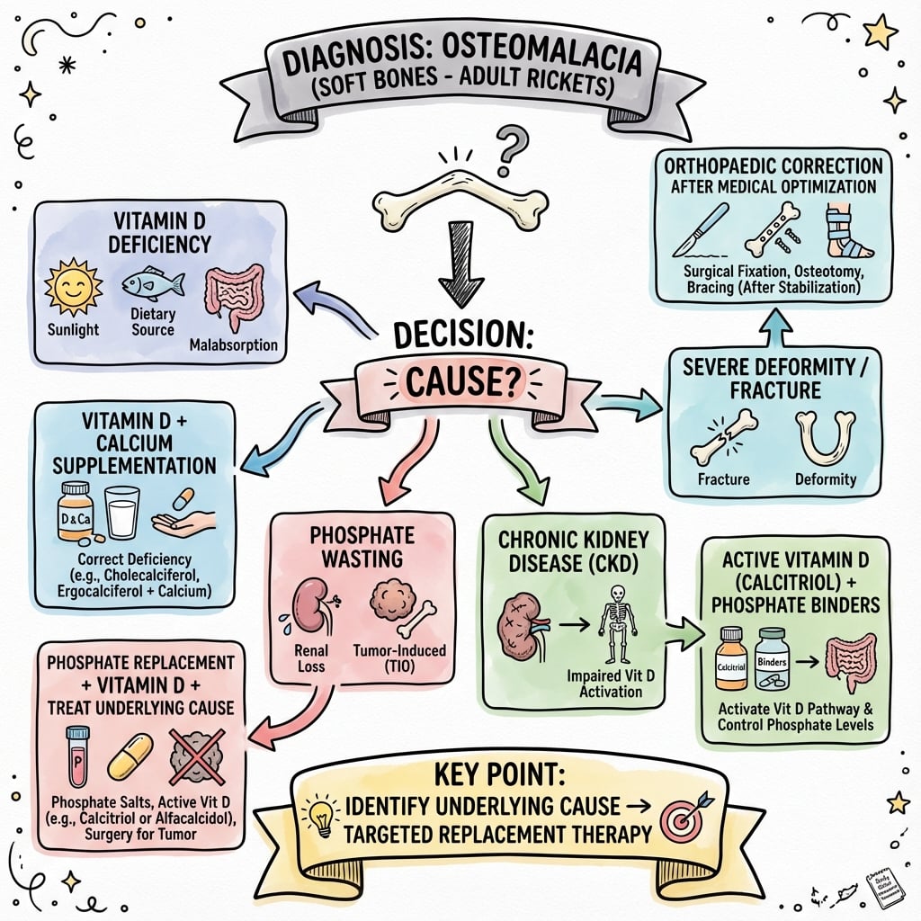
Vitamin D Replacement Protocol
Treatment Phases
- Cholecalciferol (vitamin D3) 50,000 IU weekly for 6-8 weeks
- Oral calcium 1000-1500 mg daily (divided doses with meals)
- Cholecalciferol 4000-6000 IU daily for 8-12 weeks
- Cholecalciferol 800-2000 IU daily
- Calcium 1000-1200 mg daily (dietary plus supplements)
- Recheck 25-OH vitamin D at 3 months - target 75-100 nmol/L
- Calcium and phosphate at 1, 3, 6 months then annually
- PTH and alkaline phosphatase - should normalize by 6 months
- Annual 25-OH vitamin D to ensure maintenance
In severe, prolonged osteomalacia with marked secondary hyperparathyroidism, rapid vitamin D and calcium replacement can cause hungry bone syndrome - profound hypocalcemia and hypophosphatemia as demineralized skeleton avidly takes up minerals. Monitor calcium closely in first 2 weeks. May require IV calcium gluconate if symptomatic.
Orthopaedic Implications
Pathological Fractures
- Femoral neck - bilateral, often at sites of Looser zones
- Proximal femur - subtrochanteric, intertrochanteric
- Pelvis - pubic rami, sacrum
- Ribs - multiple, painful
- Vertebrae - compression fractures
- Optimize medical management FIRST - vitamin D and calcium replacement
- Prophylactic fixation for impending fractures (Looser zones greater than 50% cortical width, symptomatic)
- Fracture fixation with caution - bone is soft, screw purchase poor
- Longer immobilization than normal fractures - delayed healing
Soft bone = poor screw purchase. Consider:
- Augmentation with cement in proximal femur fractures
- Longer plates with more screws for load distribution
- Locking plates to minimize screw toggle in soft bone
- Protected weight-bearing for 3-6 months (longer than normal)
- Aggressive vitamin D replacement perioperatively to accelerate healing
Arthroplasty Considerations
- Screen all arthroplasty candidates for vitamin D deficiency
- Optimize vitamin D greater than 75 nmol/L before elective surgery
- Correct calcium and phosphate abnormalities
- Risk of periprosthetic fracture during insertion (especially press-fit stems)
- Poor bone quality may favor cemented fixation
- Careful reaming and broaching to avoid fracture
- Delayed osseointegration of uncemented implants
- Risk of aseptic loosening if vitamin D not repleted
- Periprosthetic fracture risk with minimal trauma
- Continue vitamin D and calcium indefinitely
Prognosis and Outcomes
Expected Response to Treatment
- Calcium and phosphate normalize by 4-12 weeks
- PTH decreases by 3-6 months (may take longer if severe)
- Alkaline phosphatase declines by 6-12 months (may initially rise as bone heals)
- Bone pain improves by 6-12 weeks
- Muscle weakness reverses by 3-6 months
- Looser zones heal by 6-12 months (radiographic evidence of mineralization)
- Fracture risk decreases once vitamin D greater than 50 nmol/L
- Severe, prolonged deficiency - may have permanent skeletal deformities
- Uncontrolled underlying cause (malabsorption, chronic kidney disease)
- Non-compliance with supplementation
- Oncogenic osteomalacia with unresectable tumor
Guidelines, Registries & Global Practice
Global Epidemiology
- Vitamin D deficiency is one of the most prevalent micronutrient deficiencies worldwide, but its consequences vary by latitude, skin pigmentation, diet, sun exposure and food-fortification policy.
- Nutritional osteomalacia remains common in South Asia, the Middle East and North Africa, driven by limited effective UVB exposure (concealing dress, high latitude in winter, air pollution) and low dietary calcium/vitamin D.
- In high-income countries, frank osteomalacia is now mostly seen in the institutionalized elderly, malabsorption (coeliac disease, IBD, post-bariatric surgery), chronic kidney/liver disease, and dark-skinned migrants at higher latitudes.
Side-by-Side Guideline Comparison
- Deficiency threshold
- 25-OH vitamin D below 50 nmol/L (20 ng/mL)
- Replacement / target
- 50,000 IU weekly x8 wk then 1500-2000 IU/day; target above 75 nmol/L in at-risk
- Deficiency threshold
- Below 25 nmol/L = deficient; 25-50 = inadequate
- Replacement / target
- Loading approx 300,000 IU over 6-10 wk, then 800-2000 IU/day maintenance
- Deficiency threshold
- Below 30 nmol/L at risk of deficiency
- Replacement / target
- Lower population targets (50 nmol/L adequate for most) - less aggressive than Endocrine Society
- Deficiency threshold
- Below 30 nmol/L = deficient; 30-50 = insufficient
- Replacement / target
- Calcium AND vitamin D together; emphasises dietary calcium where intake low
- Key controversy: the Endocrine Society favours a higher individual target (above 75 nmol/L) for at-risk patients, whereas the IOM and several national bodies regard about 50 nmol/L as adequate for the general population. For symptomatic/biopsy-proven osteomalacia all bodies agree on therapeutic replacement plus calcium.
Registry & Outcome Notes
- There is no dedicated osteomalacia registry; relevant arthroplasty registries (NJR, AJRR, AOANJRR, Swedish/Norwegian) track revision and periprosthetic fracture, where poor bone quality and metabolic bone disease are recognised contributors to early failure and periprosthetic fracture.
High- vs Limited-Resource Practice
- High-resource: routine 25-OH vitamin D, PTH, FGF23 assays; functional imaging (68Ga-DOTATATE PET/CT, whole-body MRI) for TIO; access to burosumab and calcitriol.
- Limited-resource: diagnosis often clinical/radiographic and biochemical (calcium, phosphate, ALP); calcium plus vitamin D remains the cost-effective cornerstone and the global consensus stresses combined calcium-vitamin D because dietary calcium deficiency alone can cause osteomalacia/rickets even with adequate vitamin D.
Oncogenic Osteomalacia
- Rare FGF23-secreting mesenchymal tumours require functional then anatomical imaging (68Ga-DOTATATE PET/CT, whole-body MRI, selective venous FGF23 sampling) for localisation. Surgical excision is curative; burosumab is the option for unresectable/unlocalised disease.
Controversies & Areas of Uncertainty
Disagreement persists between bodies that target above 75 nmol/L for at-risk individuals (Endocrine Society) and those satisfied with about 50 nmol/L for the population (IOM, several national bodies). No high-quality trial defines the ideal target specifically for osteomalacia healing.
The 2016 global consensus highlighted that dietary calcium deficiency alone can cause rickets/osteomalacia even with adequate vitamin D, and that calcium-only or vitamin D-only therapy may be inadequate. The relative contribution varies by region and diet.
The often-quoted "fix Looser zones over 50% of cortical width" rule is expert convention extrapolated from impending-fracture (Mirels-type) reasoning, not a validated osteomalacia-specific threshold. Decisions remain individualized (site, symptoms, response to medical therapy).
Cemented fixation is widely preferred for poor bone quality, but high-quality comparative data specifically in osteomalacic bone are lacking; recommendations are extrapolated from osteoporotic and elderly cohorts.
Burosumab improves phosphate, histology and fractures in TIO, but is from small open-label cohorts without comparators, and access/cost vary widely. Surgical cure remains first-line when the tumour is localisable.
Associations between low vitamin D and PJI/poorer outcomes are observational; no RCT confirms that universal preoperative screening-and-treat improves hard arthroplasty outcomes, though correction is low-risk and biologically plausible.
MCQ Practice Points
Q: A patient presents with bone pain, low calcium (2.0 mmol/L), low phosphate (0.6 mmol/L), elevated alkaline phosphatase (450 U/L), and low 25-OH vitamin D (20 nmol/L). What is the most likely diagnosis? A: Osteomalacia due to vitamin D deficiency. The classic biochemical pattern is low calcium and phosphate (reduced absorption), elevated alkaline phosphatase (osteoblast activity trying to mineralize osteoid), and low 25-OH vitamin D. This distinguishes it from osteoporosis (normal biochemistry) and primary hyperparathyroidism (elevated calcium).
Q: What are Looser zones and where are they most commonly seen? A: Looser zones (pseudofractures) are radiolucent bands perpendicular to the cortex, representing insufficiency fractures that fail to heal due to defective mineralization. They are bilateral, symmetric, and pathognomonic for osteomalacia. Common sites (mnemonic PURFNS): Pubic rami, Ulna (distal), Ribs (lateral), Femoral neck (medial), tibia (proximal-medial), Scapula (axillary border).
Q: What is the appropriate vitamin D replacement regimen for severe deficiency (25-OH vitamin D less than 25 nmol/L)? A: Cholecalciferol 50,000 IU weekly for 6-8 weeks, followed by maintenance 800-2000 IU daily. Alternative: 4000-6000 IU daily for 8-12 weeks. Always add calcium 1000-1500 mg daily. Recheck 25-OH vitamin D at 3 months - target greater than 75 nmol/L for bone health.
Q: A patient has hypophosphatemic osteomalacia with normal 25-OH vitamin D but elevated FGF23. What is the diagnosis and treatment? A: Tumor-induced osteomalacia (oncogenic osteomalacia). FGF23-secreting tumors (typically benign mesenchymal) cause renal phosphate wasting and inhibit 1-alpha hydroxylation of vitamin D. Diagnosis requires whole-body imaging (MRI, octreotide PET) to locate tumor. Treatment: surgical excision is curative - biochemistry normalizes within 24-48 hours. If tumor not found: high-dose phosphate plus calcitriol, or emerging anti-FGF23 antibody (burosumab).
Q: Why should you screen for vitamin D deficiency before elective arthroplasty? A: Vitamin D deficiency is common in arthroplasty candidates (over 65% have insufficient or low 25-OH vitamin D in some series) and is a modifiable risk factor. Deficiency is associated with periprosthetic joint infection (25-OH vitamin D is significantly lower in PJI than in primary arthroplasty or aseptic loosening), and preoperative repletion reduces bacterial burden in experimental models. Vitamin D is essential for bone healing, osseointegration of implants, and immune function, so screen and correct deficiency before elective surgery.
Q: What is hungry bone syndrome and when does it occur in osteomalacia treatment? A: Hungry bone syndrome occurs when rapid vitamin D and calcium replacement in severe, prolonged osteomalacia causes profound hypocalcemia and hypophosphatemia as the demineralized skeleton avidly takes up minerals. Risk factors: marked secondary hyperparathyroidism, severe deficiency (less than 25 nmol/L), prolonged disease. Monitor calcium closely in first 2 weeks of treatment. May require IV calcium gluconate if symptomatic tetany develops.
Exam Viva Scenarios
Practise clinical reasoning and management decisions out loud
“A 68-year-old woman from a nursing home presents with diffuse bone pain and difficulty rising from a chair. She has had two low-energy pubic ramus fractures in the past year. Blood tests show calcium 2.0 mmol/L (normal 2.2-2.6), phosphate 0.6 mmol/L (normal 0.8-1.5), alkaline phosphatase 450 U/L (normal less than 120), 25-OH vitamin D 18 nmol/L. What is your diagnosis and management?”
“A 45-year-old man presents with progressive bone pain and multiple fractures over 3 years. He has severe hypophosphatemia (0.4 mmol/L), low 1,25-OH vitamin D, normal 25-OH vitamin D (65 nmol/L), and elevated FGF23. Radiographs show multiple Looser zones. What is your diagnosis and how do you investigate and manage this?”
“A 72-year-old woman with known osteomalacia (on vitamin D replacement for 3 months) presents with a displaced femoral neck fracture after a fall. She has a visible Looser zone on the contralateral femoral neck. How do you manage this patient?”
Key Pathophysiology
- Defective mineralization of osteoid (vs osteoporosis = reduced bone mass)
- Vitamin D deficiency leads to reduced calcium and phosphate absorption
- Accumulation of unmineralized osteoid causes soft, deformable bones
- Secondary hyperparathyroidism worsens phosphate wasting
Classic Biochemistry
- Low or low-normal calcium
- Low phosphate
- Elevated alkaline phosphatase (markedly)
- 25-OH vitamin D less than 25 nmol/L (severe deficiency)
- Elevated PTH (secondary hyperparathyroidism)
Clinical Triad
- Diffuse bone pain (worse with weight-bearing)
- Proximal myopathy (waddling gait, difficulty rising from chair)
- Pathological fractures (minimal trauma, weight-bearing bones)
- Skeletal deformities (leg bowing, vertebral compression in chronic cases)
Looser Zones (Pathognomonic)
- Radiolucent bands perpendicular to cortex
- Bilateral, symmetric, no periosteal reaction
- Sites: Pubic rami, Ribs, Femoral neck, Scapula, proximal tibia, distal Ulna (PURFNS)
- Represent stress fractures that fail to heal due to poor mineralization
Treatment Protocol
- Loading: Cholecalciferol 50,000 IU weekly for 6-8 weeks
- Maintenance: 800-2000 IU daily plus calcium 1000-1500 mg
- Target 25-OH vitamin D greater than 75 nmol/L
- Monitor calcium, phosphate, PTH, ALP at 1, 3, 6 months
- Oncogenic osteomalacia: surgical excision of FGF23-secreting tumor (curative)
Orthopaedic Pearls
- Screen all arthroplasty patients for vitamin D deficiency preoperatively
- Soft bone = poor screw purchase - consider cemented fixation, longer plates, cement augmentation
- Prophylactic fixation for Looser zones greater than 50% cortical width
- Delayed healing - protected weight-bearing for 3-6 months
- Hungry bone syndrome risk with rapid replacement in severe deficiency
Evidence Base and Key Studies
Fracture Prevention with Vitamin D Supplementation (Landmark Meta-Analysis)
- Meta-analysis of double-blind RCTs in adults aged 60 and over (5 RCTs hip fracture n=9294; 7 RCTs nonvertebral n=9820)
- Cholecalciferol 700-800 IU daily reduced hip fracture by 26% (RR 0.74) and any nonvertebral fracture by 23% (RR 0.77)
- Low-dose 400 IU daily showed NO significant benefit (hip RR 1.15, nonvertebral RR 1.03)
- Effect dependent on adequate dose and achieved 25-OH vitamin D level
Evaluation, Treatment & Prevention of Vitamin D Deficiency
- Deficiency defined as 25-OH vitamin D below 50 nmol/L (20 ng/mL); insufficiency 52-72 nmol/L (21-29 ng/mL)
- For deficiency: 50,000 IU vitamin D weekly for 8 weeks (or 6000 IU daily), then maintenance 1500-2000 IU daily
- Malabsorption, obesity and anticonvulsant/glucocorticoid use require 2-3 times higher doses
- Routine population screening not recommended; test at-risk groups (institutionalized, malabsorption, dark skin at high latitude)