Spinal Cord Injury Without Radiographic Abnormality | Pediatric Spine Trauma
- Definition: Clinical symptoms of traumatic spinal cord injury with NORMAL plain radiographs and CT scans.
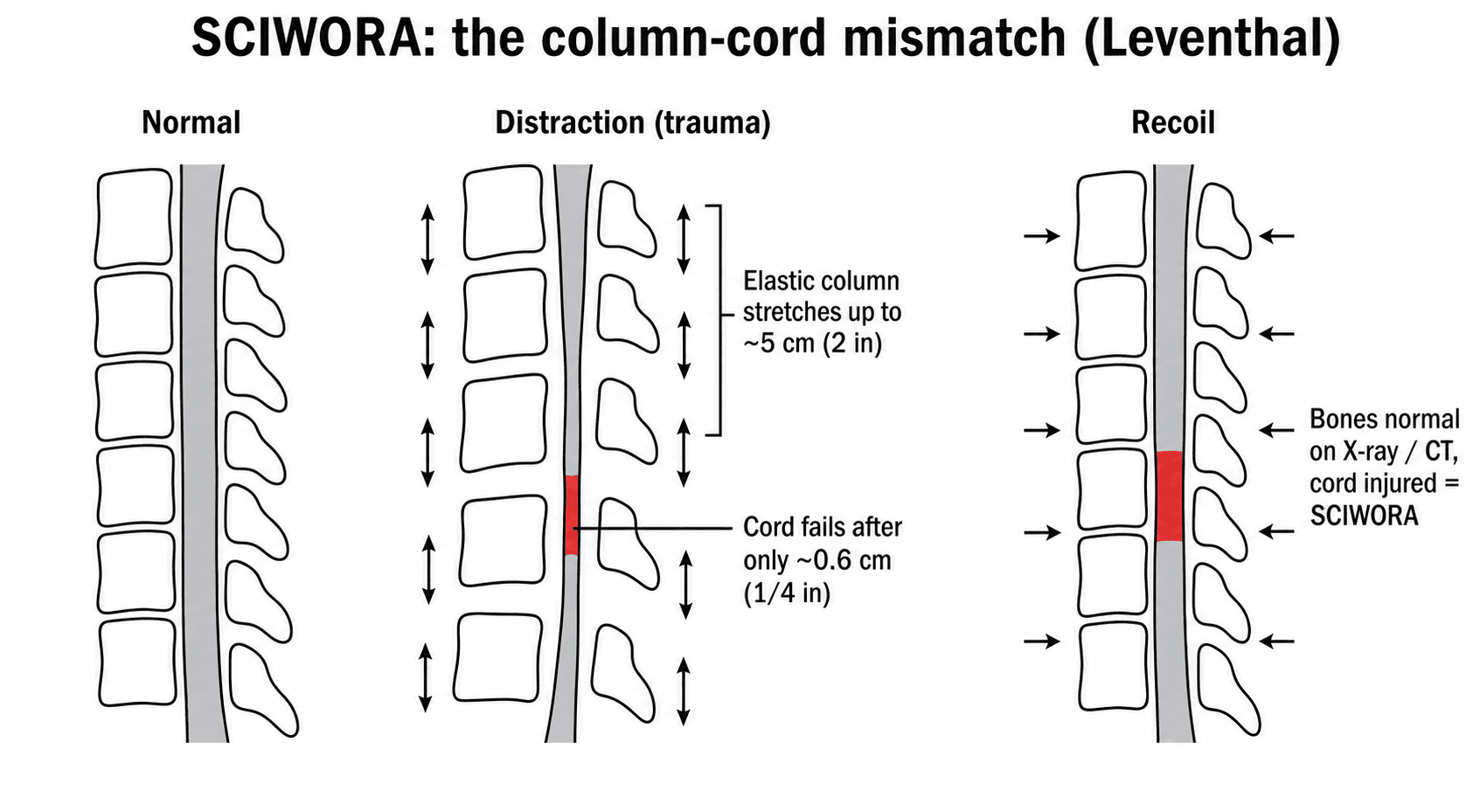
- Mechanism: Hyperflexion/Hyperextension. The pediatric spine is elastic (cartilage/ligaments) and can stretch 2 inches; the cord tears after 1/4 inch.
- Presentation: Range from transient paresthesia ('stingers') to complete quadriplegia. Recall bias: 50% have delayed onset of neuro deficits.
- Investigation: MRI is MANDATORY for any child with transient neuro symptoms or persistent neck pain after trauma.
- Steroids: The use of High Dose Methylprednisolone (NASCIS) is CONTROVERSIAL and generally NOT recommended in current guidelines (AANS/CNS).
- “The spine stretches more than the spinal cord (Leventhal's Rule).
- “Normal X-ray/CT DOES NOT rule out spinal cord injury in a child.
- “Recurrent symptoms? Think instability or missed breakdown.
- “Immobilization (Rigid Collar) is the mainstay of treatment.
False Reassurance. CT scans only show bone. In children, the injury is often purely ligamentous/discal or direct cord contusion without fracture.
The Lucid Interval. Up to 50% of children have a delay between trauma and paralysis (hours to 4 days). Warning signs: Paresthesia, Lhermitte's.
Elasticity Mismatch. Vertebral column = Elastic. Spinal Cord = Brittle. The column stretches and snaps back; the cord snaps.
High Risk. Children with SCIWORA are at high risk of recurrent injury if not immobilized, even if initial MRI is normal.
- Pediatric (SCIWORA)
- Normal (By definition)
- Adult SCI
- Fracture/Dislocation common
- Pediatric (SCIWORA)
- Distraction / Hyperflexion
- Adult SCI
- Axial Load / Burst
- Pediatric (SCIWORA)
- Ligamentous Laxity + Large Head
- Adult SCI
- Stiff Spine + Degeneration
- Pediatric (SCIWORA)
- Long segment edema (Pencil line)
- Adult SCI
- Focal contusion
ELASTICSCIWORA Risk (Why Kids?)
Hook:The pediatric spine is ELASTIC, the cord is not.
Overview and Epidemiology
- SCIWORA
- Typically less than 8 years
- Fracture-Dislocation
- Adolescents (greater than 12)
- SCIWORA
- Hyperflexion/Distraction
- Fracture-Dislocation
- Axial Load / Direct Blow
- SCIWORA
- Ligamentous stretch, Cord injury
- Fracture-Dislocation
- Bony failure, Ligament rupture
- SCIWORA
- Normal
- Fracture-Dislocation
- Abnormal (Fracture/Subluxation)
- SCIWORA
- Collar (90%)
- Fracture-Dislocation
- Surgery (50%)
- SCIWORA
- Neuromuscular Scoliosis
- Fracture-Dislocation
- Kyphosis / Arthritis
SCIWORA (Spinal Cord Injury Without Radiographic Abnormality) was defined by Pang and Wilberger in 1982. It refers to objective signs of myelopathy in the presence of NORMAL plain radiographs and CT scans. Note: With the advent of MRI, we now often see the "abnormality" in the soft tissues, but the term persists.
- Age: Predominantly children less than 8 years old. The elasticity decreases with age, so true SCIWORA becomes rarer in adolescents.
- Incidence: Accounts for 20-30% of pediatric spinal cord injuries. It is the most common pattern of spinal cord injury in young children.
- Region: Cervical spine is most commonly involved (Upper cervical in very young, Lower cervical in older kids). Thoracic SCIWORA is rare and often associated with high-velocity distraction (lap-belt injury).
Pang and Wilberger's original series (1982) was landmark because it shifted the focus from "fractures" to "neurology". Before this, many children were dismissed as malingerers or "hysterical" because X-rays were normal. They identified that 52% of these children had a delayed onset of severe paralysis, typically occurring between 30 minutes and 4 days after injury. This lead to the cardinal rule: Treat the symptoms, not the X-ray.
It is vital to distinguish these two entities, which often coexist in severe SCIWORA.
- Spinal Shock: A physiological loss of all spinal cord function caudal to the level of injury (flaccid paralysis, areflexia). It is transient (24-48 hours usually). Recovery is heralded by the return of the Bulbocavernosus Reflex.
- Neurogenic Shock: A hemodynamic phenomenon resulting from loss of sympathetic tone (T6 and above).
- Triad: Hypotension, Bradycardia, Peripheral Vasodilation (Warm peripheries).
- Treatment: Fluids + Inotropes (Noradrenaline/Dopamine) to maintain perfusion. Atropine for severe bradycardia.
Any child with neck pain and a normal X-ray requires careful clearance. Do not dismiss transient symptoms.
Pathophysiology and Mechanisms
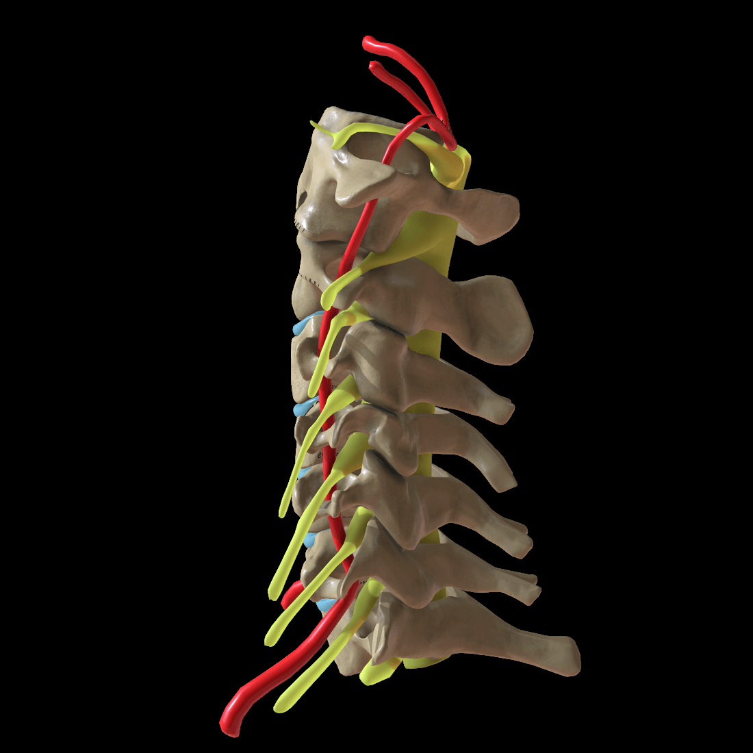
The pediatric cervical spine has unique biomechanical properties that predispose to SCIWORA:
- Ligamentous Laxity: Generalized joint hypermobility allows for excessive intersegmental motion.
- Horizontal Facet Joints: The facet joints in children are oriented more horizontally (flatter) compared to the vertical orientation in adults. This allows for significant AP translation (sliding) without fracture or dislocation.
- Large Head-to-Body Ratio: The head is disproportionately large and heavy. In young children, the fulcrum of motion is at C1-C2 (Upper Cervical), whereas in adults it is C5-C6. This places the upper cervical cord at highest risk in toddlers.
- Uncinate Processes: The uncinate processes (Joints of Luschka) are undeveloped and flat in children (they ossify and heighten by age 10). This lack of bony side-walls offers less resistance to lateral and rotational forces.
- Anterior Wedging: The vertebral bodies are wedge-shaped anteriorly, facilitating hyper-flexion.
- Blood Supply: The spinal cord blood supply is tenuous, particularly the anterior spinal artery. Watershed zones (T4-T8) are vulnerable, but in cervical SCIWORA, the mechanism is often traction injury to the penetrating vessels.
- Vampire Bite Sign: Describes the tiny paired hyperintensities on axial MRI, representing disruption of the central sulcal arteries. This finding specifically predicts poor motor recovery (LMN injury).
The spinal column can be distracted (stretched) up to 2 inches without structural failure (due to elastic ligaments and discs). However, the spinal cord ruptures after only 1/4 inch of distraction.
- Trauma: Hyperextension or Distraction.
- Action: The column stretches, tractioning the cord to failure.
- Recoil: The column snaps back to normal alignment.
- Result: The cord is injured, but the X-ray/CT looks perfect.
Patterns of Cord Injury:
- Concussion: Transient dysfunction, rapid recovery.
- Contusion/Edema: Structural change, variable recovery.
- Infarction: Vampire bite sign (vascular disruption).
- Transection: Complete loss.
Classification
Pang Classification (2004)
Based on MRI findings and highly predictive of outcome.
- Type I: Normal MRI (Neural and Extraneural).
- Outcome: Excellent. 100% full recovery.
- Type II: Abnormal MRI.
- IIa: Edema / Minimal Hemorrhage (Less than 50% of cord).
- Outcome: Good.
- IIb: Major Hemorrhage / Transection (Greater than 50% of cord).
- Outcome: Poor. Permanent paralysis likely.
- IIa: Edema / Minimal Hemorrhage (Less than 50% of cord).
Note: Purely extraneural findings (ligament injury only) are sometimes classified separately as "Unstable Spine" rather than SCIWORA.
ASIA Impairment Scale and Neurological Classification
The clinical assessment refers to the ASIA exam; the ASIA Impairment Scale (AIS) is the standardised grading of spinal-cord-injury severity used to communicate completeness and prognosis. It rests on the International Standards for Neurological Classification of Spinal Cord Injury (ISNCSCI): testing 10 key muscles and 28 dermatomes (light touch and pin-prick) on each side, plus voluntary anal contraction and S4-S5 sensation. Any motor or sensory function in the lowest sacral segments ("sacral sparing") defines an incomplete injury.
- Definition
- No motor or sensory function in the sacral segments S4-S5
- Significance
- Complete injury; worst prognosis
- Definition
- Sensory but no motor function preserved below the level, including S4-S5
- Significance
- Incomplete; sacral sparing present
- Definition
- Motor preserved below the level; more than half of key muscles below are grade under 3
- Significance
- Incomplete, weak
- Definition
- Motor preserved below the level; at least half of key muscles below are grade 3 or more
- Significance
- Incomplete, functional
- Definition
- Motor and sensory normal in a patient with a prior documented deficit
- Significance
- Recovered
The single most important distinction is complete (AIS A) versus incomplete. Test S4-S5: any voluntary anal contraction, perianal sensation or deep anal pressure means sacral sparing and an incomplete injury, which carries a far better prognosis. Assess this only after spinal shock has resolved (return of the bulbocavernosus reflex).
Incomplete Spinal Cord Syndromes
When the injury is incomplete, the pattern of deficit localises the damage within the cord and predicts recovery. These syndromes are classic exam material, and the anterior cord pattern in particular underlies the anterior-spinal-artery injury described above.
- Deficit pattern
- Upper limbs weaker than lower limbs (distal worst), variable sensory and bladder involvement
- Prognosis / note
- Commonest; hyperextension; generally good but incomplete recovery
- Deficit pattern
- Loss of motor and pain/temperature below the lesion; dorsal columns (proprioception/vibration) preserved
- Prognosis / note
- Anterior spinal artery territory; worst prognosis of the incomplete syndromes
- Deficit pattern
- Ipsilateral motor and proprioception/vibration loss; contralateral pain/temperature loss
- Prognosis / note
- Best prognosis; often penetrating or rotational injury
- Deficit pattern
- Loss of proprioception and vibration with preserved power and pain/temperature
- Prognosis / note
- Rare; produces sensory ataxia
Central cord (hands worse than legs) is the commonest pattern and the anterior cord syndrome the worst-prognosis one, while Brown-Sequard has the best. The anterior cord pattern - motor and pain/temperature lost, dorsal columns spared - is the clinical face of the anterior-spinal-artery (vampire-bite) injury seen in SCIWORA.
Clinical Assessment
- Mechanism: MVC, Fall, Sports.
- Transient Symptoms: "Stingers", "Burning hands", "Electric shocks" (Lhermitte's).
- Delay: Ask specifically about the time between injury and onset of weakness (can be delayed in 50%).
- Neurology is Key: ASIA Exam classification.
- Motor: Weakness (often bilateral).
- Sensory: Level to pin prick and light touch.
- Reflexes: Hyper-reflexia (upper motor neuron) or absent (Spinal shock).
- Tone: Flaccid initially (Shock) then Spasticity later.
- Autonomic: Priapism, bradycardia (Neurogenic shock/Hypotension).
It is crucial to differentiate SCIWORA from other causes of acute weakness:
- Transverse Myelitis: Often viral prodrome, slower onset (hours to days), fever. MRI shows enhancement.
- Guillain-Barre Syndrome: Ascending paralysis, areflexia, normal MRI spine. Lumbar puncture shows albuminocytologic dissociation.
- Spinal Cord Tumor: Insidious onset, night pain.
- Conversion Disorder: Inconsistent exam, "Hoover's sign" positive. Diagnosis of exclusion.
- Brachial Plexus Injury: Unilateral, lower motor neuron signs only (root level).
- Inconsistent history.
- Delayed presentation (parents waited days).
- Multiple fractures or bruises in different stages of healing.
- SCIWORA in a non-ambulatory infant (e.g. "fell from couch").
Investigations

Protocol:
-
Plain Radiographs: AP, Lateral, Odontoid. (Often normal). Look for prevertebral soft tissue swelling (greater than 6mm at C2), ADI widening, or subtle kyphosis.
-
CT Cervical Spine: To rule out occult fracture. (Usually normal in SCIWORA). Indicated if plain films are inadequate or suspicion of bony injury is high.
-
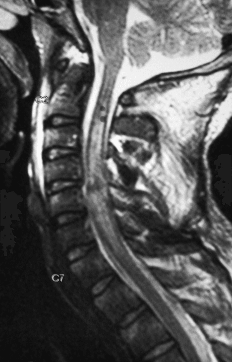
MRI (The Gold Standard): Must be performed URGENTLY (within 24-48 hours) to detect signal changes.
- Sequences: T2 Weighted (best for edema and ligament disruption), STIR (highlights edema in soft tissues), Gradient Echo/GRE (detects hemorrhage), DWI (early infarction).
- Findings: Extraneural (ligament disruption, disc herniation, epidural hematoma) or Intraneural (edema, hemorrhage, transection).
Blood tests are usually normal. Consider metabolic workup if etiology weak (e.g. transverse myelitis differential).
Advanced Imaging Considerations
- DTI (Diffusion Tensor Imaging): Emerging research tool. May show tract disruption even when T2 is normal.
- fMRI: Evaluation of cortical reorganization in chronic cases.
- Electrophysiology (SSEP/MEP): Mandatory if surgery is planned. Can help prognosticate in comatose patients.
Management Algorithm
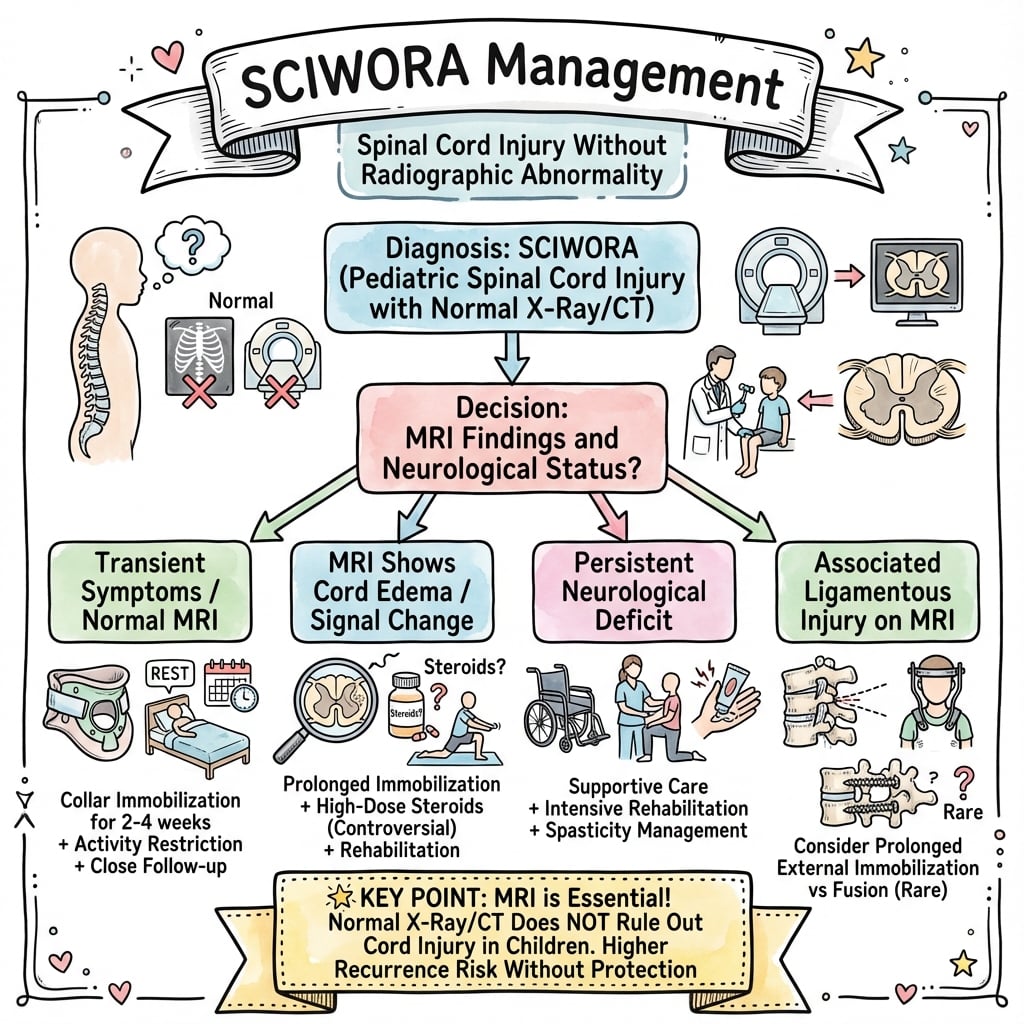
The Mainstay of Treatment
The primary goal is to prevent recurrent injury to the vulnerable cord.
- Device: Rigid Collar (Miami J or Aspen).
- Duration: Typically 12 weeks.
- Clearance: Flexion/Extension X-rays at 12 weeks to prove stability before clearing collar.
Compliance is the biggest challenge in this age group.
ISPINEManagement Priorities
Hook:I-SPINE protocol for SCIWORA.
Surgical Technique
Halo-Thoracic Vest Application
For unstable injuries in young children where collars fail.
- Pins: Use MORE pins (6-8) to distribute load.
- Torque: LOWER torque (2-4 in-lbs in toddlers, vs 8 in-lbs in adults).
- Placement: Position pins appropriately.
- Avoid: Temporal fossa (Squamous bone is paper thin).
- Vest: Custom fit. Check for skin breakdown.
Pins must be hand-tightened only to avoid penetration.
Surgical Challenges in Pediatric Spine
- Small Anatomy: Pedicles in children under 8 are miniscule (often less than 3-4mm), making screw fixation risky.
- Growth Potential: Fusing the spine arrests vertical growth. Rule of thumb: Fusing C1-C2 loses very little height, but subaxial fusion results in loss of 0.07mm per segment per year (negligible). However, it induces "Crankshaft" phenomenon if posterior fusion only is done in growing scoliosis (less relevant here).
- Fusion Rates: Massive healing potential. Non-union is rare, but overgrowth can occur.
Complications
- Recovery Potential
- 100%
- Long Term
- Full return to activity potential
- Recovery Potential
- Good (greater than 75%)
- Long Term
- Most walk independently
- Recovery Potential
- Fair (50%)
- Long Term
- Variable deficits
- Recovery Potential
- Poor (less than 10%)
- Long Term
- Permanent paralysis likely
- Recovery Potential
- Zero
- Long Term
- Permanent complete injury
Postoperative Care
Recovery Protocol
- ICU admission (monitor respiratory status if high C-spine).
- Maintain MAP greater than 85 (Perfusion).
- Rigid immobilization.
- Mobilize in collar.
- Aggressive Physiotherapy (ROM, Strengthening).
- Bowel/Bladder regimen if affected.
- Dynamic X-rays (Flex/Ext).
- MRI repeat (resolution of edema?).
- Wean collar if stable.
- Contraindicated: If persistent deficit or instability.
- Allowed: If full recovery, stable spine, and normal MRI.
- Log Rolling: Strict spinal precautions until cleared.
- Skin Care: Regular collar care (changing liners) to prevent occipital and mandibular pressure ulcers (high risk in children with thin skin).
- Blood Pressure: Avoid hypotension. Maintain MAP greater than 85mmHg (adult targets) or age-appropriate equivalent (e.g. greater than 70-75mmHg) to ensure cord perfusion.
- Bladder: Intermittent catheterization if retention present.
- Spasticity Management: Baclofen, Botox injections for contractures.
- Scoliosis Surveillance: 98% of children with SCI before skeletal maturity will develop neuromuscular scoliosis. Require 6-monthly spine X-rays until maturity.
- Psychological Support: PTSD is common in both the child and parents (guilt).
Outcomes
- Type I (Normal MRI): Nearly all recover fully, but recurrence risk exists if not immobilized.
- Type II (Edema): Variable. Short segment edema recovers well. Long segment does poorly.
- Recurrence: A "Second Hit" within 2 weeks is often more severe than the first. This is why the collar is non-negotiable.
- Mortality: High in upper cervical transections (Respiratory arrest).
NEHPang's Prognosis
Hook:NEH: Normal to Edema to Hemorrhage (Worse outcomes).
Guidelines, Registries & Global Practice
Global epidemiology:
- SCIWORA accounts for roughly 6-19% of paediatric and 9-14% of adult spinal injuries across reported series (Szwedowski 2014), and is the predominant cord-injury pattern in young children.
- The cervical spine dominates (around 46% of cases in nationwide US data), with upper cervical injury concentrated in children under 8 and lower cervical/sport-related injury in adolescents (Knox 2016).
- Mechanism shifts with age and region: motor vehicle collisions predominate in young children, contact sport in adolescents, and diving, falls and road trauma are leading causes in many limited-resource settings.
Side-by-side guidance:
- Imaging stance
- MRI for the obtunded child or persistent symptoms with normal CT; CT not sufficient to clear
- Steroids in SCI
- Recommend AGAINST routine high-dose methylprednisolone
- Imaging stance
- Maintain collar until reliable exam or MRI clearance in obtunded patients
- Steroids in SCI
- Steroids not endorsed as standard of care
- Imaging stance
- Low threshold for MRI in children; minimise unnecessary CT radiation
- Steroids in SCI
- No routine steroids; supportive cord-perfusion focus
- Imaging stance
- MRI is the reference standard for cord and ligamentous assessment
- Steroids in SCI
- Decision individualised; routine use discouraged
- There is no dedicated implant registry for SCIWORA (it is largely a non-operative, soft-tissue diagnosis). Evidence is therefore driven by national trauma and paediatric admission databases (e.g. US HCUP-KID) and specialist paediatric spinal unit series rather than arthroplasty-style registries.
- Neuromuscular scoliosis surveillance to skeletal maturity is the key longitudinal data point: a high proportion of children injured before maturity develop progressive deformity and need indefinite follow-up.
- High-resource settings: urgent MRI, paediatric ICU cord-perfusion targets, and management within a specialist paediatric spinal unit are standard.
- Limited-resource settings: where urgent MRI is unavailable, the safe default is to maintain rigid immobilisation and transfer to a centre with MRI; the collar must never be cleared on a normal CT alone. Clinical vigilance for delayed deterioration substitutes for advanced imaging.
- Transport: stabilisation and rigid immobilisation before transfer are mandatory worldwide; retrieval delay should never prompt premature collar removal.
Prevention Strategies
- Child restraints: appropriate car seats with rear-facing use kept as long as the seat allows markedly reduce cervical distraction forces in crashes — the single most effective prevention in young children.
- Sport: contact and collision sports (rugby codes, American football, wrestling, diving) demand strict "no return to play" after any transient cord symptom ('stinger', burning hands) until cleared; neck-strengthening and safe-tackle programmes reduce risk.
- Recreational hazards: trampolines and diving into shallow water are recurrent causes of severe cervical trauma in children; supervision and age-appropriate restrictions are advised.
Controversies and Areas of Uncertainty
With universal MRI, most "SCIWORA" now has visible cord or ligamentous signal change. Some authors argue the term is obsolete and propose SCIWORET (without radiographic evidence of trauma) or simply MRI-based grading. Examiners may probe whether you understand that the concept (normal X-ray/CT, abnormal cord) still matters even if the label is debated.
The traditional 12-week collar derives from Pang's instability concerns, but there is no high-level evidence for the exact duration. Many units shorten immobilisation when the MRI is normal and the spine is dynamically stable, balancing recurrence risk against collar-related skin breakdown and deconditioning.
High-dose methylprednisolone (NASCIS II) showed benefit only in a post-hoc 8-hour subgroup in adults. AANS/CNS guidelines recommend against routine use, and paediatric data are absent — yet some centres still consider it case-by-case, making this a live area of disagreement.
The ideal window for prognostic MRI (24-72 hours, to capture peak oedema/haemorrhage) and the added value of diffusion tensor imaging over standard T2/GRE remain research questions rather than settled practice.
Viva Scenarios
Clinical Decision Scenarios
Practise clinical reasoning and management decisions out loud
“A 6-year-old is intubated after a high-speed MVC. GCS 3T. Polytrauma. CT Cervical Spine is reported as normal by the registrar.”
Clinical Decision Scenarios
Practise clinical reasoning and management decisions out loud
“A 7-year-old boy tackled in rugby. Had 'burning hands' for 5 minutes. Now asymptomatic. X-rays normal.”
“Child fell from slide 2 days ago. Neck pain. Now presents with arm weakness and stumbling.”
“Parents of a child with SCIWORA ask if he will walk again. MRI shows a dark spot in the cord.”
MCQ Practice Points
Q: How much can the pediatric spinal column stretch before injury? A: Up to 2 inches. The cord ruptures at 1/4 inch. This specific mismatch causes SCIWORA.
Q: What MRI finding carries the worst prognosis in SCIWORA? A: Intramedullary Hemorrhage (and Transection). Associated with permanent complete injury.
Q: What is the current recommendation for Steroids (Methylprednisolone) in pediatric SCI? A: Not recommended/Optional. Level 1 evidence suggests risks (infection/GI bleed) outweigh benefits.
Q: Which level is most commonly affected in young children (less than 8)? A: Upper Cervical (C1-C2). Large head, fulcrum is higher. In older children (greater than 8), it moves to Lower Cervical.
Q: What percentage of patients with SCIWORA present with delayed symptoms? A: Up to 50%. This can be up to 4 days post-injury.
Definition
- Spinal Cord Injury
- Without Radiographic Abnormality
- Normal X-ray / CT
- MRI findings common (Edema)
Pathophysiology
- Elastic Column (2 inches)
- Brittle Cord (1/4 inch)
- Hyperextension / Distraction
- Recoil Injury
Pang Class (MRI)
- Type I: Normal MRI (Best)
- Type IIa: Edema (Good)
- Type IIb: Hemorrhage (Poor)
- Prognostic Value: High
- Cord Transection: Worst
Management Rules
- Rigid Collar (12 Weeks)
- MRI Mandatory for symptoms
- No Steroids (Usually)
- Avoid Sports (3-6 Months)
- Treat symptoms not X-ray
Key Stats
- Age: Less than 8 (Peak)
- Delayed Onset: 50% cases
- Recurrence: High Risk
- Region: Upper C-Spine
- Triad: Hypotension, Bradycardia, Warm
Evidence Base
Defining SCIWORA (Original Description)
- Coined the term SCIWORA — traumatic myelopathy with normal plain films and tomography, attributed to flexion, hyperextension, longitudinal distraction and ischaemia.
- Children younger than 8 years sustained more serious neurological damage and a larger proportion of upper cervical cord lesions than older children.
- 52% of children had delayed onset of paralysis up to 4 days after injury, most recalling transient paraesthesia, numbness or subjective paralysis.
- Recommended cervical immobilisation plus delayed dynamic films to exclude late instability; long-term prognosis was grim for complete and severe lesions.
Delayed Deterioration and Warning Symptoms
- 55 children with SCIWORA: 10 upper cervical, 33 lower cervical, 12 thoracic injuries; 22 complete or severe and 33 mild lesions.
- All but one of the 22 children with profound deficits were younger than 8 years; younger children were more likely to have severe upper cervical lesions.
- 15 children had delayed onset of deficit; 9 of these had transient warning symptoms (paraesthesia, subjective paralysis, Lhermitte phenomenon) 30 minutes to 4 days before deterioration.
- 8 children sustained a second SCIWORA 3 days to 10 weeks after the first, implying incipient instability from the index injury.