
Official YouTube channel
Free orthopaedic teaching, straight from the OrthoVellum channel.
Focused, exam-ready explainers that turn OrthoVellum's written teaching into moving images — high-yield orthopaedics for FRCS (Tr & Orth), FRACS and board candidates, and a fast refresher for clinicians. Every video below is from the official channel, so this page always mirrors the latest uploads. Nothing else appears here.
- Videos
- 211
- Channel
- @orthovellum
- Updates
- Automatic
Live from the official channel
youtube.com/@orthovellum
New uploads sync within the hour
About the channel
One channel, no noise
The OrthoVellum YouTube channel is our own teaching library — concise, cinematic explainers built from the same evidence-based content as the written platform. This page lists only those official uploads; you will never see third-party videos here. Watch an explainer to lock in a concept, then click through to the full written topic to go deep.
OrthoVellum
youtube.com/@orthovellum
Latest from the channel
211 videos

6:13
Why Your Arch Collapses — Talonavicular Arthritis Explained
Why Your Arch Collapses — Talonavicular Arthritis Explained ⏱ Chapters 0:00 Intro 0:51 The Keystone 1:23 Ball meets socket 1:49 The numbers 2:13 Grade It, Prove It 2:43 The grade decides 3:06 Read the standing film 3:33 Fuse It Right 4:10 One joint or three 4:42 Two screws, crossed 5:09 What lasts 5:41 Recap & sources The keystone joint, the half-motion rule, and fuse-it-right decisions In this OrthoVellum master lecture • Keystone joint: half the hindfoot motion lives here • Grades 1–2 brace six months; grade 3 fuses • Standing films: beaking, under 2mm, under 60% covered • Isolated spares cartilage; triple for pan-disease • Crossed screws, bleeding bone, 6–8 weeks dark • 85–90% unite; neighbours bill at 20–30% Full clinical topic and references: https://orthovellum.com/topics/talonavicular-arthritis Image credits • Popelka et al. — CC BY 2.0 — https://europepmc.org/article/PMC/2837861#1471-2474-11-38-5 • Laurent et al. — CC BY 4.0 — https://europepmc.org/article/PMC/13258229#jcm-15-03991-g003 • Popelka et al. — CC BY 2.0 — https://europepmc.org/article/PMC/2837861#1471-2474-11-38-3 • Gorbachova et al. — CC BY 4.0 — https://europepmc.org/article/PMC/8703616#jcm-10-05848-g006 • Lin et al. — CC BY 4.0 — https://europepmc.org/article/PMC/12786061#diagnostics-16-00018-g002 Educational only — not individual medical advice. #orthopaedics #orthopedics #medicaleducation #orthovellum Orthovellum.com
11 Sept 2026

5:15
Scaphoid Nonunion — Why the Proximal Pole Dies — Quick Revision (exam review)
The 5-minute revision cut of the full OrthoVellum lecture: Scaphoid Nonunion — Why the Proximal Pole Dies. Just the teaching core — every diagram and every rule, no intro, no recap. Watch the full lecture on the OrthoVellum channel for the complete build-up, and visit Orthovellum.com for the written topic with references. Educational only — not individual medical advice. Always consult a qualified clinician. #Orthopaedics #Orthopedics #MedEd #OrthoVellum #OrthopaedicSurgery
10 Sept 2026

16:26
Pelvic Fractures — Stop the Bleeding First
Pelvic fractures can cause life‑threatening haemorrhage before the fracture pattern is even named. The first priority is to apply a binder over the greater trochanters to reduce pelvic volume and control bleeding. Assess posterior‑ring stability and contamination to guide definitive management. ⏱ Chapters 0:00 Intro 0:54 Read the image 1:59 EXPLAINER 2:34 EXPLAINER 3:05 EXPLAINER 4:17 Read the image 4:47 EXPLAINER 7:11 EXPLAINER 7:47 EXPLAINER 8:44 Read the image 9:17 EXPLAINER 9:58 EXPLAINER 12:13 EXPLAINER 12:48 EXPLAINER 13:13 Read the image 13:46 EXPLAINER 14:48 EXPLAINER 16:03 Recap & sources This video walks through the critical early steps in managing pelvic ring injuries: recognising haemodynamic instability, correct binder placement, initiating haemorrhage control pathways, and evaluating posterior‑ring integrity using the four‑anchor approach (force, posterior ring, volume, contamination). It explains why posterior‑ring injury dictates stability and treatment, outlines the Denis zone system for sacral fractures, and highlights associated injuries that change the sequence of care. 🦴 IN THIS VIDEO YOU'LL LEARN • How to position a pelvic binder correctly over the greater trochanters • The four‑anchor decision framework for pelvic fractures: force, posterior ring, volume, contamination • How to assess posterior‑ring stability using sacral Denis zones and SI joint integrity • When to activate massive transfusion protocols and angioembolisation for haemorrhage control • How open pelvic injuries alter management priorities and require multidisciplinary input • Key clinical signs that indicate pelvic bleeding versus other sources of shock • Why posterior‑ring disruption is the treatment‑defining finding in pelvic ring injuries • How to document neurological and genitourinary exams before catheterisation or transfer • The role of CT imaging after immediate resuscitation and binder application 📌 KEY TAKEAWAYS ✔️ Binder over the greater trochanters, not the iliac crests, reduces pelvic volume instantly ✔️ Posterior‑ring injury, not anterior displacement, dictates the need for fixation ✔️ Haemorrhage control comes before fracture classification in unstable patients ✔️ Open pelvic fractures demand early antibiotics, debridement and urology/colorectal involvement ✔️ Document sacral Denis zones to predict neurological risk and guide screw safety ✔️ A non‑responder to resuscitation needs immediate haemorrhage‑control measures, not a perfect Tile or Young‑Burgess label 👩⚕️ WHO THIS IS FOR This video is designed for orthopaedic trainees, medical students, clinicians managing trauma, and interested patients who want to understand the urgent priorities in pelvic fracture care. 📚 RELATED TOPICS: Open book pelvis · Sacral fracture Denis zones · Pelvic binder application · Angioembolisation in pelvic trauma · Urethral injury assessment · Tile and Young‑Burgess classification 🔗 More free orthopaedic teaching at Orthovellum.com — subscribe for weekly videos. 🎨 CREDITS • Complex pelvic fracture case report, PMC10492189 — CC BY 4.0 — https://pmc.ncbi.nlm.nih.gov/articles/PMC10492189/ • Diagnostics (MDPI) 2022 — CC BY 4.0 — https://pmc.ncbi.nlm.nih.gov/articles/PMC8870907/ • OrthoVellum Medical Education Team — Original OrthoVellum educational illustration • Cerevisae — CC BY-SA 4.0 — https://commons.wikimedia.org/wiki/File:Pelvis_AP_view_showing_fracture_of_the_left_ischium_and_left_acetabular_wall.jpg • Valerio et al., Cureus — CC BY 4.0 — https://europepmc.org/article/PMC/PMC13268618#cureus-0018-00000108982-i09 • Gewiess et al. — CC BY 4.0 — https://europepmc.org/article/PMC/PMC11663159 • Keel MJ et al., Eur J Trauma Emerg Surg (PMC3495274) — CC BY — https://www.ncbi.nlm.nih.gov/pmc/articles/PMC3495274/ • Girgis et al., Journal of Clinical Medicine — CC BY 4.0 — https://europepmc.org/article/PMC/PMC11856568#jcm-14-01289-g004 • Chen H et al., BMC Musculoskelet Disord — Open Access (CC BY) — https://openi.nlm.nih.gov/detailedresult?img=PMC3750865 • Girgis et al., Journal of Clinical Medicine — CC BY 4.0 — https://europepmc.org/article/PMC/PMC11856568#jcm-14-01289-g003 ⚠️ DISCLAIMER: Educational only — not individual medical advice. Always consult a qualified clinician for diagnosis and treatment. #pelvicfractures #pelvicrodinjuries #bindertrauma #haemorrhagecontrol #posteriorring #sacralfracture #traumaortho #emergencymedicine #fracturemanagement #orthoeducation #tilenotation #youngburgess #openbookpelvis #angiography #neurologicalassessment
10 Sept 2026
7:27
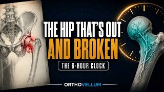
The Hip That's Out AND Broken — Fracture-Dislocations Explained
The Hip That's Out AND Broken — Fracture-Dislocations Explained ⏱ Chapters 0:00 Intro 1:18 The Anatomy Of The Emergency 2:09 Pipkin IV On 3D CT — The Head Fracture Plus… 2:57 Reduce, Then Prove It 3:49 Non-Concentric After Reduction — An… 4:24 The Operation And The Aftermath 5:13 The Nerve And The Blood Supply 5:59 The Long Arc 6:42 Recap & sources The six-hour clock, Thompson-Epstein, Pipkin, and the nerve behind the joint In this OrthoVellum master lecture • Reduce as an emergency — 12 hours multiplies necrosis odds 5.6 times • Exclude a femoral neck fracture before traction • Thompson-Epstein names the fracture; Pipkin sorts the head by the fovea • Two or three gentle attempts — then open reduction • Post-reduction CT is mandatory: concentricity, loose bodies, fractures • Sciatic nerve: 10–20% — document before and after Full clinical topic and references: https://orthovellum.com/topics/hip-fracture-dislocations Educational only — not individual medical advice. #orthopaedics #orthopedics #medicaleducation #orthovellum Orthovellum.com
10 Sept 2026

10:17
The Talus Fell Out — Total Talar Dislocation Explained
The Talus Fell Out — Total Talar Dislocation Explained ⏱ Chapters 0:00 Intro 1:15 A Bone With No Margin For Error 2:07 Count The Joints 2:48 Closed Tri-Articular Dislocation — AP And… 3:16 Reduce, Then Look 4:20 Before And After Reduction — The Associated… 5:05 The Number Everyone Quotes Is Wrong 6:45 Reimplanted Talus, Protected By External… 7:50 Surveillance And Salvage 8:43 Tibiocalcaneal Arthrodesis After Talar Loss… 9:30 Recap & sources Urgent reduction, the honest necrosis numbers, and never discarding the bone In this OrthoVellum master lecture • Reduce within hours — skin necrosis does not wait for imaging • Count the joints: all three, or it is not this injury • One gentle closed attempt; failure means open reduction • CT after reduction — most carry talar fractures needing fixation • Never discard the extruded talus: debride, reimplant, pin and span • Necrosis honestly: 4 of 21 reported — follow-up too short to trust Full clinical topic and references: https://orthovellum.com/topics/total-talar-dislocation Educational only — not individual medical advice. #orthopaedics #orthopedics #medicaleducation #orthovellum Orthovellum.com
10 Sept 2026

6:57
Why Your Finger Locks and Clicks — Trigger Finger Explained
Why Your Finger Locks and Clicks — Trigger Finger Explained ⏱ Chapters 0:00 Intro 0:50 The Size Mismatch 1:21 The pulley map 1:46 The numbers 2:14 Grade It, Prove It 2:50 The grade decides 3:20 When it is not trigger finger 3:54 Shrink It Or Open It 4:34 The sheath, not the tendon 5:15 Ten minutes, wide awake 5:47 What lasts 6:24 Recap & sources The A1 pulley, the Green grades, and the inject-or-release decision In this OrthoVellum master lecture • A1 pulley stenosis: nodule catches, thumb and ring lead • Green grades 1–4 set splint, injection, or release • Inject the sheath, never the tendon; repeat at most once • Divide A1 completely; A2 and A4 are never released • Diabetics: a third succeed — offer surgery sooner • 56% recur, 18% need surgery; children resolve alone Full clinical topic and references: https://orthovellum.com/topics/trigger-finger Image credits • Jengojan S et al. (PMC11480152) — CC BY 4.0 — https://pmc.ncbi.nlm.nih.gov/articles/PMC11480152/ • Lee YK et al. (PMC4616507) — CC BY 4.0 — https://pmc.ncbi.nlm.nih.gov/articles/PMC4616507/ • Sederberg M et al. (PMC11889526) — CC BY 4.0 — https://pmc.ncbi.nlm.nih.gov/articles/PMC11889526/ • Mafi P et al. (PMC12455252) — CC BY 4.0 — https://pmc.ncbi.nlm.nih.gov/articles/PMC12455252/ • Ryskalin L et al. (PMC12843178) — CC BY 4.0 — https://pmc.ncbi.nlm.nih.gov/articles/PMC12843178/ Educational only — not individual medical advice. #orthopaedics #orthopedics #medicaleducation #orthovellum Orthovellum.com
10 Sept 2026

6:48
Acute Compartment Syndrome — Diagnosis Before Damage
Acute Compartment Syndrome — Diagnosis Before Damage ⏱ Chapters 0:00 Intro 0:24 BOAST · COMPARTMENT SYNDROME 2025 0:47 BOAST · COMPARTMENT SYNDROME 2025 1:25 BOAST · COMPARTMENT SYNDROME 2025 1:46 BOAST · COMPARTMENT SYNDROME 2025 2:14 BOAST · COMPARTMENT SYNDROME 2025 2:41 BOAST · COMPARTMENT SYNDROME 2025 3:08 BOAST · COMPARTMENT SYNDROME 2025 3:39 BOAST · COMPARTMENT SYNDROME 2025 4:03 BOAST 2025 · AAOS 2025 4:15 BOAST · COMPARTMENT SYNDROME 2025 4:43 BOAST · COMPARTMENT SYNDROME 2025 5:12 Anatomical Reference—Not A Tissue-Viability… 5:51 BOAST · COMPARTMENT SYNDROME 2025 6:12 BOAST 2025 · AAOS 2025 6:25 BOAST · COMPARTMENT SYNDROME 2025 Serial examination, delta pressure, and complete fasciotomy In this OrthoVellum master lecture Full clinical topic and references: https://orthovellum.com/topics/compartment-syndrome Image credits • Mumford 1910 · Volkmann contracture · public domain · historical sequela, not a tissue-viability test — Public Domain — https://commons.wikimedia.org/wiki/File:The_practice_of_surgery_(1910)_(14756898256).jpg • BMC musculoskeletal disorders — cc by — https://europepmc.org/article/PMC/PMC11716319 • PloS one — cc by — https://europepmc.org/article/PMC/PMC13016300 • Toxins — cc by — https://europepmc.org/article/PMC/PMC13030228 • Life (Basel, Switzerland) — cc by — https://europepmc.org/article/PMC/PMC12735328 • Frontiers in bioengineering and biotechnology — cc by — https://europepmc.org/article/PMC/PMC12872933 • OrthoVellum original educational illustration — Original OrthoVellum educational illustration • OrthoVellum original clinical teaching animation — Original OrthoVellum educational animation Educational only — not individual medical advice. #orthopaedics #orthopedics #medicaleducation #orthovellum Orthovellum.com
10 Sept 2026

9:20
The Ankle That Won't Reduce — Bosworth Fracture-Dislocation
The Ankle That Won't Reduce — Bosworth Fracture-Dislocation ⏱ Chapters 0:00 Intro 1:29 The Lock 2:28 Axial CT: The Fibula Locked Between… 3:45 Recognise It Before You Pull 4:48 After A Failed Attempt: Overlap And… 5:48 The Operation 6:52 Residual Clear-Space Widening Corrected… 7:50 Postoperative CT: The Fibula Centred In The… 8:32 Recap & sources The fibula locked behind the tibia, the view that proves it, and the operation that frees it In this OrthoVellum master lecture • Irreducible by bony block — the fibula locked behind the posterior tubercle • The triad: external rotation, axilla sign, the reduction that bounces back • Stop when it locks — repeated attempts damage the joint • Judge reduction on the lateral view; the AP can lie • Posterolateral window: unhook, fix, stabilise the syndesmosis • The endpoint is the fibula centred in the incisura on CT Full clinical topic and references: https://orthovellum.com/topics/bosworth-fracture-dislocations Educational only — not individual medical advice. #orthopaedics #orthopedics #medicaleducation #orthovellum Orthovellum.com
10 Sept 2026
10:00
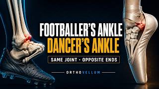
Footballer's Ankle vs Dancer's Ankle — Ankle Impingement Explained
Footballer's Ankle vs Dancer's Ankle — Ankle Impingement Explained ⏱ Chapters 0:00 Intro 1:18 Two Ends Of The Same Joint 2:22 Mechanism 3:11 Through the scope 3:42 Prove It 4:39 Symptomatic or incidental 5:34 The pathway 6:23 Fix It 7:27 The portal map is a nerve map 8:36 The numbers 9:10 Recap & sources Positional pain, the tests that prove it, and when arthroscopy earns its place In this OrthoVellum master lecture • Mechanical diagnosis: positional, reproducible, end-range pain • Dorsiflexion pain: spurs or post-sprain meniscoid lesion • Plantarflexion pain: os trigonum or Stieda, with the FHL beside it • Marrow oedema separates symptomatic from incidental • Three to six months conservative before the scope • Portals are a nerve map: SPN, sural, tibial Full clinical topic and references: https://orthovellum.com/topics/ankle-impingement-syndromes Educational only — not individual medical advice. #orthopaedics #orthopedics #medicaleducation #orthovellum Orthovellum.com
10 Sept 2026

8:51
Complex Distal Femur Fixation — Why Plates Fail — Quick Revision (exam review)
The 9-minute revision cut of the full OrthoVellum lecture: Complex Distal Femur Fixation — Why Plates Fail. Just the teaching core — every diagram and every rule, no intro, no recap. Watch the full lecture on the OrthoVellum channel for the complete build-up, and visit Orthovellum.com for the written topic with references. Educational only — not individual medical advice. Always consult a qualified clinician. #Orthopaedics #Orthopedics #MedEd #OrthoVellum #OrthopaedicSurgery
8 Sept 2026

7:52
Anterior Shoulder Instability — Bone Loss Changes the Operation — Quick Revision (exam review)
The 8-minute revision cut of the full OrthoVellum lecture: Anterior Shoulder Instability — Bone Loss Changes the Operation. Just the teaching core — every diagram and every rule, no intro, no recap. Watch the full lecture on the OrthoVellum channel for the complete build-up, and visit Orthovellum.com for the written topic with references. Educational only — not individual medical advice. Always consult a qualified clinician. #Orthopaedics #Orthopedics #MedEd #OrthoVellum #OrthopaedicSurgery
8 Sept 2026
4:40
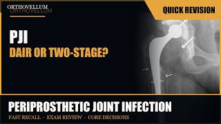
Periprosthetic Joint Infection — Biofilm Changes Everything — Quick Revision (exam review)
The 5-minute revision cut of the full OrthoVellum lecture: Periprosthetic Joint Infection — Biofilm Changes Everything. Just the teaching core — every diagram and every rule, no intro, no recap. Watch the full lecture on the OrthoVellum channel for the complete build-up, and visit Orthovellum.com for the written topic with references. Educational only — not individual medical advice. Always consult a qualified clinician. #Orthopaedics #Orthopedics #MedEd #OrthoVellum #OrthopaedicSurgery
8 Sept 2026

5:21
Cervical Myelopathy — The Cord Under Pressure — Quick Revision (exam review)
The 5-minute revision cut of the full OrthoVellum lecture: Cervical Myelopathy — The Cord Under Pressure. Just the teaching core — every diagram and every rule, no intro, no recap. Watch the full lecture on the OrthoVellum channel for the complete build-up, and visit Orthovellum.com for the written topic with references. Educational only — not individual medical advice. Always consult a qualified clinician. #Orthopaedics #Orthopedics #MedEd #OrthoVellum #OrthopaedicSurgery
8 Sept 2026

4:03
Pilon Fractures — The Soft-Tissue Clock — Quick Revision (exam review)
The 4-minute revision cut of the full OrthoVellum lecture: Pilon Fractures — The Soft-Tissue Clock. Just the teaching core — every diagram and every rule, no intro, no recap. Watch the full lecture on the OrthoVellum channel for the complete build-up, and visit Orthovellum.com for the written topic with references. Educational only — not individual medical advice. Always consult a qualified clinician. #Orthopaedics #Orthopedics #MedEd #OrthoVellum #OrthopaedicSurgery
8 Sept 2026

6:53
Patellar Instability — The Geometry Behind Recurrence — Quick Revision (exam review)
The 7-minute revision cut of the full OrthoVellum lecture: Patellar Instability — The Geometry Behind Recurrence. Just the teaching core — every diagram and every rule, no intro, no recap. Watch the full lecture on the OrthoVellum channel for the complete build-up, and visit Orthovellum.com for the written topic with references. Educational only — not individual medical advice. Always consult a qualified clinician. #Orthopaedics #Orthopedics #MedEd #OrthoVellum #OrthopaedicSurgery
8 Sept 2026

4:53
Neck of Femur Fractures — Fix or Replace? — Quick Revision (exam review)
The 5-minute revision cut of the full OrthoVellum lecture: Neck of Femur Fractures — Fix or Replace?. Just the teaching core — every diagram and every rule, no intro, no recap. Watch the full lecture on the OrthoVellum channel for the complete build-up, and visit Orthovellum.com for the written topic with references. Educational only — not individual medical advice. Always consult a qualified clinician. #Orthopaedics #Orthopedics #MedEd #OrthoVellum #OrthopaedicSurgery
8 Sept 2026

5:26
Rotator Cuff Tears — Repairability, Force Couples, Failure — Quick Revision (exam review)
The 5-minute revision cut of the full OrthoVellum lecture: Rotator Cuff Tears — Repairability, Force Couples, Failure. Just the teaching core — every diagram and every rule, no intro, no recap. Watch the full lecture on the OrthoVellum channel for the complete build-up, and visit Orthovellum.com for the written topic with references. Educational only — not individual medical advice. Always consult a qualified clinician. #Orthopaedics #Orthopedics #MedEd #OrthoVellum #OrthopaedicSurgery
8 Sept 2026

4:50
Cauda Equina Syndrome — The Decompression Decision — Quick Revision (exam review)
The 5-minute revision cut of the full OrthoVellum lecture: Cauda Equina Syndrome — The Decompression Decision. Just the teaching core — every diagram and every rule, no intro, no recap. Watch the full lecture on the OrthoVellum channel for the complete build-up, and visit Orthovellum.com for the written topic with references. Educational only — not individual medical advice. Always consult a qualified clinician. #Orthopaedics #Orthopedics #MedEd #OrthoVellum #OrthopaedicSurgery
8 Sept 2026
7:23
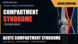
Acute Compartment Syndrome — Diagnosis Before Damage — Quick Revision (exam review)
The 7-minute revision cut of the full OrthoVellum lecture: Acute Compartment Syndrome — Diagnosis Before Damage. Just the teaching core — every diagram and every rule, no intro, no recap. Watch the full lecture on the OrthoVellum channel for the complete build-up, and visit Orthovellum.com for the written topic with references. Educational only — not individual medical advice. Always consult a qualified clinician. #Orthopaedics #Orthopedics #MedEd #OrthoVellum #OrthopaedicSurgery
8 Sept 2026

4:34
Femoral Head Osteonecrosis — Preserve or Replace? — Quick Revision (exam review)
The 5-minute revision cut of the full OrthoVellum lecture: Femoral Head Osteonecrosis — Preserve or Replace?. Just the teaching core — every diagram and every rule, no intro, no recap. Watch the full lecture on the OrthoVellum channel for the complete build-up, and visit Orthovellum.com for the written topic with references. Educational only — not individual medical advice. Always consult a qualified clinician. #Orthopaedics #Orthopedics #MedEd #OrthoVellum #OrthopaedicSurgery
8 Sept 2026

3:56
Periprosthetic Femoral Fractures — Fix or Revise? — Quick Revision (exam review)
The 4-minute revision cut of the full OrthoVellum lecture: Periprosthetic Femoral Fractures — Fix or Revise?. Just the teaching core — every diagram and every rule, no intro, no recap. Watch the full lecture on the OrthoVellum channel for the complete build-up, and visit Orthovellum.com for the written topic with references. Educational only — not individual medical advice. Always consult a qualified clinician. #Orthopaedics #Orthopedics #MedEd #OrthoVellum #OrthopaedicSurgery
8 Sept 2026
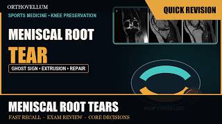
4:36
Meniscal Root Tears — The Hoop-Stress Failure — Quick Revision (exam review)
The 5-minute revision cut of the full OrthoVellum lecture: Meniscal Root Tears — The Hoop-Stress Failure. Just the teaching core — every diagram and every rule, no intro, no recap. Watch the full lecture on the OrthoVellum channel for the complete build-up, and visit Orthovellum.com for the written topic with references. Educational only — not individual medical advice. Always consult a qualified clinician. #Orthopaedics #Orthopedics #MedEd #OrthoVellum #OrthopaedicSurgery
8 Sept 2026

5:34
Open Fractures — Antibiotics, Debridement, Coverage — Quick Revision (clinical review)
The 6-minute revision cut of the full OrthoVellum lecture: Open Fractures — Antibiotics, Debridement, Coverage. Just the teaching core — every diagram and every rule, no intro, no recap. Watch the full lecture on the OrthoVellum channel for the complete build-up, and visit Orthovellum.com for the written topic with references. Educational only — not individual medical advice. Always consult a qualified clinician. #Orthopaedics #Orthopedics #MedEd #OrthoVellum #OrthopaedicSurgery
8 Sept 2026

8:37
Scapholunate Injury — From Dynamic Gap to DISI
Scapholunate Injury — From Dynamic Gap to DISI ⏱ Chapters 0:00 Intro 0:49 SPECTRUM 1:44 RADIOGRAPHY 2:38 IMAGING PATHWAY 3:36 EWAS ARTHROSCOPY 4:26 EXPLAINER 5:03 EXPLAINER 6:00 EXPLAINER 6:58 SLAC STAGING 7:29 SALVAGE DECISION 8:02 EXPLAINER Provoke occult instability, stage ligament and cartilage, repair what is repairable, and choose reconstruction or salvage from reducibility In this OrthoVellum master lecture Full clinical topic and references: https://orthovellum.com/topics/scapholunate-dissociation Image credits • Tischler BT et al., Insights into Imaging (PMC4141341) — CC BY — https://www.ncbi.nlm.nih.gov/pmc/articles/PMC4141341/ Educational only — not individual medical advice. #orthopaedics #orthopedics #medicaleducation #orthovellum Orthovellum.com
5 Sept 2026
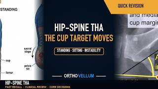
8:35
The Hip-Spine Relationship in THA — A Moving Cup Target — Quick Revision (clinical review)
The 9-minute revision cut of the full OrthoVellum lecture: The Hip-Spine Relationship in THA — A Moving Cup Target. Just the teaching core — every diagram and every rule, no intro, no recap. Watch the full lecture on the OrthoVellum channel for the complete build-up, and visit Orthovellum.com for the written topic with references. Educational only — not individual medical advice. Always consult a qualified clinician. #Orthopaedics #Orthopedics #MedEd #OrthoVellum #OrthopaedicSurgery
5 Sept 2026

8:03
Peroneal Tendon Tears and Instability — Groove, Retinaculum — Quick Revision (clinical review)
The 8-minute revision cut of the full OrthoVellum lecture: Peroneal Tendon Tears and Instability — Groove, Retinaculum, Repair. Just the teaching core — every diagram and every rule, no intro, no recap. Watch the full lecture on the OrthoVellum channel for the complete build-up, and visit Orthovellum.com for the written topic with references. Educational only — not individual medical advice. Always consult a qualified clinician. #Orthopaedics #Orthopedics #MedEd #OrthoVellum #OrthopaedicSurgery
5 Sept 2026

9:11
The Hip-Spine Relationship in THA — A Moving Cup Target
The Hip-Spine Relationship in THA — A Moving Cup Target ⏱ Chapters 0:00 Intro 0:48 EXPLAINER 1:24 EXPLAINER 1:51 TWO AXES 2:22 EXPLAINER 2:55 FUNCTIONAL IMAGING 3:27 EXPLAINER 3:57 STIFF SPINE 4:28 EXPLAINER 5:25 IMPINGEMENT PATHWAY 5:51 EXPLAINER 6:31 EXPLAINER 7:03 HIP OR SPINE FIRST? 7:36 EXPLAINER 8:30 PREVENTION SEQUENCE Measure standing and seated pelvic behaviour, identify stiffness and imbalance, and plan component orientation for the positions the patient actually uses In this OrthoVellum master lecture Full clinical topic and references: https://orthovellum.com/topics/hip-spine-relationship-tha Image credits • Nevit Dilmen, annotated by Mikael Häggström — CC BY-SA 3.0 — https://commons.wikimedia.org/wiki/File:Lateral_X-ray_of_lumbar_spine_spondylosis.jpg • Mikael Häggström — CC0 — https://commons.wikimedia.org/wiki/File:Acetabular_inclination_of_hip_prosthesis.jpg • Silverjonny — Public domain — https://commons.wikimedia.org/wiki/File:Wiki_post-op.jpg • Frontiers in Bioengineering 2023 (PMC10665526) — CC BY — https://www.ncbi.nlm.nih.gov/pmc/articles/PMC10665526/ • J Clin Med (MDPI) 2023 (PMC10219306) — CC BY — https://www.ncbi.nlm.nih.gov/pmc/articles/PMC10219306/ • Malaysian orthopaedic journal — cc by — https://europepmc.org/article/PMC/PMC13133578 • Advances in orthopedics — cc by — https://europepmc.org/article/PMC/PMC6230414 • Yon et al., Medicina (2025), CC BY 4.0 — CC BY 4.0 — https://europepmc.org/article/PMC/PMC11766848#medicina-61-00112-g004 • Al-Humairi et al., Cureus (2025), CC BY 4.0 — CC BY 4.0 — https://europepmc.org/article/PMC/PMC12318351#cureus-0017-00000087263-i02 • Kim et al., Medicina (2024), CC BY 4.0 — CC BY 4.0 — https://europepmc.org/article/PMC/PMC11123322#medicina-60-00680-g003 • Rehman et al., Cureus (2025), CC BY 4.0 — CC BY 4.0 — https://europepmc.org/article/PMC/PMC12521857#cureus-0017-00000092335-i01 • Kim et al., Medicina (2024), CC BY 4.0 — CC BY 4.0 — https://europepmc.org/article/PMC/PMC11123322#medicina-60-00680-g001 • Mariani P et al. - Case Rep Orthop (CC-BY 4.0) — CC BY — https://www.ncbi.nlm.nih.gov/pmc/articles/PMC4258342/ • Yeo AK et al. - Patient Saf Surg (CC-BY 4.0) — CC BY — https://www.ncbi.nlm.nih.gov/pmc/articles/PMC4658799/ • Wu CC, J Orthop Surg Res — CC BY — https://www.ncbi.nlm.nih.gov/pmc/articles/PMC9258056/ • OrthoVellum — Original AI illustration Educational only — not individual medical advice. #orthopaedics #orthopedics #medicaleducation #orthovellum Orthovellum.com
5 Sept 2026

10:22
Diabetic Foot Infection — Depth, Perfusion, Debridement
Diabetic Foot Infection — Depth, Perfusion, Debridement ⏱ Chapters 0:00 Intro 0:50 EXPLAINER 1:33 EXPLAINER 2:34 BONE INFECTION 3:10 EXPLAINER 3:54 EXPLAINER 5:06 EXPLAINER 6:13 EXPLAINER 7:12 DEBRIDEMENT 7:50 EXPLAINER 8:24 ANTIMICROBIAL STEWARDSHIP 9:07 RESPONSE LOOP 10:09 EXPLAINER Diagnose infection clinically, define the depth and blood supply, obtain meaningful cultures, and control the source before the limb is lost In this OrthoVellum master lecture Full clinical topic and references: https://orthovellum.com/topics/diabetic-foot-infections Image credits • Wellcome Collection via Wikimedia Commons — CC BY 4.0 — https://commons.wikimedia.org/wiki/File:Arteries_of_the_foot;_three_figures._Lithograph_by_N.H_Jacob_Wellcome_V0008447.jpg • Milorad Dimic MD via Wikimedia Commons — CC BY-SA 3.0 — https://commons.wikimedia.org/wiki/File:Diabetic_Wound_121.jpg • Journal of Diabetes Research via Europe PMC — CC BY 3.0 — https://europepmc.org/articles/PMC4322665 • Diabetic Foot & Ankle via Europe PMC — CC BY 2.0 — https://europepmc.org/articles/PMC3805841 • International Journal of Emergency Medicine via Europe PMC — CC BY 2.0 — https://europepmc.org/articles/PMC3084168 • Insights into Imaging via Europe PMC — CC BY 4.0 — https://europepmc.org/articles/PMC5265197 • Cardiovascular and Interventional Radiology via Europe PMC — CC BY 4.0 — https://europepmc.org/articles/PMC4735259 • Wikimedia Commons — CC BY 3.0 — https://commons.wikimedia.org/wiki/File:Charcot_foot_arthropathy.jpg • OrthoVellum illustration — Original OrthoVellum educational illustration • Trials via Europe PMC — CC BY 2.0 — https://europepmc.org/articles/PMC4156638 Educational only — not individual medical advice. #orthopaedics #orthopedics #medicaleducation #orthovellum Orthovellum.com
5 Sept 2026

8:39
Peroneal Tendon Tears and Instability — Groove, Retinaculum, Repair
Peroneal Tendon Tears and Instability — Groove, Retinaculum, Repair ⏱ Chapters 0:00 Intro 0:45 EXPLAINER 1:24 EXPLAINER 1:53 IMAGING 2:42 EXPLAINER 3:09 DIAGNOSTIC PATHWAY 3:37 EXPLAINER 4:05 INSTABILITY MECHANICS 4:41 TENDON FUNCTION 5:11 EXPLAINER 5:41 TEAR ALGORITHM 6:11 EXPLAINER 6:41 FIBULAR GROOVE 7:09 EXPLAINER 8:01 CONCOMITANT MECHANICS Differentiate tendon disease from dynamic instability, restore the superior retinaculum, and reconstruct function without a rigid tear-percentage rule In this OrthoVellum master lecture Full clinical topic and references: https://orthovellum.com/topics/peroneal-tendon-tears Image credits • Skowalchik7 via Wikimedia Commons — CC BY-SA 4.0 — https://commons.wikimedia.org/wiki/File:Peroneal.jpg Educational only — not individual medical advice. #orthopaedics #orthopedics #medicaleducation #orthovellum Orthovellum.com
5 Sept 2026

10:35
VTE Prevention in Orthopaedics — Thrombosis Risk Versus Bleeding
VTE Prevention in Orthopaedics — Thrombosis Risk Versus Bleeding ⏱ Chapters 0:00 Intro 0:49 DYNAMIC RISK 2:02 NET BENEFIT 3:09 EXPLAINER 3:49 PROCEDURE-SPECIFIC PLAN 4:31 MULTIMODAL PREVENTION 5:31 EXPLAINER 6:04 AGENT SELECTION 6:54 TIMING 7:31 EXPLAINER 8:05 SAFETY MONITORING 8:43 EXPLAINER 9:51 SUSPECTED EVENT Reassess risk as the operation and patient evolve, combine mechanical and pharmacological prevention, and never let prophylaxis become unmonitored treatment In this OrthoVellum master lecture Full clinical topic and references: https://orthovellum.com/topics/venous-thromboembolism-comprehensive Image credits • Enter — CC BY-SA 4.0 — https://commons.wikimedia.org/wiki/File:MTPS_gelber_Hintergrund.jpg • James Heilman, MD — CC BY-SA 3.0 — https://commons.wikimedia.org/wiki/File:Deep_vein_thrombosis_of_the_right_leg.jpg • Easow et al. — CC BY 4.0 — https://europepmc.org/article/PMC/PMC12796808 • Ayad et al. — CC BY 4.0 — https://europepmc.org/article/PMC/PMC12602250 • Zhang et al. — CC BY 4.0 — https://europepmc.org/article/PMC/PMC13243409 • Kaneki et al. — CC BY 4.0 — https://europepmc.org/article/PMC/PMC12998537 • Htun et al. — CC BY 4.0 — https://europepmc.org/article/PMC/PMC12711594 • Wang et al. — CC BY 4.0 — https://europepmc.org/article/PMC/PMC13294096 • Velliou et al. — CC BY 4.0 — https://europepmc.org/article/PMC/PMC12842806 • Le Pennec et al. — CC BY 4.0 — https://europepmc.org/article/PMC/PMC7921798 • OrthoVellum illustration — Original OrthoVellum educational illustration Educational only — not individual medical advice. #orthopaedics #orthopedics #medicaleducation #orthovellum Orthovellum.com
5 Sept 2026

8:20
Spinal Epidural Abscess — MRI Before Neurology Is Lost
Spinal Epidural Abscess — MRI Before Neurology Is Lost ⏱ Chapters 0:00 Intro 0:40 DIAGNOSTIC TRAP 1:34 TRIAGE 2:09 NEURAL INJURY 2:56 EXPLAINER 3:33 EXPLAINER 4:03 ANTIBIOTIC TIMING 4:59 SURGICAL DECISION 5:53 EXPLAINER 6:25 EXPLAINER 7:23 MONITORING LOOP Recognise the incomplete presentation, culture before antibiotics when safe, and decompress threatened neural tissue without delay In this OrthoVellum master lecture Full clinical topic and references: https://orthovellum.com/topics/spinal-epidural-abscess Image credits • Dr Graham Beards — CC BY-SA 4.0 — https://commons.wikimedia.org/wiki/File:Staphylococcus_aureus_Gram_stain.jpg • Hellerhoff — CC BY-SA 4.0 — https://commons.wikimedia.org/wiki/File:Spondylodiscitis_LWK_34_82M_-_MR_und_CT_-_001.jpg • Dubois C, Lievens P. — CC BY 4.0 — https://pmc.ncbi.nlm.nih.gov/articles/PMC13218703/ • Ikedum I, Mapouka M, Arinze F. — CC BY 4.0 — https://pmc.ncbi.nlm.nih.gov/articles/PMC12660633/ • Jing Jing Chan and Jen Jen Oh — CC BY 4.0 — https://commons.wikimedia.org/wiki/File:MRI_of_the_lumbar_spine_with_abscess_in_the_posterior_epidural_space,_causing_cauda_equina_syndrome.jpg • Sher Z, Shah RN, Imtiaz M, Raza H, Elmahadi A, Hussain R. — CC BY 4.0 — https://pmc.ncbi.nlm.nih.gov/articles/PMC13259601/ • Bohara S, Karki S, Thapa B, Khanal N, Ghimire B, Bohara S, Pradhanang A, Sharma MR. — CC BY 4.0 — https://pmc.ncbi.nlm.nih.gov/articles/PMC12476968/ • Alani A, Taylor S, Yu C. — CC BY 4.0 — https://pmc.ncbi.nlm.nih.gov/articles/PMC12704467/ • OrthoVellum Medical Education Team — Original OrthoVellum illustration — original://orthovellum/spinal-epidural-abscess/management-algorithm Educational only — not individual medical advice. #orthopaedics #orthopedics #medicaleducation #orthovellum Orthovellum.com
5 Sept 2026

8:09
Scapholunate Injury — From Dynamic Gap to DISI — Quick Revision (clinical review)
The 8-minute revision cut of the full OrthoVellum lecture: Scapholunate Injury — From Dynamic Gap to DISI. Just the teaching core — every diagram and every rule, no intro, no recap. Watch the full lecture on the OrthoVellum channel for the complete build-up, and visit Orthovellum.com for the written topic with references. Educational only — not individual medical advice. Always consult a qualified clinician. #Orthopaedics #Orthopedics #MedEd #OrthoVellum #OrthopaedicSurgery
5 Sept 2026
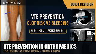
10:09
VTE Prevention in Orthopaedics — Thrombosis Risk Versus Bleeding — Quick Revision (clinical review)
The 10-minute revision cut of the full OrthoVellum lecture: VTE Prevention in Orthopaedics — Thrombosis Risk Versus Bleeding. Just the teaching core — every diagram and every rule, no intro, no recap. Watch the full lecture on the OrthoVellum channel for the complete build-up, and visit Orthovellum.com for the written topic with references. Educational only — not individual medical advice. Always consult a qualified clinician. #Orthopaedics #Orthopedics #MedEd #OrthoVellum #OrthopaedicSurgery
5 Sept 2026

8:18
Chondroblastoma — The Epiphyseal Lesion in the Young — Quick Revision (clinical review)
The 8-minute revision cut of the full OrthoVellum lecture: Chondroblastoma — The Epiphyseal Lesion in the Young. Just the teaching core — every diagram and every rule, no intro, no recap. Watch the full lecture on the OrthoVellum channel for the complete build-up, and visit Orthovellum.com for the written topic with references. Educational only — not individual medical advice. Always consult a qualified clinician. #Orthopaedics #Orthopedics #MedEd #OrthoVellum #OrthopaedicSurgery
5 Sept 2026

7:56
Spinal Epidural Abscess — MRI Before Neurology Is Lost — Quick Revision (clinical review)
The 8-minute revision cut of the full OrthoVellum lecture: Spinal Epidural Abscess — MRI Before Neurology Is Lost. Just the teaching core — every diagram and every rule, no intro, no recap. Watch the full lecture on the OrthoVellum channel for the complete build-up, and visit Orthovellum.com for the written topic with references. Educational only — not individual medical advice. Always consult a qualified clinician. #Orthopaedics #Orthopedics #MedEd #OrthoVellum #OrthopaedicSurgery
5 Sept 2026

9:47
Diabetic Foot Infection — Depth, Perfusion, Debridement — Quick Revision (clinical review)
The 10-minute revision cut of the full OrthoVellum lecture: Diabetic Foot Infection — Depth, Perfusion, Debridement. Just the teaching core — every diagram and every rule, no intro, no recap. Watch the full lecture on the OrthoVellum channel for the complete build-up, and visit Orthovellum.com for the written topic with references. Educational only — not individual medical advice. Always consult a qualified clinician. #Orthopaedics #Orthopedics #MedEd #OrthoVellum #OrthopaedicSurgery
5 Sept 2026
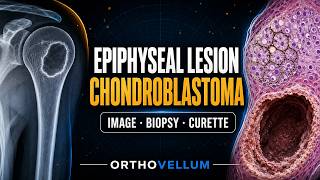
8:48
Chondroblastoma — The Epiphyseal Lesion in the Young
Chondroblastoma — The Epiphyseal Lesion in the Young ⏱ Chapters 0:00 Intro 0:49 DIAGNOSTIC FRAME 1:48 EPIPHYSEAL DIFFERENTIAL 2:46 ONCOLOGIC WORK-FLOW 3:45 OPERATIVE PRINCIPLE 4:43 EXTENDED CURETTAGE 5:11 EXPLAINER 6:11 EXPLAINER 7:12 EXPLAINER 7:46 RECURRENCE WORK-UP 8:16 EXPLAINER Recognise the matrix and oedema pattern, biopsy through the definitive approach, and curette completely without sacrificing the joint In this OrthoVellum master lecture Full clinical topic and references: https://orthovellum.com/topics/chondroblastoma Image credits • Via Wikimedia Commons — CC BY-SA 4.0 — https://commons.wikimedia.org/wiki/Category:Chondroblastoma • Case reports in orthopedics — CC BY — http://www.ncbi.nlm.nih.gov/pmc/articles/PMC4274911 • Clinical sarcoma research — CC BY — http://www.ncbi.nlm.nih.gov/pmc/articles/PMC4186744 • Journal of surgical case reports — cc by — https://europepmc.org/article/PMC/PMC13232625 • Cancers — cc by — https://europepmc.org/article/PMC/PMC11201376 • Cureus — cc by — https://europepmc.org/article/PMC/PMC13214491 • Insights into imaging — CC BY — http://www.ncbi.nlm.nih.gov/pmc/articles/PMC4263798 • Frontiers in radiology — cc by — https://europepmc.org/article/PMC/PMC13079598 • OrthoVellum Medical Education Team — Original OrthoVellum educational illustration Educational only — not individual medical advice. #orthopaedics #orthopedics #medicaleducation #orthovellum Orthovellum.com
1 Sept 2026

7:31
Acute Compartment Syndrome — Diagnosis Before Damage
Acute Compartment Syndrome — Diagnosis Before Damage ⏱ Chapters 0:00 Intro 0:48 EXPLAINER 1:28 EXPLAINER 2:10 EXPLAINER 3:14 EXPLAINER 3:50 EXPLAINER 4:30 EXPLAINER 5:11 EXPLAINER 5:49 EXPLAINER 6:19 VISUAL EXPLAINER 6:58 EXPLAINER Serial examination, delta pressure, and complete fasciotomy In this OrthoVellum master lecture Full clinical topic and references: https://orthovellum.com/topics/compartment-syndrome Image credits • Kashuk JL et al., Patient Saf Surg — CC BY — https://openi.nlm.nih.gov/detailedresult?img=PMC4338393 • Via NIH Open-i — CC BY — https://openi.nlm.nih.gov/detailedresult?img=PMC4878063 • Mumford JG, The Practice of Surgery (1910), via Wikimedia Commons — Public Domain — https://commons.wikimedia.org/wiki/File:The_practice_of_surgery_(1910)_(14756898256).jpg • Topaz M et al., Indian J Plast Surg — CC BY — https://openi.nlm.nih.gov/detailedresult?img=PMC5104701 • Toxins — cc by — https://europepmc.org/article/PMC/PMC13030228 • BMC musculoskeletal disorders — cc by — https://europepmc.org/article/PMC/PMC11716319 • PloS one — cc by — https://europepmc.org/article/PMC/PMC13016300 • OrthoVellum — OrthoVellum original asset • Life (Basel, Switzerland) — cc by — https://europepmc.org/article/PMC/PMC12735328 • Frontiers in bioengineering and biotechnology — cc by — https://europepmc.org/article/PMC/PMC12872933 Educational only — not individual medical advice. #orthopaedics #orthopedics #medicaleducation #orthovellum Orthovellum.com
1 Sept 2026
8:39
Supracondylar Humerus Fractures — Perfusion, Nerves, Pinning
A supracondylar humerus fracture is the most common elbow injury in children, where perfusion status and nerve function dictate urgent management. Assessing the pink pulseless hand, performing a thorough neurovascular exam, and planning pinning strategy based on Gartland classification are essential steps. Prompt reduction and appropriate K‑wire fixation prevent complications such as cubitus varus or nerve palsy. ⏱ Chapters 0:00 Intro 0:52 CLASSIFICATION 1:23 RADIOGRAPHS 1:51 BOAST 2020 2:22 MOTOR EXAMINATION 2:49 OPERATING PRIORITY 3:19 VASCULAR DECISION 4:09 EXPLAINER 5:06 FLUOROSCOPY 5:32 FIXATION 6:02 NERVE DEFICIT · BEFORE OR AFTER 6:33 SURVEILLANCE 7:03 MALUNION 7:33 EVIDENCE 8:07 Recap & sources This video walks through the complete approach to supracondylar humerus fractures in children, covering mechanism, Gartland classification, neurovascular assessment, radiographic evaluation, reduction principles, pinning strategies, and key complications. Understanding these elements is vital for timely decision‑making and optimal outcomes in this common paediatric trauma. 🦴 IN THIS VIDEO YOU'LL LEARN • How to assess perfusion, pulse and hand colour in the pink pulseless hand scenario • Step‑by‑step neurovascular examination, including the OK‑sign test for anterior interosseous nerve function • Interpretation of AP and true lateral radiographs: anterior humeral line, Baumann angle and fat pad sign • Application of the Gartland classification to guide treatment decisions for types I‑III • Principles of urgent closed reduction and K‑wire pinning strategies (lateral only vs crossed pins) • Recognition of complications such as cubitus varus, nerve palsy and compartment syndrome • When to explore the brachial artery versus close monitoring after reduction • Documentation requirements for neurovascular status before, intra‑ and post‑operatively 📌 KEY TAKEAWAYS ✔️ Perfusion, not pulse alone, drives urgency in supracondylar fractures ✔️ A pink, warm hand without a radial pulse still needs immediate reduction ✔️ The OK‑sign test is the reliable way to detect anterior interosseous nerve injury ✔️ Gartland type III fractures require urgent closed reduction and K‑wire fixation ✔️ Residual rotation or medial column collapse predicts cubitus varus malunion ✔️ Post‑reduction neurovascular re‑check is mandatory to catch new deficits 👩⚕️ WHO THIS IS FOR This video is designed for orthopaedic trainees, medical students, clinicians managing paediatric elbow injuries, and interested patients or caregivers seeking a clear, evidence‑based overview of supracondylar humerus fracture assessment and treatment. 📚 RELATED TOPICS: cubitus varus · brachial artery injury · anterior interosseous nerve palsy · K‑wire fixation techniques · paediatric elbow trauma · compartment syndrome in children 🔗 More free orthopaedic teaching at Orthovellum.com — subscribe for weekly videos. 🎨 CREDITS • Benoudina Samir — CC BY-SA 4.0 — CC BY-SA 4.0 — https://commons.wikimedia.org/wiki/File:Baumann%27s_Angle.jpg • Zhang M-R, Zeng X, Guo J-L, Zhao K, Hu J-H, Guan J-H (PMC13270536) — CC BY 4.0 — CC BY 4.0 — https://pmc.ncbi.nlm.nih.gov/articles/PMC13270536/ • Mostafa MF, Paralaya KN, El-Husseini MI (PMC12968072) — CC BY 4.0 — CC BY 4.0 — https://pmc.ncbi.nlm.nih.gov/articles/PMC12968072/ • Maeda Y, Okimura S, Narita Y, Katsumata A, Teramoto A (PMC12490966) — CC BY 4.0 — CC BY 4.0 — https://pmc.ncbi.nlm.nih.gov/articles/PMC12490966/ • Badawy M, Sadek SI, Hassan M, Elnagar AM, Shaheen E, Algohiny IA (PMC12781478) — CC BY 4.0 — CC BY 4.0 — https://pmc.ncbi.nlm.nih.gov/articles/PMC12781478/ • Kilborn T, Moodley H, Mears S; annotations by Häggström M — CC BY-SA 4.0 — CC BY-SA 4.0 — https://commons.wikimedia.org/wiki/File:Anterior_humeral_line.svg • Wu Y, Wen J, Hui W, Wang J, Yang F, Qiu X (PMC12967997) — CC BY 4.0 — CC BY 4.0 — https://pmc.ncbi.nlm.nih.gov/articles/PMC12967997/ • Nakagawa T, Yasui Y, Kawabata K, Sasahara J, Inui T, Nei M, Konno Y, Kawano H, Miyamoto W (PMC13259555) — CC BY 4.0 — CC BY 4.0 — https://pmc.ncbi.nlm.nih.gov/articles/PMC13259555/ • Seo KB, Kim BS, Park YG, Lim C (PMC11123191) — CC BY 4.0 — CC BY 4.0 — https://pmc.ncbi.nlm.nih.gov/articles/PMC11123191/ • Afaque SF, Jeenjwadia N, Agrawal U, Verma V (PMC12664120) — CC BY 4.0 — CC BY 4.0 — https://pmc.ncbi.nlm.nih.gov/articles/PMC12664120/ ⚠️ DISCLAIMER: Educational only — not individual medical advice. Always consult a qualified clinician for diagnosis and treatment. #supracondylarhumerusfracture #paediatricorthopaedics #elbowfracture #kwirefixation #gartlandclassification #perfusionassessment #ainervenur #cubitusvarus #brachialartery #childrensortho #fracturemanagement #reductiontechnique #postopmonitoring #orthovellum
1 Sept 2026
5:34
Meniscal Root Tears — The Hoop-Stress Failure
Meniscal Root Tears — The Hoop-Stress Failure ⏱ Chapters 0:00 Intro 0:41 EXPLAINER 1:12 EXPLAINER 1:42 EXPLAINER 2:10 EXPLAINER 2:43 EXPLAINER 3:37 EXPLAINER 4:27 EXPLAINER 5:04 EXPLAINER MRI signs, LaPrade classification, selection, repair, and alignment In this OrthoVellum master lecture Full clinical topic and references: https://orthovellum.com/topics/meniscal-root-tear Educational only — not individual medical advice. #orthopaedics #orthopedics #medicaleducation #orthovellum Orthovellum.com
1 Sept 2026

7:54
Knee Dislocation — Vascular Threat Before Ligament Reconstruction
Knee Dislocation — Vascular Threat Before Ligament Reconstruction ⏱ Chapters 0:00 Intro 0:29 VASCULAR ANATOMY 1:17 BASELINE EXAM 2:07 IRREDUCIBLE KNEE 2:58 DYSVASCULAR LIMB 4:21 NERVE MAP 5:12 LIGAMENT MAP 6:02 RECONSTRUCTION 6:49 REHABILITATION 7:14 Recap & sources Occult dislocation, ABI pathway, popliteal rescue, Schenck pattern, reconstruction timing In this OrthoVellum master lecture Full clinical topic and references: https://orthovellum.com/topics/knee-dislocation-management Image credits • Yilmaz S et al., Case Rep Orthop 2015 (PMC4546968), CC BY — CC BY — https://www.ncbi.nlm.nih.gov/pmc/articles/PMC4546968/ • Cureus — cc by — https://europepmc.org/article/PMC/PMC11344193 • Clinical case reports — cc by — https://europepmc.org/article/PMC/PMC13090157 • Cureus — cc by — https://europepmc.org/article/PMC/PMC13050584 • Annals of vascular diseases — cc by — https://europepmc.org/article/PMC/PMC11807706 • Cureus — cc by — https://europepmc.org/article/PMC/PMC12958003 • Journal of experimental orthopaedics — cc by — https://europepmc.org/article/PMC/PMC12086806 Educational only — not individual medical advice. #orthopaedics #orthopedics #medicaleducation #orthovellum Orthovellum.com
1 Sept 2026

7:23
Charcot Neuro-Osteoarthropathy — Stage, Offload, Reconstruct
Charcot Neuro-Osteoarthropathy — Stage, Offload, Reconstruct ⏱ Chapters 0:00 Intro 0:41 FIRST DECISION 1:28 EXPLAINER 2:16 WHY THE ARCH COLLAPSES 3:07 PRE-RADIOGRAPHIC DISEASE 3:54 EXPLAINER 4:41 IDENTIFYING REMISSION 5:31 MEDICATION 6:26 EXPLAINER Recognise the active neuropathic foot, protect it before certainty, and reconstruct only the limb-threatening mechanics In this OrthoVellum master lecture Full clinical topic and references: https://orthovellum.com/topics/charcot-neuroarthropathy Image credits • Medicalpal via Wikimedia Commons — CC BY-SA 4.0 — https://commons.wikimedia.org/wiki/Category:Charcot_arthropathy • Cellier, EFORT Open Reviews 2025, Figure 4 — CC BY 4.0 — https://pmc.ncbi.nlm.nih.gov/articles/PMC12139603/figure/EOR-2025-0057fig4/ • Milbourn et al, J Clin Med 2026, Figure 5 — CC BY 4.0 — https://pmc.ncbi.nlm.nih.gov/articles/PMC12986205/figure/jcm-15-01993-g005/ • Berhane et al, Foot Ankle Orthop 2025, Figure 1 — CC BY 4.0 — https://pmc.ncbi.nlm.nih.gov/articles/PMC11808755/figure/10.1177_24730114251315670-fig1/ • Milbourn et al, J Clin Med 2026, Figure 3 — CC BY 4.0 — https://pmc.ncbi.nlm.nih.gov/articles/PMC12986205/figure/jcm-15-01993-g003/ • Mascio et al, Diagnostics 2025, Figure 1 — CC BY 4.0 — https://pmc.ncbi.nlm.nih.gov/articles/PMC11940856/figure/diagnostics-15-00767-g001/ • Milbourn et al, J Clin Med 2026, Figure 4 — CC BY 4.0 — https://pmc.ncbi.nlm.nih.gov/articles/PMC12986205/figure/jcm-15-01993-g004/ • Jonathan Moore via Wikimedia Commons — CC BY 3.0 — https://commons.wikimedia.org/wiki/Category:Diabetic_foot • Via Wikimedia Commons — CC BY-SA 4.0 — https://commons.wikimedia.org/wiki/Category:Charcot_arthropathy • Embaby et al, JB JS Open Access 2026, Figure 2 — CC BY 4.0 — https://pmc.ncbi.nlm.nih.gov/articles/PMC12995262/figure/jbjsoa-11-e25.00261-g002/ Educational only — not individual medical advice. #orthopaedics #orthopedics #medicaleducation #orthovellum Orthovellum.com
1 Sept 2026
7:21
Lumbar Spinal Stenosis — Claudication, Imaging, Decompression — Quick Revision (clinical review)
The 7-minute revision cut of the full OrthoVellum lecture: Lumbar Spinal Stenosis — Claudication, Imaging, Decompression. Just the teaching core — every diagram and every rule, no intro, no recap. Watch the full lecture on the OrthoVellum channel for the complete build-up, and visit Orthovellum.com for the written topic with references. Educational only — not individual medical advice. Always consult a qualified clinician. #Orthopaedics #Orthopedics #MedEd #OrthoVellum #OrthopaedicSurgery
1 Sept 2026

6:57
Charcot Neuro-Osteoarthropathy — Stage, Offload, Reconstruct — Quick Revision (clinical review)
The 7-minute revision cut of the full OrthoVellum lecture: Charcot Neuro-Osteoarthropathy — Stage, Offload, Reconstruct. Just the teaching core — every diagram and every rule, no intro, no recap. Watch the full lecture on the OrthoVellum channel for the complete build-up, and visit Orthovellum.com for the written topic with references. Educational only — not individual medical advice. Always consult a qualified clinician. #Orthopaedics #Orthopedics #MedEd #OrthoVellum #OrthopaedicSurgery
1 Sept 2026
5:56
Femoral Head Osteonecrosis — Preserve or Replace?
Femoral Head Osteonecrosis — Preserve or Replace? ⏱ Chapters 0:00 Intro 0:48 EXPLAINER 1:23 EXPLAINER 2:13 EXPLAINER 2:40 EXPLAINER 3:18 EXPLAINER 3:50 EXPLAINER 4:14 EXPLAINER 4:28 VISUAL EXPLAINER 4:48 EXPLAINER 5:16 EXPLAINER MRI, ARCO staging, collapse risk, core decompression, and arthroplasty In this OrthoVellum master lecture Full clinical topic and references: https://orthovellum.com/topics/avascular-necrosis-hip Image credits • Bilge O et al., J Orthop Surg Res (PMC4423414) - CC-BY — CC BY — https://www.ncbi.nlm.nih.gov/pmc/articles/PMC4423414/ • Kalekar et al., Cureus 2024 (PMC11458061) - CC-BY 4.0 — CC BY — https://www.ncbi.nlm.nih.gov/pmc/articles/PMC11458061/ • OrthoVellum AI illustration — Original OrthoVellum educational illustration • Frontiers in Bioengineering 2023 (PMC10665526) — CC BY — https://www.ncbi.nlm.nih.gov/pmc/articles/PMC10665526/ • J Clin Med (MDPI) 2023 (PMC10219306) — CC BY — https://www.ncbi.nlm.nih.gov/pmc/articles/PMC10219306/ • Tonogai I et al., Case Rep Orthop (PMC4129925) — CC BY — https://www.ncbi.nlm.nih.gov/pmc/articles/PMC4129925/ • Case reports in orthopedics — cc by — https://europepmc.org/article/PMC/PMC13213072 • Diagnostics (Basel, Switzerland) — cc by — https://europepmc.org/article/PMC/PMC12839861 Educational only — not individual medical advice. #orthopaedics #orthopedics #medicaleducation #orthovellum Orthovellum.com
1 Sept 2026

5:02
Periprosthetic Femoral Fractures — Fix or Revise?
Periprosthetic Femoral Fractures — Fix or Revise? ⏱ Chapters 0:00 Intro 0:39 EXPLAINER 1:02 EXPLAINER 1:48 EXPLAINER 2:16 EXPLAINER 2:47 EXPLAINER 3:14 EXPLAINER 3:45 EXPLAINER 4:10 EXPLAINER 4:37 EXPLAINER Vancouver classification, stem stability, fixation, and revision reconstruction In this OrthoVellum master lecture Full clinical topic and references: https://orthovellum.com/topics/periprosthetic-femoral-fracture-post-thr Educational only — not individual medical advice. #orthopaedics #orthopedics #medicaleducation #orthovellum Orthovellum.com
1 Sept 2026
8:20
Terrible Triad of the Elbow — Rebuild Stability in Sequence
Terrible Triad of the Elbow — Rebuild Stability in Sequence ⏱ Chapters 0:00 Intro 0:43 EXPLAINER 1:17 EXPLAINER 2:14 EXPLAINER 3:09 PREOPERATIVE PLAN 3:51 EXPLAINER 4:26 EXPLAINER 4:53 OPERATIVE SEQUENCE 5:22 EXPLAINER 6:20 EXPLAINER 6:48 REHABILITATION 7:20 FOLLOW-UP Define the fragments, reconstruct the buttresses, repair the lateral ligament and prove a concentric functional arc In this OrthoVellum master lecture Full clinical topic and references: https://orthovellum.com/topics/terrible-triad-elbow Image credits • Orthopaedic surgery — cc by — https://europepmc.org/article/PMC/PMC10102310 • Frontiers in surgery — cc by — https://europepmc.org/article/PMC/PMC12852477 • Journal of orthopaedics and traumatology : official journal of the Italian Society of Orthopaedics and Traumatology — cc by — https://europepmc.org/article/PMC/PMC11582235 • Radiology case reports — cc by — https://europepmc.org/article/PMC/PMC12268023 • Clinical case reports — cc by — https://europepmc.org/article/PMC/PMC12578567 • International orthopaedics — cc by — https://europepmc.org/article/PMC/PMC11889027 • OrthoVellum — OrthoVellum original asset • Orthopaedic surgery — cc by — https://europepmc.org/article/PMC/PMC11216846 • Frontiers in surgery — cc by — https://europepmc.org/article/PMC/PMC12310578 Educational only — not individual medical advice. #orthopaedics #orthopedics #medicaleducation #orthovellum Orthovellum.com
1 Sept 2026

8:09
Open Fractures — Antibiotics, Debridement, Coverage
Open fractures demand immediate antibiotics and meticulous debridement to prevent infection, with soft‑tissue coverage planned from the first incision. Understanding Gustilo‑Anderson grading guides the urgency of vascular repair, flap reconstruction and fixation strategy. This lecture breaks down the emergency priorities, timing of surgery and orthoplastic principles that save limb and function. ⏱ Chapters 0:00 Intro 0:53 EXPLAINER 1:55 EXPLAINER 2:47 EXPLAINER 3:26 EXPLAINER 3:58 VISUAL EXPLAINER 4:23 EXPLAINER 5:20 EXPLAINER 5:54 EXPLAINER 6:55 EVIDENCE 7:26 Recap & sources The video explains how to assess an open fracture beyond the visible wound, initiate time‑critical antibiotics, apply the Gustilo‑Anderson classification after debridement, perform disciplined excision‑based debridement using the four Cs for muscle viability, choose fixation that preserves soft‑tissue options, and coordinate orthoplastic coverage for types IIIB and IIIC injuries. It also outlines emergency department actions such as wound photography, splinting, tetanus review and avoiding bedside washouts, providing a systematic approach that reduces infection, non‑union and amputation risk. 🦴 IN THIS VIDEO YOU'LL LEARN • How to recognise an open fracture and assess the zone of injury beyond the skin wound • The correct timing and choice of intravenous antibiotics according to BOAST guidelines • How to apply the Gustilo‑Anderson classification after operative debridement • Principles of safe debridement: excision, the four Cs for muscle viability, and appropriate irrigation • Indications for immediate versus delayed debridement based on contamination and vascular status • Fixation options that preserve soft‑tissue envelope and allow orthoplastic coverage • Coordination of orthoplastic planning for flap coverage in Gustilo type IIIB and IIIC injuries • Emergency department actions: wound photography, splinting, tetanus review and avoiding bedside washouts 📌 KEY TAKEAWAYS ✔️ Antibiotics within the hour save lives and limbs ✔️ The true Gustilo grade is known only after debridement ✔️ Debride devitalised tissue, don’t just irrigate ✔️ Plan fixation with coverage in mind from the start ✔️ Vascular injury (type IIIC) overrides all other priorities 👩⚕️ WHO THIS IS FOR This video is designed for orthopaedic trainees, medical students, junior doctors and any clinician or patient seeking a clear, evidence‑based approach to open fracture management. 📚 RELATED TOPICS: Gustilo‑Anderson classification · Orthoplastic flap reconstruction · External fixation techniques · Antibiotic prophylaxis in trauma · Compartment syndrome in open fractures · Soft‑tissue debridement endpoints 🔗 More free orthopaedic teaching at Orthovellum.com — subscribe for weekly videos. 🎨 CREDITS • Sarkar et al. — CC BY 4.0 — https://pmc.ncbi.nlm.nih.gov/articles/PMC11911500/ • Honda et al. — CC BY 4.0 — https://pmc.ncbi.nlm.nih.gov/articles/PMC13141035/ • Kim et al. — CC BY 4.0 — https://pmc.ncbi.nlm.nih.gov/articles/PMC13164335/ • Romeo et al. — CC BY 4.0 — https://pmc.ncbi.nlm.nih.gov/articles/PMC13174045/ • Georgievski et al. — CC BY 4.0 — https://pmc.ncbi.nlm.nih.gov/articles/PMC13078379/ • Goumenos et al. — CC BY 4.0 — https://pmc.ncbi.nlm.nih.gov/articles/PMC13113936/ • Ahmed et al. — CC BY 4.0 — https://pmc.ncbi.nlm.nih.gov/articles/PMC13295163/ • Kong et al. — CC BY 4.0 — https://pmc.ncbi.nlm.nih.gov/articles/PMC13249585/ • OrthoVellum — Original OrthoVellum educational illustration ⚠️ DISCLAIMER: Educational only — not individual medical advice. Always consult a qualified clinician for diagnosis and treatment. #orthopaedics #opentrauma #gustiloads #antibioticsprophylaxis #debridement #orthoplastic #fracturecare #emergencymanagement #softtissue #vascularinjury #fixationmethods #muscletests
1 Sept 2026
5:45
Acetabular Fractures — Columns, Patterns, and Fixation
Acetabular Fractures — Columns, Patterns, and Fixation ⏱ Chapters 0:00 Intro 0:46 EXPLAINER 1:34 EXPLAINER 2:20 EXPLAINER 3:18 EXPLAINER 3:54 EXPLAINER 4:41 EXPLAINER Judet–Letournel patterns, radiographs, CT, approaches, and reduction In this OrthoVellum master lecture Full clinical topic and references: https://orthovellum.com/topics/acetabular-fractures Image credits • Keel MJ et al., Eur J Trauma Emerg Surg (PMC3495274) — CC BY — https://www.ncbi.nlm.nih.gov/pmc/articles/PMC3495274/ • Gansslen A, Tonetti J, Pohlemann T, Arch Orthop Trauma Surg 2024 (PMC11576829) — CC BY — https://www.ncbi.nlm.nih.gov/pmc/articles/PMC11576829/ • Jarraya M et al., Radiol Res Pract (PMC3613077) — CC BY — https://www.ncbi.nlm.nih.gov/pmc/articles/PMC3613077/ • Butler BA, Selley RS, Cantrell CK et al., Cureus 2022 (PMC8858574) — CC BY — https://www.ncbi.nlm.nih.gov/pmc/articles/PMC8858574/ • Complex pelvic fracture case report, PMC10492189 — CC BY 4.0 — https://pmc.ncbi.nlm.nih.gov/articles/PMC10492189/ Educational only — not individual medical advice. #orthopaedics #orthopedics #medicaleducation #orthovellum Orthovellum.com
1 Sept 2026

4:54
Cauda Equina Syndrome — The Decompression Decision
Cauda Equina Syndrome — The Decompression Decision ⏱ Chapters 0:00 Intro 0:13 VISUAL EXPLAINER 0:41 EXPLAINER 1:36 EXPLAINER 2:24 EXPLAINER 3:19 EXPLAINER 3:57 EXPLAINER Red flags, MRI, bladder dysfunction, timing, and decompression In this OrthoVellum master lecture Full clinical topic and references: https://orthovellum.com/topics/cauda-equina-syndrome Image credits • Tsai TT, Lee SH, Niu CC, Lai PL, Chen LH and Chen WJ — CC BY 4.0 — https://pmc.ncbi.nlm.nih.gov/articles/PMC4714439/ • Jain M, Srivastava U, Saxena S, Singh AK and Kumar A — CC BY 4.0 — https://pmc.ncbi.nlm.nih.gov/articles/PMC2876902/ • Gao S, Shi L, Cao C, Wei J, Lv W and Li W — CC BY 4.0 — https://pmc.ncbi.nlm.nih.gov/articles/PMC12711808/ • Siddiqi I, Shay A, Ueno A, Dennison S, Wong C, Rao SC, Sweiss R and Miulli DE — CC BY 4.0 — https://pmc.ncbi.nlm.nih.gov/articles/PMC13300833/ • Sollenberger C, Wu AQ, Hoglund Z, Kathawate VG, Welch W, Ozturk A, Shin J and Judy BF — CC BY 4.0 — https://pmc.ncbi.nlm.nih.gov/articles/PMC12875885/ • OrthoVellum Medical Education Team — Original OrthoVellum educational illustration Educational only — not individual medical advice. #orthopaedics #orthopedics #medicaleducation #orthovellum Orthovellum.com
1 Sept 2026
8:27
Lumbar Spinal Stenosis — Claudication, Imaging, Decompression
Lumbar Spinal Stenosis — Claudication, Imaging, Decompression ⏱ Chapters 0:00 Intro 0:49 CLAUDICATION 1:47 ANATOMY 2:36 MRI METHOD 3:11 CLINICAL CORRELATION · THE SCAN IS NOT THE… 3:56 NONOPERATIVE CARE 4:51 SURGICAL DECISION 5:49 NORDSTEN-DS 6:50 EVIDENCE 7:52 Recap & sources Neurogenic claudication, level-specific MRI, nonoperative care, decompression and selective fusion In this OrthoVellum master lecture Full clinical topic and references: https://orthovellum.com/topics/lumbar-spinal-stenosis Image credits • Kumar BM and Munakomi S — CC BY 4.0 — https://pmc.ncbi.nlm.nih.gov/articles/PMC4608354/ • Gao B et al. Frontiers in Surgery, 2026 — CC BY 4.0 — https://pmc.ncbi.nlm.nih.gov/articles/PMC13294698/ • Jmarchn — CC BY-SA 3.0 — https://commons.wikimedia.org/wiki/File:LumbarSpinalStenosis_case1_2_7.png • Cai H et al. Brain & Spine, 2025 — CC BY 4.0 — https://pmc.ncbi.nlm.nih.gov/articles/PMC12354802/ • Li Y and Zhu C. Frontiers in Surgery, 2025 — CC BY 4.0 — https://pmc.ncbi.nlm.nih.gov/articles/PMC12588896/ • Girão MMV et al. Insights into Imaging, 2023 — CC BY 4.0 — https://pmc.ncbi.nlm.nih.gov/articles/PMC10244307/ • Khojasteh N et al. Scientific Reports, 2026 — CC BY 4.0 — https://pmc.ncbi.nlm.nih.gov/articles/PMC12921040/ • Ahn Y et al. Journal of Clinical Medicine, 2026 — CC BY 4.0 — https://pmc.ncbi.nlm.nih.gov/articles/PMC13301750/ • Honjoh K et al. Journal of Clinical Medicine, 2026 — CC BY 4.0 — https://pmc.ncbi.nlm.nih.gov/articles/PMC13207235/ Educational only — not individual medical advice. #orthopaedics #orthopedics #medicaleducation #orthovellum Orthovellum.com
1 Sept 2026

8:38
Flexor Tendon Injuries — Zones, Repair, Rehabilitation
Flexor Tendon Injuries — Zones, Repair, Rehabilitation ⏱ Chapters 0:00 Intro 1:02 BEDSIDE DIAGNOSIS 1:29 TIMING 2:00 VISUAL EXPLAINER 2:27 ZONE II 3:24 CORE REPAIR 4:16 PERIPHERAL SUTURE 6:06 TRUMBLE RCT 7:04 COMPLICATION PATTERN 7:58 Recap & sources Bedside tendon testing, Zone II anatomy, multi-strand repair, pulley management and supervised motion In this OrthoVellum master lecture Full clinical topic and references: https://orthovellum.com/topics/flexor-tendon-repair-techniques Image credits • Indian journal of orthopaedics — cc by — https://europepmc.org/article/PMC/PMC2739469 • Archives of orthopaedic and trauma surgery — cc by — https://europepmc.org/article/PMC/PMC9925601 • Life (Basel, Switzerland) — cc by — https://europepmc.org/article/PMC/PMC12941832 • Cureus — cc by — https://europepmc.org/article/PMC/PMC12994093 • Yang W et al., Clinics (Sao Paulo, Brazil) — CC BY — https://europepmc.org/article/PMC/PMC5629735 • OrthoVellum — OrthoVellum original asset Educational only — not individual medical advice. #orthopaedics #orthopedics #medicaleducation #orthovellum Orthovellum.com
1 Sept 2026

5:32
Rotator Cuff Tears — Repairability, Force Couples, Failure
Rotator Cuff Tears — Repairability, Force Couples, Failure ⏱ Chapters 0:00 Intro 0:28 EXPLAINER 1:03 EXPLAINER 1:27 EXPLAINER 2:08 EXPLAINER 2:52 EXPLAINER 3:31 EXPLAINER 4:32 EXPLAINER Tear pattern, muscle quality, force couples, repair strategy, and failure In this OrthoVellum master lecture Full clinical topic and references: https://orthovellum.com/topics/rotator-cuff-tears Image credits • InjuryMap via Wikimedia Commons — CC BY-SA 4.0 — https://commons.wikimedia.org/wiki/File:Rotator_cuff_injury.svg • Agnollitto PM et al., Radiol Bras — CC BY — https://www.ncbi.nlm.nih.gov/pmc/articles/PMC4770398/ • Neto N et al., Insights Imaging — CC BY — https://www.ncbi.nlm.nih.gov/pmc/articles/PMC4805620/ • Universitaetsspital Zuerich via Wikimedia Commons — CC BY-SA 3.0 — https://commons.wikimedia.org/wiki/File:2_MRI._Complete_tear_and_rupture_of_the_supraspinatus_tendon._Wide_transmural_damage_(2.4_x_2.4_cm)..jpg • OrthoVellum AI illustration — Original OrthoVellum educational illustration Educational only — not individual medical advice. #orthopaedics #orthopedics #medicaleducation #orthovellum Orthovellum.com
1 Sept 2026

7:35
Reverse Shoulder Arthroplasty — Center of Rotation, Stability, Failure
Reverse Shoulder Arthroplasty — Center of Rotation, Stability, Failure ⏱ Chapters 0:00 Intro 0:46 CORE CONCEPT 1:10 VISUAL EXPLAINER 1:34 FROM CUFF FAILURE TO DELTOID ELEVATION 2:24 INDICATION 3:18 PREOPERATIVE MAP 4:07 DESIGN TRADE-OFF 4:55 INSTABILITY 5:47 PAINFUL REVERSE 6:41 REVISION WORK-UP Build deltoid-powered elevation, preserve rotation where possible, and diagnose failure by mechanism In this OrthoVellum master lecture Full clinical topic and references: https://orthovellum.com/topics/reverse-total-shoulder-arthroplasty Image credits • Atraumatic scapular body fractures after reverse total shoulder arthroplasty: a case series — CC BY 4.0 — https://europepmc.org/article/PMC/11065732#gr5 • Navigation in reverse total shoulder arthroplasty: a retrospective study — CC BY 4.0 — https://europepmc.org/article/PMC/13091041#gr2 • Functional Outcomes After Reverse Total Shoulder Arthroplasty: Comparing Grammont and Lateralized Prosthesis Designs — CC BY 4.0 — https://europepmc.org/article/PMC/13028952#cureus-0018-00000105919-i04 • Navigation in reverse total shoulder arthroplasty: a retrospective study — CC BY 4.0 — https://europepmc.org/article/PMC/13091041#gr1 • Primary and Revision Reverse Shoulder Arthroplasty Using Custom-Made 3D-Printed Baseplates for Severe Multiplanar Glenoid Bone Defects: A Retrospective Study of Clinical and Radiographic Outcomes — CC BY 4.0 — https://europepmc.org/article/PMC/12429269#jcm-14-06153-g001 • Stress shielding in stemmed reverse shoulder arthroplasty: an updated review — CC BY 4.0 — https://europepmc.org/article/PMC/11415035#sicotj-10-37-fig3 • Surgical decisions on implant-related parameters can enhance knowledge transfer for glenoid bone grafting in primary reverse shoulder arthroplasty: a scoping review of heterogeneity sources — CC BY 4.0 — https://europepmc.org/article/PMC/11457808#EOR-23-0128fig4 • Glenoid morphology in patients undergoing reverse total shoulder arthroplasty due to fracture — CC BY 4.0 — https://europepmc.org/article/PMC/12263477#402_2025_5977_Fig7_HTML • KChri75 — CC BY-SA 4.0 — https://commons.wikimedia.org/wiki/File:Right_RSP_Monoblock.jpg • Functional Outcomes After Reverse Total Shoulder Arthroplasty: Comparing Grammont and Lateralized Prosthesis Designs — CC BY 4.0 — https://europepmc.org/article/PMC/13028952#cureus-0018-00000105919-i03 Educational only — not individual medical advice. #orthopaedics #orthopedics #medicaleducation #orthovellum Orthovellum.com
1 Sept 2026
8:16
TKA Alignment and Gap Balancing — Mechanical to Functional
TKA Alignment and Gap Balancing — Mechanical to Functional ⏱ Chapters 0:00 Intro 0:48 EVIDENCE 1:45 MECHANICAL ALIGNMENT 2:39 TWO REFERENCES 3:32 GAP MISMATCH 4:28 CORRECTION LOGIC 5:25 PERSONALISED ALIGNMENT 6:21 SOFT-TISSUE BALANCE 7:16 FINAL VERIFICATION Choose an alignment target, preserve the joint line, and solve the gap mismatch without trading it for instability In this OrthoVellum master lecture Full clinical topic and references: https://orthovellum.com/topics/tka-alignment-options Educational only — not individual medical advice. #orthopaedics #orthopedics #medicaleducation #orthovellum Orthovellum.com
1 Sept 2026

7:48
TKA Alignment and Gap Balancing — Mechanical to Functional — Quick Revision (clinical review)
The 8-minute revision cut of the full OrthoVellum lecture: TKA Alignment and Gap Balancing — Mechanical to Functional. Just the teaching core — every diagram and every rule, no intro, no recap. Watch the full lecture on the OrthoVellum channel for the complete build-up, and visit Orthovellum.com for the written topic with references. Educational only — not individual medical advice. Always consult a qualified clinician. #Orthopaedics #Orthopedics #MedEd #OrthoVellum #OrthopaedicSurgery
1 Sept 2026

9:11
Osteochondritis Dissecans of the Knee — Stability Decides Treatment
Osteochondritis Dissecans of the Knee — Stability Decides Treatment ⏱ Chapters 0:00 Intro 0:52 CORE DECISION 1:50 HEALING POTENTIAL 2:46 EXPLAINER 3:31 EXPLAINER 4:25 STABLE JUVENILE OCD 5:02 DRILLING 5:58 UNSTABLE OCD 6:31 EXPLAINER 7:30 EXPLAINER 8:21 RETURN TO IMPACT Separate healing potential from mechanical instability, preserve viable progeny, and restore bone before cartilage In this OrthoVellum master lecture Full clinical topic and references: https://orthovellum.com/topics/osteochondritis-dissecans-knee Image credits • Kanne TR et al. (PMC12459983) — CC BY 4.0 — CC BY 4.0 — https://pmc.ncbi.nlm.nih.gov/articles/PMC12459983/ • Kanto R et al. (PMC4714482) — CC BY 4.0 — CC BY 4.0 — https://pmc.ncbi.nlm.nih.gov/articles/PMC4714482/ • O’Brien S et al. (PMC12801032) — CC BY 4.0 — CC BY 4.0 — https://pmc.ncbi.nlm.nih.gov/articles/PMC12801032/ • Lim HC et al. (PMC3177932) — CC BY 2.0 — CC BY 2.0 — https://pmc.ncbi.nlm.nih.gov/articles/PMC3177932/ • Konarski W et al. (PMC11049496) — CC BY 4.0 — CC BY 4.0 — https://pmc.ncbi.nlm.nih.gov/articles/PMC11049496/ • Rosenberg SI et al. (PMC13241550) — CC BY 4.0 — CC BY 4.0 — https://pmc.ncbi.nlm.nih.gov/articles/PMC13241550/ • Nguyen JC et al. (PMC13198500) — CC BY 4.0 — CC BY 4.0 — https://pmc.ncbi.nlm.nih.gov/articles/PMC13198500/ Educational only — not individual medical advice. #orthopaedics #orthopedics #medicaleducation #orthovellum Orthovellum.com
1 Sept 2026

7:27
Flexor Tendon Injuries — Zones, Repair, Rehabilitation — Quick Revision (clinical review)
The 7-minute revision cut of the full OrthoVellum lecture: Flexor Tendon Injuries — Zones, Repair, Rehabilitation. Just the teaching core — every diagram and every rule, no intro, no recap. Watch the full lecture on the OrthoVellum channel for the complete build-up, and visit Orthovellum.com for the written topic with references. Educational only — not individual medical advice. Always consult a qualified clinician. #Orthopaedics #Orthopedics #MedEd #OrthoVellum #OrthopaedicSurgery
1 Sept 2026

7:07
Reverse Shoulder Arthroplasty — Center of Rotation, Stability — Quick Revision (clinical review)
The 7-minute revision cut of the full OrthoVellum lecture: Reverse Shoulder Arthroplasty — Center of Rotation, Stability, Failure. Just the teaching core — every diagram and every rule, no intro, no recap. Watch the full lecture on the OrthoVellum channel for the complete build-up, and visit Orthovellum.com for the written topic with references. Educational only — not individual medical advice. Always consult a qualified clinician. #Orthopaedics #Orthopedics #MedEd #OrthoVellum #OrthopaedicSurgery
1 Sept 2026

4:58
Neck of Femur Fractures — Fix or Replace?
Neck of Femur Fractures — Fix or Replace? ⏱ Chapters 0:00 Intro 0:42 EXPLAINER 1:16 EXPLAINER 1:58 EXPLAINER 2:27 EXPLAINER 3:19 EXPLAINER 4:08 EXPLAINER Displacement, blood supply, fixation, hemiarthroplasty, and total hip replacement In this OrthoVellum master lecture Full clinical topic and references: https://orthovellum.com/topics/neck-of-femur-fracture Image credits • Frontiers in Bioengineering 2023 (PMC10665526) — CC BY — https://www.ncbi.nlm.nih.gov/pmc/articles/PMC10665526/ • J Clin Med (MDPI) 2023 (PMC10219306) — CC BY — https://www.ncbi.nlm.nih.gov/pmc/articles/PMC10219306/ • Tonogai I et al., Case Rep Orthop (PMC4129925) — CC BY — https://www.ncbi.nlm.nih.gov/pmc/articles/PMC4129925/ • Case reports in orthopedics — cc by — https://europepmc.org/article/PMC/PMC13213072 • Diagnostics (Basel, Switzerland) — cc by — https://europepmc.org/article/PMC/PMC12839861 • Mariani P et al. - Case Rep Orthop (CC-BY 4.0) — CC BY — https://www.ncbi.nlm.nih.gov/pmc/articles/PMC4258342/ • Yeo AK et al. - Patient Saf Surg (CC-BY 4.0) — CC BY — https://www.ncbi.nlm.nih.gov/pmc/articles/PMC4658799/ Educational only — not individual medical advice. #orthopaedics #orthopedics #medicaleducation #orthovellum Orthovellum.com
1 Sept 2026

7:01
Patellar Instability — The Geometry Behind Recurrence
Patellar Instability — The Geometry Behind Recurrence ⏱ Chapters 0:00 Intro 0:18 VISUAL EXPLAINER 0:39 EXPLAINER 1:19 EXPLAINER 2:06 EXPLAINER 2:32 EXPLAINER 2:52 VISUAL EXPLAINER 3:22 EXPLAINER 4:20 EXPLAINER 5:05 EXPLAINER 5:50 EXPLAINER 6:23 EXPLAINER Trochlear dysplasia, patella alta, TT–TG, MPFL, and surgical planning In this OrthoVellum master lecture Full clinical topic and references: https://orthovellum.com/topics/patellofemoral-instability Image credits • James Heilman, MD — CC BY-SA 4.0 — https://commons.wikimedia.org/wiki/File:PetellardislocationChildMark.png • Mikael Häggström, M.D. — CC0 — https://commons.wikimedia.org/wiki/File:X-ray_of_patellar_subluxation.jpg • OrthoVellum AI illustration — Original OrthoVellum educational illustration • Yilmaz S et al., Case Rep Orthop 2015 (PMC4546968), CC BY — CC BY — https://www.ncbi.nlm.nih.gov/pmc/articles/PMC4546968/ • Henry Vandyke Carter, Gray's Anatomy (1918), via Wikimedia Commons — Public Domain — https://commons.wikimedia.org/wiki/Category:Gray%27s_Anatomy_plates • Cureus — cc by — https://europepmc.org/article/PMC/PMC11344193 • Clinical case reports — cc by — https://europepmc.org/article/PMC/PMC13090157 • OrthoVellum — OrthoVellum original asset Educational only — not individual medical advice. #orthopaedics #orthopedics #medicaleducation #orthovellum Orthovellum.com
1 Sept 2026

4:07
Pilon Fractures — The Soft-Tissue Clock
Pilon Fractures — The Soft-Tissue Clock ⏱ Chapters 0:00 Intro 0:39 EXPLAINER 1:12 EXPLAINER 1:50 EXPLAINER 2:38 EXPLAINER 3:20 EXPLAINER CT mapping, staged fixation, articular reconstruction, and wound risk In this OrthoVellum master lecture Full clinical topic and references: https://orthovellum.com/topics/pilon-fractures Image credits • Vasiliadis ES et al. (PMC2751749) — CC BY 2.0 — CC BY 2.0 — https://pmc.ncbi.nlm.nih.gov/articles/PMC2751749/ • Luther L et al. (PMC12733445) — CC BY 4.0 — CC BY 4.0 — https://pmc.ncbi.nlm.nih.gov/articles/PMC12733445/ • Badavath RR et al. (PMC11281882) — CC BY 4.0 — CC BY 4.0 — https://pmc.ncbi.nlm.nih.gov/articles/PMC11281882/ • Touloupakis G et al. (PMC10103912) — CC BY 3.0 — CC BY 3.0 — https://pmc.ncbi.nlm.nih.gov/articles/PMC10103912/ • Li D et al. (PMC7519531) — CC BY 4.0 — CC BY 4.0 — https://pmc.ncbi.nlm.nih.gov/articles/PMC7519531/ Educational only — not individual medical advice. #orthopaedics #orthopedics #medicaleducation #orthovellum Orthovellum.com
1 Sept 2026

5:28
Cervical Myelopathy — The Cord Under Pressure
Cervical Myelopathy — The Cord Under Pressure ⏱ Chapters 0:00 Intro 0:29 EXPLAINER 1:07 EXPLAINER 1:36 EXPLAINER 2:13 EXPLAINER 2:41 EXPLAINER 3:33 EXPLAINER 3:55 EXPLAINER 4:26 EXPLAINER 5:04 EXPLAINER Cord compression, long-tract signs, MRI, alignment, and decompression strategy In this OrthoVellum master lecture Full clinical topic and references: https://orthovellum.com/topics/cervical-myelopathy Image credits • Orthopaedic Surgery — CC BY 4.0 — https://europepmc.org/article/PMC/PMC11787972 • Journal of Clinical Medicine — CC BY 4.0 — https://europepmc.org/article/PMC/PMC12940946 • Journal of Clinical Medicine — CC BY 4.0 — https://europepmc.org/article/PMC/PMC13026389 • Journal of Clinical Medicine — CC BY 4.0 — https://europepmc.org/article/PMC/PMC12842165 • Journal of Clinical Medicine — CC BY 4.0 — https://europepmc.org/article/PMC/PMC12842453 • Cureus — CC BY 4.0 — https://europepmc.org/article/PMC/PMC13184407 Educational only — not individual medical advice. #orthopaedics #orthopedics #medicaleducation #orthovellum Orthovellum.com
1 Sept 2026

5:55
Scaphoid Nonunion — Why the Proximal Pole Dies
Scaphoid Nonunion — Why the Proximal Pole Dies ⏱ Chapters 0:00 Intro 0:41 EXPLAINER 1:15 EXPLAINER 2:11 EXPLAINER 3:10 EXPLAINER 3:58 EXPLAINER 4:48 EXPLAINER 5:17 Recap & sources Blood supply, humpback deformity, CT union, graft choice, and fixation In this OrthoVellum master lecture • Use CT to define displacement and humpback deformity • Treat proximal-pole perfusion as a biological constraint • Choose fixation and graft around the failure mechanism Full clinical topic and references: https://orthovellum.com/topics/scaphoid-fractures Image credits • Jarraya M et al., Radiol Res Pract (PMC3613077) — CC BY — https://www.ncbi.nlm.nih.gov/pmc/articles/PMC3613077/ • Diagnostics (MDPI) (PMC11545356) — CC BY — https://www.ncbi.nlm.nih.gov/pmc/articles/PMC11545356/ • Life (MDPI) (PMC10381215) — CC BY — https://www.ncbi.nlm.nih.gov/pmc/articles/PMC10381215/ Educational only — not individual medical advice. #orthopaedics #orthopedics #medicaleducation #orthovellum Orthovellum.com
1 Sept 2026

4:44
Periprosthetic Joint Infection — Biofilm Changes Everything
Periprosthetic Joint Infection — Biofilm Changes Everything ⏱ Chapters 0:00 Intro 0:39 EXPLAINER 1:29 DIAGNOSTIC PROBABILITY 2:17 TREATMENT STRATEGY 3:09 DIAGNOSTIC ORDER 3:55 EXPLAINER Diagnosis, DAIR, implant exchange, antibiotics, and revision strategy In this OrthoVellum master lecture Full clinical topic and references: https://orthovellum.com/topics/periprosthetic-joint-infection Image credits • Arthroplasty today — CC BY 4.0 — https://europepmc.org/article/PMC/PMC13089072 • Lima et al., Interdiscip Perspect Infect Dis 2013 (PMC3760112), CC BY 4.0 — CC BY 4.0 — https://www.ncbi.nlm.nih.gov/pmc/articles/PMC3760112/ • Cureus — CC BY 4.0 — https://europepmc.org/article/PMC/PMC13267528 • Frontiers in cellular and infection microbiology — CC BY 4.0 — https://europepmc.org/article/PMC/PMC13110994 • Frontiers in surgery — CC BY 4.0 — https://europepmc.org/article/PMC/PMC13236635 • Journal of clinical medicine — CC BY 4.0 — https://europepmc.org/article/PMC/PMC12841734 Educational only — not individual medical advice. #orthopaedics #orthopedics #medicaleducation #orthovellum Orthovellum.com
31 Aug 2026

8:26
Anterior Shoulder Instability — Bone Loss Changes the Operation
Anterior Shoulder Instability — Bone Loss Changes the Operation ⏱ Chapters 0:00 Intro 1:49 RECURRENCE CHANGES THE PROBLEM 2:34 MEASURE BEFORE YOU OPERATE 3:44 ENGAGEMENT RISK 4:54 OPERATION FOLLOWS GEOMETRY 5:39 ADDITIVE PROCEDURES 6:46 A Failed Stabilisation Is A Diagnosis, Not… 7:34 OPERATIVE BLUEPRINT 7:58 Recap & sources Bankart lesions, bipolar bone loss, the glenoid track, remplissage, and Latarjet In this OrthoVellum master lecture • Quantify glenoid and humeral bone loss • Use the glenoid track to predict engagement • Match Bankart, remplissage, or bone block to the mechanism Full clinical topic and references: https://orthovellum.com/topics/anterior-shoulder-instability Image credits • James Heilman, MD — CC BY-SA 4.0 — https://commons.wikimedia.org/wiki/File:AnterDisAP.png • Cooke SJ et al. - Sports Med Arthrosc Rehabil Ther Technol — CC BY 4.0 — https://openi.nlm.nih.gov/detailedresult?img=PMC2654032 • Spiegl UJ et al., BMC Musculoskelet Disord — CC BY — https://www.ncbi.nlm.nih.gov/pmc/articles/PMC3813982/ • OrthoVellum Medical Education Team — Original OrthoVellum educational illustration • Zinner MA, Neufeld EV, Goodwillie AD, Journal of Clinical Medicine, 2024 — CC BY 4.0 — https://pmc.ncbi.nlm.nih.gov/articles/PMC11679992/ • Zhang Z et al., Arthroscopy Techniques, 2025 — CC BY 4.0 — https://pmc.ncbi.nlm.nih.gov/articles/PMC12800990/ • Ruiz JA et al., JSES Reviews, Reports, and Techniques, 2026 — CC BY 4.0 — https://pmc.ncbi.nlm.nih.gov/articles/PMC13264342/ • Marra RM, McDowell C, Turnbull J, Cureus, 2026 — CC BY 4.0 — https://pmc.ncbi.nlm.nih.gov/articles/PMC13265081/ • OrthoVellum — OrthoVellum original asset Educational only — not individual medical advice. #orthopaedics #orthopedics #medicaleducation #orthovellum Orthovellum.com
30 Aug 2026

7:18
Tibial Plateau Fractures — The Joint Surface Is the Fracture
Tibial Plateau Fractures — The Joint Surface Is the Fracture ⏱ Chapters 0:00 Intro 0:18 Axial Load Turns The Joint Surface Into The… 1:22 PATTERN CHANGES PRIORITY 2:27 DECISION ORDER 3:14 THE SOFT-TISSUE CLOCK 3:35 OPERATIVE SEQUENCE 3:58 Elevate, Support, Then Raft Beneath The… 5:01 COLUMN-SPECIFIC FIXATION 6:04 Failure Begins Before The Radiograph Looks… 6:29 OPERATIVE BLUEPRINT 6:53 Recap & sources CT mapping, soft-tissue timing, articular reconstruction, raft fixation, and alignment In this OrthoVellum master lecture • Map the fragments and columns with CT • Let the soft tissues determine timing • Restore congruity, alignment, slope, and support Full clinical topic and references: https://orthovellum.com/topics/tibial-plateau-fractures Image credits • OrthoVellum — OrthoVellum original asset Educational only — not individual medical advice. #orthopaedics #orthopedics #medicaleducation #orthovellum Orthovellum.com
29 Aug 2026

9:28
Complex Distal Femur Fixation — Why Plates Fail
Complex Distal Femur Fixation — Why Plates Fail ⏱ Chapters 0:00 Intro 0:22 Read The Fracture In Three Dimensions 1:10 Restore The Axis Before Locking The… 1:59 HOFFA · LETENNEUR 2:49 IMPLANT CHOICE 5:02 MEDIAL COLUMN 6:26 OPERATIVE SEQUENCE 7:39 Nonunion Is A System Failure, Not A Single… 8:06 The Broken Plate Is The Final Event 8:30 OPERATIVE BLUEPRINT 8:57 Recap & sources Alignment, Hoffa fragments, working length, medial support, and construct failure In this OrthoVellum master lecture • Restore alignment before locking • Control the articular block directly • Design working length and medial support Full clinical topic and references: https://orthovellum.com/topics/distal-femur-fractures Image credits • BMC Surgery — CC BY — https://europepmc.org/article/PMC/PMC9531427 • Frontiers in Surgery — CC BY — https://europepmc.org/article/PMC/PMC13270536 • SMARTT — CC BY — http://www.ncbi.nlm.nih.gov/pmc/articles/PMC3475009 • Wu CC, J Orthop Surg Res — CC BY — https://www.ncbi.nlm.nih.gov/pmc/articles/PMC9258056/ • Cureus — CC BY — https://europepmc.org/article/PMC/PMC13157617 • OrthoVellum — OrthoVellum original asset Educational only — not individual medical advice. #orthopaedics #orthopedics #medicaleducation #orthovellum Orthovellum.com
28 Aug 2026

2:44
Basic Orthopaedic Surgical Instruments — Name, Use, Risk — Quick Revision (exam review)
The 3-minute revision cut of the full OrthoVellum lecture: Basic Orthopaedic Surgical Instruments — Name, Use, Risk. Just the teaching core — every diagram and every rule, no intro, no recap. Watch the full lecture on the OrthoVellum channel for the complete build-up, and visit Orthovellum.com for the written topic with references. Educational only — not individual medical advice. Always consult a qualified clinician. #Orthopaedics #Orthopedics #MedEd #OrthoVellum #OrthopaedicSurgery
27 Aug 2026

14:57
Basic Orthopaedic Surgical Instruments — Name, Use, Risk
Basic Orthopaedic Surgical Instruments — Name, Use, Risk ⏱ Chapters 0:00 Intro 1:25 EXPLAINER 1:53 EXPLAINER 2:16 EXPLAINER 2:33 Read the image 3:05 EXPLAINER 4:15 EXPLAINER 5:08 EXPLAINER 5:39 EXPLAINER 5:55 Read the image 6:40 EXPLAINER 7:05 EXPLAINER 8:50 EXPLAINER 9:18 Read the image 9:47 EXPLAINER 10:04 EXPLAINER 10:24 EXPLAINER 12:08 EXPLAINER 12:56 Read the image 13:24 EXPLAINER 13:43 EXPLAINER 14:32 Recap & sources A practical hand-tray guide for students and theatre beginners In this OrthoVellum master lecture • Classify an unfamiliar instrument by function and design • Match teeth and jaw pattern to the tissue • Drilling, tapping, measuring and driving form a sequence • Always see and protect the tip of a retractor or lever • Cleaning precedes sterilisation, and sharps safety is a team process Full clinical topic and references: https://orthovellum.com/topics/surgical-instruments Image credits • Armin — Public domain — https://commons.wikimedia.org/wiki/File%3AMayo_surgical_scissors.jpg • Phalinn Ooi — CC BY 2.0 — https://commons.wikimedia.org/wiki/File%3APneumatic_surgical_drill.jpg • Wellcome Collection — CC BY 4.0 — https://commons.wikimedia.org/wiki/File%3ABone_forceps_and_necrosis_forceps%2C_ancient_and_modern._Wellcome_M0000161.jpg • دماوندی — CC0 — https://commons.wikimedia.org/wiki/File%3AVolkmann_Retractor_2.jpg • Phyo WP, CC BY-SA 4.0, via Wikimedia Commons — CC BY-SA 4.0 — https://commons.wikimedia.org/wiki/File:Debakey_forceps.jpg • آرمین — CC0 — https://commons.wikimedia.org/wiki/File%3AMiler-Sen_retractors_01.JPG • Wellcome Collection — CC BY 4.0 — https://commons.wikimedia.org/wiki/File%3ABrain_surgery%2C_20th_century._Wellcome_M0010592.jpg • Netha Hussain — CC BY-SA 3.0 — https://commons.wikimedia.org/wiki/File%3AFarabeauf_periosteal_elevator.jpg • Scandinavian Journal of Trauma, Resuscitation and Emergency Medicine — CC BY 2.0 — https://europepmc.org/article/PMC/PMC2650680 • MacPiek — CC BY 3.0 — https://commons.wikimedia.org/wiki/File%3AWeitlaner_Retraktor.jpg • Medicina (Kaunas, Lithuania) — CC BY 4.0 — https://europepmc.org/article/PMC/PMC12843139 • Community eye health — CC BY 4.0 — https://europepmc.org/article/PMC/PMC10762709 • Dentistry journal — CC BY 4.0 — https://europepmc.org/article/PMC/PMC13298274 • Knee surgery, sports traumatology, arthroscopy : official journal of the ESSKA — CC BY 4.0 — https://europepmc.org/article/PMC/PMC10089991 • OrthoVellum surgical illustration — Original AI illustration Educational only — not individual medical advice. #orthopaedics #orthopedics #medicaleducation #orthovellum Orthovellum.com
27 Aug 2026

4:23
Calcaneal Fractures — Anatomy, X‑Rays and Classification — Quick Revision (exam review)
The 4-minute revision cut of the full OrthoVellum lecture: Calcaneal Fractures — Anatomy, X‑Rays and Classification. Just the teaching core — every diagram and every rule, no intro, no recap. Watch the full lecture on the OrthoVellum channel for the complete build-up, and visit Orthovellum.com for the written topic with references. Educational only — not individual medical advice. Always consult a qualified clinician. #Orthopaedics #Orthopedics #MedEd #OrthoVellum #OrthopaedicSurgery
27 Aug 2026

17:36
Calcaneal Fractures — Anatomy, X‑Rays and Classification
Calcaneal Fractures — Anatomy, X‑Rays and Classification ⏱ Chapters 0:00 Intro 1:35 EXPLAINER 1:58 Read the image 2:26 EXPLAINER 3:03 EXPLAINER 4:21 EXPLAINER 5:26 Read the image 5:53 EXPLAINER 7:07 EXPLAINER 8:23 EXPLAINER 9:01 EXPLAINER 9:30 EXPLAINER 9:52 Read the image 10:45 EXPLAINER 12:06 EXPLAINER 12:43 EXPLAINER 13:26 EXPLAINER 14:41 Read the image 15:06 EXPLAINER 16:01 EXPLAINER 17:12 Recap & sources Böhler angle, Gissane angle, Essex-Lopresti and Sanders CT In this OrthoVellum master lecture • Böhler angle measures loss of calcaneal height • The axial view shows heel width and varus • Essex-Lopresti describes the secondary fracture pattern • Sanders classification is based on coronal CT through the posterior facet • Skin threat and patient factors can outweigh the classification label Full clinical topic and references: https://orthovellum.com/topics/calcaneal-fractures Image credits • Feng Y et al., BMC Musculoskelet Disord (PMC4946135) — CC BY 4.0 — https://openi.nlm.nih.gov/detailedresult?img=PMC4946135 • Rev Bras Ortop 2025 (PMC12307035) — CC BY 4.0 — https://openi.nlm.nih.gov/ • Open-i / NIH (PMC5242061) — CC BY 4.0 — https://openi.nlm.nih.gov/detailedresult?img=PMC5242061 • OrthoVellum illustration — OrthoVellum original Educational only — not individual medical advice. #orthopaedics #orthopedics #medicaleducation #orthovellum Orthovellum.com
27 Aug 2026

4:11
Spinal Tuberculosis — Pott’s Disease Explained — Quick Revision (exam review)
The 4-minute revision cut of the full OrthoVellum lecture: Spinal Tuberculosis — Pott’s Disease Explained. Just the teaching core — every diagram and every rule, no intro, no recap. Watch the full lecture on the OrthoVellum channel for the complete build-up, and visit Orthovellum.com for the written topic with references. Educational only — not individual medical advice. Always consult a qualified clinician. #Orthopaedics #Orthopedics #MedEd #OrthoVellum #OrthopaedicSurgery
27 Aug 2026
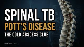
20:10
Spinal Tuberculosis — Pott’s Disease Explained
Spinal Tuberculosis — Pott’s Disease Explained ⏱ Chapters 0:00 Intro 1:52 EXPLAINER 2:40 EXPLAINER 3:25 EXPLAINER 3:55 Read the image 4:26 EXPLAINER 6:28 EXPLAINER 7:27 EXPLAINER 7:46 EXPLAINER 8:09 Read the image 8:56 EXPLAINER 9:36 EXPLAINER 11:07 EXPLAINER 12:29 EXPLAINER 13:22 EXPLAINER 14:03 EXPLAINER 14:43 EXPLAINER 17:01 Read the image 17:40 EXPLAINER 18:10 EXPLAINER 18:43 EXPLAINER 19:46 Recap & sources MRI patterns, tissue diagnosis, chemotherapy and when to decompress In this OrthoVellum master lecture • Spinal TB often spreads across adjacent vertebrae beneath the longitudinal ligament • MRI defines abscess, epidural disease and cord compression • Tissue diagnosis should precede empiric assumptions when feasible • Multidrug chemotherapy is the foundation of treatment • Progressive neurology, instability and deformity are major surgical indications Full clinical topic and references: https://orthovellum.com/topics/tuberculosis-spine-potts Image credits • Benoit Prieur — CC0 — https://commons.wikimedia.org/wiki/File:Mal_de_Pott_au_mus%C3%A9e_Testut-Latarjet.JPG • Salih AF, Malik S. — CC BY 4.0 — https://pmc.ncbi.nlm.nih.gov/articles/PMC12720216/ • Ringshausen FC, Tannapfel A, Nicolas V, Weber A, Duchna HW, Schultze-Werninghaus G, Rohde G. — CC BY 2.0 — https://pmc.ncbi.nlm.nih.gov/articles/PMC2784744/ • Arteaga DD, Arasaratnam RJ, Armitige LY, Storey DF. — CC BY 4.0 — https://pmc.ncbi.nlm.nih.gov/articles/PMC12282389/ • Chowdhury D, Chowdhury AH, Diba MI, Taiwo DD. — CC BY 4.0 — https://pmc.ncbi.nlm.nih.gov/articles/PMC12698498/ • OrthoVellum Medical Education Team — Original OrthoVellum illustration — original://orthovellum/tuberculosis-spine-potts/pathology • OrthoVellum Medical Education Team — Original OrthoVellum illustration — original://orthovellum/tuberculosis-spine-potts/management Educational only — not individual medical advice. #orthopaedics #orthopedics #medicaleducation #orthovellum Orthovellum.com
27 Aug 2026
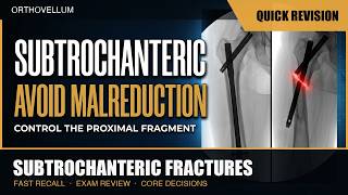
4:10
Subtrochanteric Fractures — Avoid Malreduction — Quick Revision (exam review)
The 4-minute revision cut of the full OrthoVellum lecture: Subtrochanteric Fractures — Avoid Malreduction. Just the teaching core — every diagram and every rule, no intro, no recap. Watch the full lecture on the OrthoVellum channel for the complete build-up, and visit Orthovellum.com for the written topic with references. Educational only — not individual medical advice. Always consult a qualified clinician. #Orthopaedics #Orthopedics #MedEd #OrthoVellum #OrthopaedicSurgery
27 Aug 2026

18:28
Subtrochanteric Fractures — Avoid Malreduction
Subtrochanteric Fractures — Avoid Malreduction ⏱ Chapters 0:00 Intro 1:47 EXPLAINER 2:28 EXPLAINER 3:01 EXPLAINER 3:41 Read the image 4:16 EXPLAINER 6:07 EXPLAINER 7:20 EXPLAINER 7:34 Read the image 8:20 EXPLAINER 8:43 EXPLAINER 9:08 EXPLAINER 11:51 EXPLAINER 12:04 Read the image 12:34 EXPLAINER 13:15 EXPLAINER 14:35 EXPLAINER 15:59 Read the image 16:35 EXPLAINER 17:12 EXPLAINER 18:04 Recap & sources Control the proximal fragment before the nail locks the deformity in In this OrthoVellum master lecture • Muscle forces explain the predictable deformity • Reduction must be achieved before guidewire and reaming • A lateral start point creates varus • Percutaneous tools are planned, not rescue improvisations • Final fluoroscopy must confirm length, rotation and both-plane alignment Full clinical topic and references: https://orthovellum.com/topics/subtrochanteric-fractures Image credits • Tonogai I et al., Case Rep Orthop (PMC4129925) — CC BY — https://www.ncbi.nlm.nih.gov/pmc/articles/PMC4129925/ • Case reports in orthopedics — cc by — https://europepmc.org/article/PMC/PMC13213072 • Diagnostics (Basel, Switzerland) — cc by — https://europepmc.org/article/PMC/PMC12839861 • Malaysian orthopaedic journal — cc by — https://europepmc.org/article/PMC/PMC13133578 • OrthoVellum — OrthoVellum original asset • Advances in orthopedics — cc by — https://europepmc.org/article/PMC/PMC6230414 • Orthopaedic surgery — cc by — https://europepmc.org/article/PMC/PMC10925515 • Yon et al., Medicina (2025), CC BY 4.0 — CC BY 4.0 — https://europepmc.org/article/PMC/PMC11766848#medicina-61-00112-g004 • Al-Humairi et al., Cureus (2025), CC BY 4.0 — CC BY 4.0 — https://europepmc.org/article/PMC/PMC12318351#cureus-0017-00000087263-i01 • Lee et al., Journal of Clinical Medicine (2026), CC BY 4.0 — CC BY 4.0 — https://europepmc.org/article/PMC/PMC13258431#jcm-15-04375-g001 Educational only — not individual medical advice. #orthopaedics #orthopedics #medicaleducation #orthovellum Orthovellum.com
27 Aug 2026

15:37
Complex Distal Femur Fractures — Fixation That Survives
Complex Distal Femur Fractures — Fixation That Survives ⏱ Chapters 0:00 Intro 1:32 EXPLAINER 2:12 EXPLAINER 2:31 EXPLAINER 3:26 EXPLAINER 3:49 Read the image 4:49 EXPLAINER 5:30 EXPLAINER 6:49 EXPLAINER 7:28 EXPLAINER 7:59 EXPLAINER 8:38 Read the image 9:02 EXPLAINER 9:44 EXPLAINER 9:54 EXPLAINER 10:31 EXPLAINER 11:48 EXPLAINER 12:51 Read the image 13:18 EXPLAINER 14:16 Read the image 15:12 Recap & sources Length, alignment, Hoffa fragments and construct stiffness In this OrthoVellum master lecture • CT is essential for Hoffa and articular fragments • Restore limb alignment before locking the construct • Bridge comminution with sensible plate length and working length • Medial instability may need additional support • Nonunion analysis must include biology, infection and mechanics Full clinical topic and references: https://orthovellum.com/topics/distal-femur-fractures Image credits • BMC surgery — cc by — https://europepmc.org/article/PMC/PMC9531427 • Cureus — cc by — https://europepmc.org/article/PMC/PMC13157617 • OrthoVellum — OrthoVellum original asset • Frontiers in surgery — cc by — https://europepmc.org/article/PMC/PMC13270536 • Sports medicine, arthroscopy, rehabilitation, therapy & technology : SMARTT — CC BY — http://www.ncbi.nlm.nih.gov/pmc/articles/PMC3475009 • Wu CC, J Orthop Surg Res — CC BY — https://www.ncbi.nlm.nih.gov/pmc/articles/PMC9258056/ Educational only — not individual medical advice. #orthopaedics #orthopedics #medicaleducation #orthovellum Orthovellum.com
27 Aug 2026

3:10
Complex Distal Femur Fractures — Fixation That Survives — Quick Revision (exam review)
The 3-minute revision cut of the full OrthoVellum lecture: Complex Distal Femur Fractures — Fixation That Survives. Just the teaching core — every diagram and every rule, no intro, no recap. Watch the full lecture on the OrthoVellum channel for the complete build-up, and visit Orthovellum.com for the written topic with references. Educational only — not individual medical advice. Always consult a qualified clinician. #Orthopaedics #Orthopedics #MedEd #OrthoVellum #OrthopaedicSurgery
27 Aug 2026

4:05
DHS vs IM Nail — Intertrochanteric Fractures — Quick Revision (exam review)
The 4-minute revision cut of the full OrthoVellum lecture: DHS vs IM Nail — Intertrochanteric Fractures. Just the teaching core — every diagram and every rule, no intro, no recap. Watch the full lecture on the OrthoVellum channel for the complete build-up, and visit Orthovellum.com for the written topic with references. Educational only — not individual medical advice. Always consult a qualified clinician. #Orthopaedics #Orthopedics #MedEd #OrthoVellum #OrthopaedicSurgery
26 Aug 2026

15:47
DHS vs IM Nail — Intertrochanteric Fractures
DHS vs IM Nail — Intertrochanteric Fractures ⏱ Chapters 0:00 Intro 1:46 EXPLAINER 2:29 EXPLAINER 3:04 Read the image 4:16 EXPLAINER 4:59 EXPLAINER 6:12 EXPLAINER 6:22 Read the image 7:32 EXPLAINER 8:16 EXPLAINER 9:32 EXPLAINER 10:08 EXPLAINER 11:00 Read the image 11:25 EXPLAINER 12:19 EXPLAINER 13:05 EXPLAINER 13:45 EXPLAINER 14:22 Read the image 15:23 Recap & sources Choose the implant from stability, the lateral wall and fracture geometry In this OrthoVellum master lecture • Stable geometry can be treated with a dynamic hip screw • Reverse obliquity and lateral-wall failure favour a cephalomedullary nail • Reduction quality matters more than brand choice • Varus and poor head-element position cause cut-out • Treat osteoporosis, frailty and falls alongside the fracture Full clinical topic and references: https://orthovellum.com/topics/intertrochanteric-fractures Image credits • Frontiers in Bioengineering 2023 (PMC10665526) — CC BY — https://www.ncbi.nlm.nih.gov/pmc/articles/PMC10665526/ • J Clin Med (MDPI) 2023 (PMC10219306) — CC BY — https://www.ncbi.nlm.nih.gov/pmc/articles/PMC10219306/ • Yon et al., Medicina (2025), CC BY 4.0 — CC BY 4.0 — https://europepmc.org/article/PMC/PMC11766848#medicina-61-00112-g004 • Al-Humairi et al., Cureus (2025), CC BY 4.0 — CC BY 4.0 — https://europepmc.org/article/PMC/PMC12318351#cureus-0017-00000087263-i01 • Lee et al., Journal of Clinical Medicine (2026), CC BY 4.0 — CC BY 4.0 — https://europepmc.org/article/PMC/PMC13258431#jcm-15-04375-g001 • Wu CC, J Orthop Surg Res — CC BY — https://www.ncbi.nlm.nih.gov/pmc/articles/PMC9258056/ • Al-Humairi et al., Cureus (2025), CC BY 4.0 — CC BY 4.0 — https://europepmc.org/article/PMC/PMC12318351#cureus-0017-00000087263-i02 • Yon et al., Medicina (2025), CC BY 4.0 — CC BY 4.0 — https://europepmc.org/article/PMC/PMC11766848#medicina-61-00112-g001 • Tonogai I et al., Case Rep Orthop (PMC4129925) — CC BY — https://www.ncbi.nlm.nih.gov/pmc/articles/PMC4129925/ • Mariani P et al. - Case Rep Orthop (CC-BY 4.0) — CC BY — https://www.ncbi.nlm.nih.gov/pmc/articles/PMC4258342/ • Yeo AK et al. - Patient Saf Surg (CC-BY 4.0) — CC BY — https://www.ncbi.nlm.nih.gov/pmc/articles/PMC4658799/ Educational only — not individual medical advice. #orthopaedics #orthopedics #medicaleducation #orthovellum Orthovellum.com
26 Aug 2026

3:34
Calcaneal Fractures — Operative Techniques — Quick Revision (exam review)
The 4-minute revision cut of the full OrthoVellum lecture: Calcaneal Fractures — Operative Techniques. Just the teaching core — every diagram and every rule, no intro, no recap. Watch the full lecture on the OrthoVellum channel for the complete build-up, and visit Orthovellum.com for the written topic with references. Educational only — not individual medical advice. Always consult a qualified clinician. #Orthopaedics #Orthopedics #MedEd #OrthoVellum #OrthopaedicSurgery
26 Aug 2026

19:55
Calcaneal Fractures — Operative Techniques
Calcaneal Fractures — Operative Techniques ⏱ Chapters 0:00 Intro 1:39 EXPLAINER 2:11 EXPLAINER 2:53 EXPLAINER 3:17 Read the image 3:52 EXPLAINER 5:29 EXPLAINER 6:52 EXPLAINER 7:37 EXPLAINER 7:58 Read the image 8:26 EXPLAINER 9:20 EXPLAINER 10:26 EXPLAINER 12:02 EXPLAINER 12:54 Read the image 13:27 EXPLAINER 13:59 EXPLAINER 14:17 EXPLAINER 15:32 EXPLAINER 17:15 EXPLAINER 17:35 Read the image 17:58 EXPLAINER 18:25 EXPLAINER 18:52 EXPLAINER 19:31 Recap & sources Restore the posterior facet without losing the skin In this OrthoVellum master lecture • CT planning starts with the posterior facet and sustentaculum • Soft-tissue condition determines operative timing • Correct tuberosity varus, height and width before final fixation • The approach trades exposure against wound risk • Subtalar arthritis and wound breakdown dominate long-term failure Full clinical topic and references: https://orthovellum.com/topics/calcaneal-fractures Image credits • Feng Y et al., BMC Musculoskelet Disord (PMC4946135) — CC BY 4.0 — https://openi.nlm.nih.gov/detailedresult?img=PMC4946135 • Rev Bras Ortop 2025 (PMC12307035) — CC BY 4.0 — https://openi.nlm.nih.gov/ • Open-i / NIH (PMC5242061) — CC BY 4.0 — https://openi.nlm.nih.gov/detailedresult?img=PMC5242061 • OrthoVellum illustration — OrthoVellum original Educational only — not individual medical advice. #orthopaedics #orthopedics #medicaleducation #orthovellum Orthovellum.com
26 Aug 2026

3:12
Orthopaedic Trauma for Medical Students — The First Hour — Quick Revision (exam review)
The 3-minute revision cut of the full OrthoVellum lecture: Orthopaedic Trauma for Medical Students — The First Hour. Just the teaching core — every diagram and every rule, no intro, no recap. Watch the full lecture on the OrthoVellum channel for the complete build-up, and visit Orthovellum.com for the written topic with references. Educational only — not individual medical advice. Always consult a qualified clinician. #Orthopaedics #Orthopedics #MedEd #OrthoVellum #OrthopaedicSurgery
26 Aug 2026
16:25
Orthopaedic Trauma for Medical Students — The First Hour
Orthopaedic Trauma for Medical Students — The First Hour ⏱ Chapters 0:00 Intro 1:39 EXPLAINER 2:17 EXPLAINER 3:05 EXPLAINER 3:27 Read the image 4:06 EXPLAINER 5:23 EXPLAINER 7:03 EXPLAINER 7:32 EXPLAINER 8:04 EXPLAINER 8:44 EXPLAINER 10:09 EXPLAINER 10:53 Read the image 11:17 EXPLAINER 11:56 EXPLAINER 13:15 EXPLAINER 13:50 EXPLAINER 15:04 Read the image 16:01 Recap & sources ABCDE, shock, splints and when definitive fixation must wait In this OrthoVellum master lecture • ABCDE outranks the dramatic fracture • Every injured limb needs documented skin, vascular and nerve findings • Images include the joint above and below • Definitive fixation depends on physiology, not enthusiasm • Repeat assessment catches deterioration after the first survey Full clinical topic and references: https://orthovellum.com/topics/polytrauma-management Image credits • Dhar SA et al. via J Trauma Manag Outcomes via Open-i (NIH) (Open Access (CC BY)) — CC BY — https://pmc.ncbi.nlm.nih.gov/articles/PMC2253507/ • von Rüden C et al. via J Orthop Surg Res via Open-i (NIH) (Open Access (CC BY)) — CC BY — https://pmc.ncbi.nlm.nih.gov/articles/PMC4335365/ • OrthoVellum — OrthoVellum • Hellerhoff via Wikimedia Commons — CC BY-SA 3.0 — https://commons.wikimedia.org • Uzun M et al., Adv Orthop 2014 (PMC4273594), CC BY — CC BY — https://www.ncbi.nlm.nih.gov/pmc/articles/PMC4273594/ • Wu CC, J Orthop Surg Res — CC BY — https://www.ncbi.nlm.nih.gov/pmc/articles/PMC9258056/ • Trauma case reports — cc by — https://europepmc.org/article/PMC/PMC13141035 • Kulshrestha et al., Indian Journal of Orthopaedics, via PubMed Central — CC BY — https://pmc.ncbi.nlm.nih.gov/articles/PMC2740348/ • EFORT open reviews — CC BY 4.0 — https://europepmc.org/article/PMC/PMC13240633 • Complex pelvic fracture case report, PMC10492189 — CC BY 4.0 — https://pmc.ncbi.nlm.nih.gov/articles/PMC10492189/ • Jalo via Wikimedia Commons — CC BY 4.0 — https://commons.wikimedia.org/wiki/File:A_fracture_of_the_tibia_and_fibula.jpg Educational only — not individual medical advice. #orthopaedics #orthopedics #medicaleducation #orthovellum Orthovellum.com
26 Aug 2026

16:26
Pelvic Fractures — Stop the Bleeding First
Pelvic Fractures — Stop the Bleeding First ⏱ Chapters 0:00 Intro 0:54 Read the image 1:59 EXPLAINER 2:34 EXPLAINER 3:05 EXPLAINER 4:17 Read the image 4:47 EXPLAINER 7:11 EXPLAINER 7:47 EXPLAINER 8:44 Read the image 9:17 EXPLAINER 9:58 EXPLAINER 12:13 EXPLAINER 12:48 EXPLAINER 13:13 Read the image 13:46 EXPLAINER 14:48 EXPLAINER 16:03 Recap & sources Binder position, haemorrhage control and posterior-ring stability In this OrthoVellum master lecture • In pelvic trauma, bleeding and physiology come before classification • The binder belongs over the greater trochanters • Posterior-ring disruption determines mechanical instability • Haemorrhage control may require packing, embolisation or both • Definitive fixation restores the posterior ring before completing the front Full clinical topic and references: https://orthovellum.com/topics/pelvic-ring-injuries Image credits • Complex pelvic fracture case report, PMC10492189 — CC BY 4.0 — https://pmc.ncbi.nlm.nih.gov/articles/PMC10492189/ • Diagnostics (MDPI) 2022 — CC BY 4.0 — https://pmc.ncbi.nlm.nih.gov/articles/PMC8870907/ • OrthoVellum Medical Education Team — Original OrthoVellum educational illustration • Cerevisae — CC BY-SA 4.0 — https://commons.wikimedia.org/wiki/File:Pelvis_AP_view_showing_fracture_of_the_left_ischium_and_left_acetabular_wall.jpg • Valerio et al., Cureus — CC BY 4.0 — https://europepmc.org/article/PMC/PMC13268618#cureus-0018-00000108982-i09 • Gewiess et al. — CC BY 4.0 — https://europepmc.org/article/PMC/PMC11663159 • Keel MJ et al., Eur J Trauma Emerg Surg (PMC3495274) — CC BY — https://www.ncbi.nlm.nih.gov/pmc/articles/PMC3495274/ • Girgis et al., Journal of Clinical Medicine — CC BY 4.0 — https://europepmc.org/article/PMC/PMC11856568#jcm-14-01289-g004 • Chen H et al., BMC Musculoskelet Disord — Open Access (CC BY) — https://openi.nlm.nih.gov/detailedresult?img=PMC3750865 • Girgis et al., Journal of Clinical Medicine — CC BY 4.0 — https://europepmc.org/article/PMC/PMC11856568#jcm-14-01289-g003 Educational only — not individual medical advice. #orthopaedics #orthopedics #medicaleducation #orthovellum Orthovellum.com
26 Aug 2026

3:55
Pelvic Fractures — Stop the Bleeding First — Quick Revision (exam review)
The 4-minute revision cut of the full OrthoVellum lecture: Pelvic Fractures — Stop the Bleeding First. Just the teaching core — every diagram and every rule, no intro, no recap. Watch the full lecture on the OrthoVellum channel for the complete build-up, and visit Orthovellum.com for the written topic with references. Educational only — not individual medical advice. Always consult a qualified clinician. #Orthopaedics #Orthopedics #MedEd #OrthoVellum #OrthopaedicSurgery
26 Aug 2026

7:59
Atlanto-Occipital Dislocation — The Injury You Must Not Traction — Quick Revision (exam review)
The 8-minute revision cut of the full OrthoVellum lecture: Atlanto-Occipital Dislocation — The Injury You Must Not Traction. Just the teaching core — every diagram and every rule, no intro, no recap. Watch the full lecture on the OrthoVellum channel for the complete build-up, and visit Orthovellum.com for the written topic with references. Educational only — not individual medical advice. Always consult a qualified clinician. #Orthopaedics #Orthopedics #MedEd #OrthoVellum #OrthopaedicSurgery
26 Aug 2026

14:02
Atlanto-Occipital Dislocation — The Injury You Must Not Traction
Atlanto-Occipital Dislocation — The Injury You Must Not Traction ⏱ Chapters 0:00 Intro 0:32 Why The Junction Fails 2:18 Why The Junction Fails: Read The Image 2:54 Diagnosis On CT And MRI 5:32 Diagnosis On CT And MRI: Read The Image 6:06 The Resuscitation Trap 8:33 The Resuscitation Trap: Read The Image 9:17 Definitive Stabilisation 12:47 Definitive Stabilisation: Read The Image 13:14 Atlanto-Occipital Dislocation — Core… 13:39 Recap & sources Recognise craniocervical dissociation before movement becomes fatal In this OrthoVellum master lecture • AOD is primarily a ligamentous craniocervical injury • CT measurements must be interpreted together • Normal pulses or a reassuring plain film do not exclude it • Do not apply traction to a distraction injury • Definitive treatment is occipitocervical stabilisation Full clinical topic and references: https://orthovellum.com/topics/atlanto-occipital-dislocation Image credits • Dave BR et al. — CC BY 4.0 — https://pmc.ncbi.nlm.nih.gov/articles/PMC12857585/figure/FIG6/ • Hillis M et al. — CC BY 4.0 — https://pmc.ncbi.nlm.nih.gov/articles/PMC13053567/figure/F3/ • Lee SH et al. — CC BY 4.0 — https://pmc.ncbi.nlm.nih.gov/articles/PMC9744818/figure/Fig1/ • Kwon JH et al. — CC BY 4.0 — https://pmc.ncbi.nlm.nih.gov/articles/PMC11727652/figure/jcm-13-07639-g001/ Educational only — not individual medical advice. #orthopaedics #orthopedics #medicaleducation #orthovellum Orthovellum.com
26 Aug 2026

10:07
Baker's Cyst (Popliteal Synovial Cyst) — 10-Min Quick Revision
The 10-minute revision cut of the full OrthoVellum lecture: Baker's Cyst — The One-Way Valve. Just the teaching core — every diagram and every rule, no intro, no recap. Watch the full lecture on the OrthoVellum channel for the complete build-up, and visit Orthovellum.com for the written topic with references. Educational only — not individual medical advice. Always consult a qualified clinician. #Orthopaedics #Orthopedics #MedEd #OrthoVellum #OrthopaedicSurgery
23 Aug 2026

11:17
Baker's Cyst Explained: The One-Way Valve & Why Excision Fails
Baker's cyst, also known as a popliteal cyst, functions as a one-way valve that traps synovial fluid from the knee joint. Understanding this mechanism explains why treating the cyst alone leads to high recurrence rates. This video breaks down the anatomy, pathophysiology, and clinical implications for trainees and clinicians. ⏱ Chapters 0:00 Intro 0:42 The Bursa And The Valve 2:05 The Communicating Neck: Between Mg And Sm 3:25 Clinical Presentation & Rupture 5:23 Cyst Rupture: Fluid Extravasation Into Calf 5:56 Imaging The Fluid And The Joint 7:09 MRI: Communicating Peduncle And Loose Bodies 7:50 Treatment: Address The Cause 9:15 Arthroscopic Valve Enlargement Vs Open… 9:55 Baker'S Cyst — The Core Principles 10:53 Recap & sources This episode explores the gastrocnemius-semimembranosus bursa as the true source of a Baker's cyst, details the one-way valve mechanism described by Lindgren and Rauschning, and demonstrates how underlying meniscal tears or osteoarthritis drive cyst formation. We cover clinical signs such as Foucher's sign, rupture mimicking DVT (pseudothrombophlebitis), imaging findings, and why addressing intra-articular pathology is essential to prevent recurrence. 🦴 IN THIS VIDEO YOU'LL LEARN • How the gastrocnemius-semimembranosus bursa communicates with the knee joint via a one-way valve • Why intra‑articular pressure from meniscal tears or osteoarthritis inflates the cyst • How to recognise Foucher's sign and its variation with knee extension and flexion • Differentiating a ruptured Baker's cyst from deep‑vein thrombosis using clinical clues • Interpreting ultrasound and MRI findings that show the cyst’s neck and associated knee pathology • Why isolated cyst excision fails and recurrence rates approach 63 % without treating the joint • Key indications for conservative management versus referral for surgical intervention • The role of crescent sign and crescentic ecchymosis in confirming pseudothrombophlebitis • How treating the underlying knee pathology leads to cyst resolution 📌 KEY TAKEAWAYS ✔️ Baker's cyst is almost always secondary to intra‑articular knee disease ✔️ The one‑way valve traps fluid, making the cyst a pressure‑dependent marker ✔️ Foucher's sign – cyst firmer in extension – helps confirm the diagnosis ✔️ Rupture mimics DVT; always exclude thrombosis with Doppler ultrasound ✔️ Treat the knee pathology, not the cyst, to avoid recurrence ✔️ Imaging (US/MRI) should assess both cyst and joint for meniscal tear or arthritis 👩⚕️ WHO THIS IS FOR Designed for orthopaedic trainees, medical students, clinicians, and curious patients who want a clear, evidence‑based understanding of Baker's cyst pathophysiology and management. 📚 RELATED TOPICS: Meniscal tear · Knee osteoarthritis · Popliteal artery aneurysm · Pseudothrombophlebitis · Synovial chondromatosis · Posterior cruciate ligament injury 🔗 More free orthopaedic teaching at Orthovellum.com — subscribe for weekly videos. 🎨 CREDITS • Original Orthovellum production. ⚠️ DISCLAIMER: Educational only — not individual medical advice. Always consult a qualified clinician for diagnosis and treatment. #BakersCyst #PoplitealCyst #OneWayValve #FouchersSign #Pseudothrombophlebitis #MeniscalTear #KneeOsteoarthritis #UltrasoundKnee #MRImaging #OrthopaedicEducation #JointEffusion #RecurrenceRisk #GastrocnemiusSemimembranosus #IntraArticularPathology #Orthovellum
23 Aug 2026

11:09
Internal Impingement — The Thrower's Shoulder — Quick Revision (exam review)
The 11-minute revision cut of the full OrthoVellum lecture: Internal Impingement — The Thrower's Shoulder. Just the teaching core — every diagram and every rule, no intro, no recap. Watch the full lecture on the OrthoVellum channel for the complete build-up, and visit Orthovellum.com for the written topic with references. Educational only — not individual medical advice. Always consult a qualified clinician. #Orthopaedics #Orthopedics #MedEd #OrthoVellum #OrthopaedicSurgery
19 Aug 2026

12:46
Internal Impingement — The Thrower's Shoulder
Internal impingement, often called the thrower's shoulder, describes the normal contact between the posterosuperior rotator cuff and labrum in the ABER position that becomes painful when posterior capsule contracture creates GIRD. This video explains how a tight posterior capsule shifts the humeral head, increases contact force, and leads to undersurface cuff tears and labral fraying. Learn the pathophysiology, diagnostic clues, and evidence‑based management. ⏱ Chapters 0:00 Intro 0:30 The Contact That Is Normal 1:40 The Contact, On MRI And Under The Scope 3:13 Measuring The Thrower 5:10 Measure The Arc — Scapula Held Down 6:33 Rehab First 7:41 Undersurface Tear — Measure The Residual… 8:16 Surgery As Salvage 9:24 Tighten — Or Preserve? 10:24 Repair Without Completing The Tear 11:44 Recap & sources This episode breaks down the mechanics of internal impingement in overhead athletes, detailing the normal ABER contact, the role of posterior capsule contracture and GIRD, imaging findings on MRI and arthroscopy, and the stepwise treatment algorithm from conservative stretching to surgical debridement or repair. Understanding these concepts is essential for clinicians managing shoulder pain in throwers and for anyone seeking to prevent progression to full‑thickness tears. 🦴 IN THIS VIDEO YOU'LL LEARN • How normal posterosuperior cuff‑labrum contact occurs in the ABER position • Why posterior capsule contracture produces GIRD and alters humeral head translation • How to measure glenohumeral internal rotation deficit and apply the 20‑25° threshold • The difference between adaptive external‑rotation gain and pathologic total‑arc loss • MRI and arthroscopic features that identify articular‑sided cuff tears and labral fraying • First‑line conservative strategies including posterior capsule stretching and scapular stabilization • Indications for arthroscopic debridement versus repair in progressive disease • How SICK scapula syndrome coexists and influences rehabilitation • The distinction between posterosuperior and anterosuperior internal impingement variants 📌 KEY TAKEAWAYS ✔️ Internal impingement is a normal ABER contact that becomes painful only with posterior capsule tightness. ✔️ GIRD over 20‑25° signals pathologic contracture; total‑arc loss predicts injury better than IR loss alone. ✔️ Conservative posterior capsule stretching and scapular work for 3‑6 months resolves most cases. ✔️ Arthroscopic debridement is reserved for failed conservative care; repair is needed for full‑thickness tears. ✔️ Assess for SICK scapula; addressing scapular dyskinesis is essential to prevent recurrence. ✔️ Differentiate from external (subacromial) impingement and anterosuperior variants by pain position. 👩⚕️ WHO THIS IS FOR This video is designed for orthopaedic trainees, medical students, physiotherapists, and clinicians who manage shoulder injuries, as well as interested patients and athletes seeking to understand the thrower's shoulder. 📚 RELATED TOPICS: Posterior capsule stretching techniques · SICK scapula syndrome · SLAP lesions and labral repair · Rotator cuff articular‑sided tears · External (subacromial) impingement · Humeral retroversion in overhead athletes 🔗 More free orthopaedic teaching at Orthovellum.com — subscribe for weekly videos. 🎨 CREDITS • Original Orthovellum production. ⚠️ DISCLAIMER: Educational only — not individual medical advice. Always consult a qualified clinician for diagnosis and treatment. #internalimpingement #throwersshoulder #GIRD #ABERposition #posteriorscapsule #shoulderpain #rotatorcufftear #labralfray #scapulardyskinesis #sickscapula #orthopaedics #sportsmedicine #physiotherapy #arthroscopy #conservativetreatment
19 Aug 2026
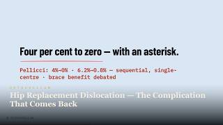
10:54
Hip Replacement Dislocation — The Complication That Comes Back
Hip replacement dislocation is a feared complication that often recurs, driven by impingement and limited jump distance. Understanding the stability ladder helps surgeons and trainees restore stability after a dislocation event. This video breaks down the mechanics, risk factors, and management strategies. ⏱ Chapters 0:00 Intro 0:26 Why Hips Dislocate 1:26 The Dislocated THA — Read It Before You Pull 2:44 The First Dislocation 3:46 The Posterior Repair — Rotators Back To Bone 4:51 The Recurrent Dislocator 5:38 The Functional Zone — Standing And Sitting… 6:54 Buying Stability Back 7:56 Dual Mobility — Read Its Films Differently 8:36 The Precaution Clock 9:57 Recap & sources The video explains why hips dislocate after arthroplasty, detailing the roles of prosthetic impingement, jump distance, and component positioning. It covers posterior dislocation mechanics, clinical presentation, reduction technique, neurovascular assessment, and prevention strategies including head size, dual mobility, and soft‑tissue repair. Recognising these factors is essential to reduce recurrence and improve patient outcomes. 🦴 IN THIS VIDEO YOU'LL LEARN • How prosthetic neck‑on‑cup impingement creates a fulcrum that leads to dislocation • The concept of jump distance and how head size, liner coverage and dual mobility increase stability • Why posterior dislocation accounts for 75‑80% of cases and its typical limb position • Key steps in safe closed reduction, including neurovascular examination before and after • How to interpret AP pelvis radiographs to assess cup inclination, version and impingement • The influence of the Lewinnek safe zones and combined anteversion on stability • Prevention strategies: restoring offset, using 36 mm or larger heads, and dual mobility in high‑risk patients • Recognising when recurrent dislocation warrants revision surgery and which constructs improve stability 📌 KEY TAKEAWAYS ✔️ Impingement is the trigger; jump distance is the stability reserve that prevents escape ✔️ Posterior dislocation presents with flexion, adduction, internal rotation and a shortened limb ✔️ A head size of 36 mm or larger significantly lowers dislocation risk ✔️ Meticulous posterior soft‑tissue repair and offset restoration are critical for stability ✔️ Dual mobility constructs provide a large effective head and high jump distance for high‑risk patients ✔️ Recurrent dislocation after the first event often requires revision to restore stability 👩⚕️ WHO THIS IS FOR This video is useful for orthopaedic trainees, medical students, practising clinicians and interested patients who want to understand hip replacement dislocation, its causes and how to prevent recurrence. 📚 RELATED TOPICS: Component version analysis · Dual mobility arthroplasty · Lewinnek safe zones · Jump distance and head size · Posterior soft‑tissue repair · Revision strategies for recurrent instability 🔗 More free orthopaedic teaching at Orthovellum.com — subscribe for weekly videos. 🎨 CREDITS • Original Orthovellum production. ⚠️ DISCLAIMER: Educational only — not individual medical advice. Always consult a qualified clinician for diagnosis and treatment. #hipreplacement #dislocation #impingement #jumpdistance #stabilityladder #arthroplasty #orthopaedics #jointreplacement #posteriordislocation #dualmobility #headsize #softtissue #reduction #neurovascular #revision
17 Aug 2026
10:48
MRI Cartilage Assessment — Grading the Surface You Cannot Heal — Quick Revision (exam review)
The 11-minute revision cut of the full OrthoVellum lecture: MRI Cartilage Assessment — Grading the Surface You Cannot Heal. Just the teaching core — every diagram and every rule, no intro, no recap. Watch the full lecture on the OrthoVellum channel for the complete build-up, and visit Orthovellum.com for the written topic with references. Educational only — not individual medical advice. Always consult a qualified clinician. #Orthopaedics #Orthopedics #MedEd #OrthoVellum #OrthopaedicSurgery
17 Aug 2026

12:47
MRI Cartilage Assessment — Grading the Surface You Cannot Heal
MRI cartilage assessment is the cornerstone for grading the avascular articular surface that cannot heal, guiding repair planning and follow‑up. This video walks you through the optimal MRI sequences, the modified Outerbridge and MOCART grading systems, and how compositional techniques such as T2 mapping and dGEMRIC reveal biochemical changes before any morphological defect is visible. ⏱ Chapters 0:00 Intro 0:38 The Tissue And Its Limits 1:19 Normal Protocol Vs Advanced Medial Disease 2:09 The Sequences 3:27 Coronal 3D Dess — The Isotropic Measuring… 4:59 Grading What You See 5:58 Grade 4 Defect — Multiplanar, With… 7:26 Measuring Repair 8:16 T1Rho Colour Maps — Composition Before… 9:01 Mocart At Two Years — Marlovits, 26 Knees 9:48 The Cartilage Repair Clock 11:31 Recap & sources The episode covers normal articular cartilage thickness (2‑4 mm) and its zonal variation, why the tissue’s limited healing capacity makes early detection vital, and the step‑by‑step MRI protocol — from routine PD‑FS and 3D gradient‑echo sequences to direct MR arthrography. It explains how to apply the modified Outerbridge classification (Grades 0‑4) and the MOCART score for postoperative repair, and highlights what compositional MRI sees first, namely early T2‑mapping and dGEMRIC changes that precede macroscopic cartilage loss. 🦴 IN THIS VIDEO YOU'LL LEARN • Identify the normal thickness range of articular cartilage and its variation across femoral condyle, patellar surface and tibial plateau. • Recognise the layered appearance of deep radial and superficial transitional zones on high‑resolution MRI. • Explain why routine PD fat‑suppressed sequences have limited sensitivity for partial‑thickness chondral defects. • Describe the advantages of 3D gradient‑echo sequences (SPGR, FLASH, DESS) for isotropic thickness mapping and surface assessment. • Detail how direct MR arthrography with dilute gadolinium improves detection of subtle cartilage surface lesions. • Apply the modified Outerbridge classification (Grades 0‑4) to MRI findings and correlate with ICRS depth thresholds. • Interpret the six components of the MOCART score (fill, integration, surface, signal, bone interface, effusion) for postoperative cartilage repair. • Understand how compositional MRI techniques — T2 mapping and dGEMRIC — detect early biochemical cartilage degeneration before morphological changes appear. 📌 KEY TAKEAWAYS ✔️ Cartilage cannot heal spontaneously; even tiny defects progress toward osteoarthritis. ✔️ Standard knee MRI misses up to 70 % of partial‑thickness lesions — dedicated sequences are essential. ✔️ Modified Outerbridge Grades 0‑4 provide a simple, reproducible MRI grading framework. ✔️ MOCART score quantifies repair quality across fill, integration, surface, signal, bone and effusion. ✔️ T2 mapping and dGEMRIC reveal cartilage biochemical injury months before visible thinning or fissuring. ✔️ 3 Tesla field strength combined with thin isotropic 3D GRE offers the best cartilage‑fluid contrast for accurate measurement. 👩⚕️ WHO THIS IS FOR This video is designed for orthopaedic trainees, medical students, clinicians managing joint pathology, and interested patients who want to understand how MRI evaluates articular cartilage and informs treatment decisions. 📚 RELATED TOPICS: Osteochondral defect imaging · T2 mapping of cartilage · dGEMRIC for glycosaminoglycan assessment · MOCART score application post‑repair · 3D DESS thickness mapping · Direct MR arthrography technique 🔗 More free orthopaedic teaching at Orthovellum.com — subscribe for weekly videos. 🎨 CREDITS • Original Orthovellum production. ⚠️ DISCLAIMER: Educational only — not individual medical advice. Always consult a qualified clinician for diagnosis and treatment. #orthopaedics #mricartilage #outerbridge #mocart #t2mapping #dgemric #3tesla #pdfs #mrarthrography #chondraldefect #osteoarthritis #kneeimaging #cartilagerepair #quantitativemri #musculoskeletalimaging
17 Aug 2026
11:42
Hamstring Injuries — The Muscle That Tears at Full Speed
Hamstring injuries strike the biceps femoris long head at the exact moment it works hardest – during terminal swing when the muscle lengthens under an eccentric load up to two and a half times body weight. This biomechanical peak explains why eighty per cent of tears occur here, setting the stage for a high reinjury risk if rehabilitation is inadequate. ⏱ Chapters 0:00 Intro 0:32 Why The Hamstring Tears 1:38 High-Grade Biceps Femoris — Oedema Around A… 2:59 Grading What You See 3:41 The Published Bamic Key — The Suffix… 5:01 The Reinjury Problem 5:54 Criterion-Based Continuum — Five Phases To… 7:23 The Displaced Avulsion 8:22 Chronic Avulsion — Heterotopic Ossification… 9:05 The Rehabilitation Ladder 10:33 Recap & sources This video explores the anatomy, mechanism, and classification of hamstring injuries, highlighting why the biceps femoris long head is most vulnerable, how sprint‑type and stretch‑type mechanisms differ, and what MRI features indicate a central‑tendon lesion. It explains the British Athletics Muscle Injury Classification (BAMIC), outlines surgical indications for proximal avulsion, and details evidence‑based prevention and return‑to‑sport criteria that reduce reinjury risk. 🦴 IN THIS VIDEO YOU'LL LEARN • Identify why the biceps femoris long head is the most frequently injured hamstring muscle. • Explain the biomechanics of terminal swing and eccentric loading that precipitate tears. • Differentiate sprint‑type from stretch‑type hamstring injuries based on mechanism and prognosis. • Apply the British Athletics Muscle Injury Classification (BAMIC) and interpret the A, B, C suffixes. • Read MRI findings to assess tear location, central tendon involvement, and retraction distance. • Recognise clinical indicators for surgical repair of proximal hamstring avulsion (e.g., over 2 cm retraction). • Outline evidence‑based prevention strategies, including Nordic hamstring exercises and H:Q ratio targets. • Define return‑to‑sport criteria that minimise reinjury risk, such as Askling H‑test negativity and functional strength benchmarks. 📌 KEY TAKEAWAYS ✔️ Eighty per cent of hamstring tears involve the biceps femoris long head at its proximal musculotendinous junction. ✔️ The BAMIC ‘C’ suffix (intratendinous injury) predicts longer recovery and higher reinjury than peripheral strains of the same grade. ✔️ Sprint‑type injuries heal faster than stretch‑type lesions, even when the latter appear milder initially. ✔️ Proximal avulsion requires surgical repair when retraction exceeds 2 cm or more than two tendons are avulsed. ✔️ Reinjury rates climb to ~34 % if athletes return before meeting functional criteria such as an H:Q ratio over 0.8. ✔️ Nordic hamstring exercises reduce injury incidence by approximately 70 % when incorporated into training programmes. 👩⚕️ WHO THIS IS FOR This video is designed for orthopaedic trainees, medical students, sports clinicians, and any healthcare professional seeking a clear, evidence‑based update on hamstring muscle injuries. Interested patients and athletes will also find the explanation of mechanism, prognosis and return‑to‑sport guidance valuable. 📚 RELATED TOPICS: Proximal hamstring avulsion repair · Nordic hamstring exercise programme · Askling H‑test assessment · Hamstring‑to‑quadriceps strength ratio · BAMIC muscle injury classification · Stretch‑type versus sprint‑type hamstring injury 🔗 More free orthopaedic teaching at Orthovellum.com — subscribe for weekly videos. 🎨 CREDITS • Original Orthovellum production. ⚠️ DISCLAIMER: Educational only — not individual medical advice. Always consult a qualified clinician for diagnosis and treatment. #orthopaedics #sportsmedicine #hamstringinjury #bicepsfemoris #terminalswing #eccentricload #MRIgrading #reinjuryprvention #nordicexercises #asklingtest #proximalavulsion #hamstringrehab #HQRratio #sprinttype #stretchtype
17 Aug 2026

11:05
Fifth Metatarsal Stress Fractures — The Fracture That Refuses to Heal
Fifth metatarsal stress fractures, especially the Jones fracture at zone two, are notorious for delayed healing due to a watershed blood supply. Understanding the zone classification guides whether conservative casting or intramedullary screw fixation is appropriate. This video breaks down the anatomy, epidemiology, and management principles you need to know. ⏱ Chapters 0:00 Intro 0:31 Why The Fifth Breaks 1:42 Zone 2 On The Oblique Film — Name The Zone… 2:59 Reading The Film 3:42 Torg Staging — Chronicity Decides Healing 4:45 The Treatment Ladder 5:40 High And Lateral — The Screw That Fills The… 6:55 The Screw Tip Is A New Stress Riser 7:32 Back To Play 9:15 Cavus Or Flat — The Lateral Column Still… 9:56 Recap & sources This video explains why proximal fifth metatarsal fractures heal poorly, detailing the Lawrence‑Botte zone system, the watershed blood supply at the metaphyseal‑diaphyseal junction, and how Torg classification predicts healing potential. It contrasts conservative management of zone 1 avulsions with surgical indications for zones 2 and 3, and highlights return‑to‑sport timelines and key radiographic signs. 🦴 IN THIS VIDEO YOU'LL LEARN • Identify the three Lawrence‑Botte zones of the proximal fifth metatarsal • Differentiate a true Jones fracture (zone 2) from a tuberosity avulsion (zone 1) • Explain why zone 2 is a watershed area with poor retrograde blood flow • Recognise the radiographic features of Torg type I, II and III fractures • Determine when conservative non‑weight‑bearing casting is appropriate • Select patients who benefit from intramedullary screw fixation • Describe the role of bone grafting in chronic non‑unions • Outline return‑to‑sport expectations after surgical versus conservative treatment • Spot common pitfalls such as missing a zone 2 fracture on oblique views 📌 KEY TAKEAWAYS ✔️ Zone 1 avulsions heal reliably with conservative care ✔️ Zone 2 Jones fractures have a 25‑50 % non‑union rate without surgery ✔️ Zone 3 stress fractures carry the highest non‑union risk and often need grafting ✔️ Intramedullary screw fixation reduces healing time to 6‑8 weeks in athletes ✔️ Naming the zone before treatment prevents inappropriate management 👩⚕️ WHO THIS IS FOR This content is suited for orthopaedic trainees, medical students, clinicians managing foot injuries, and interested patients who want a clear, evidence‑based understanding of fifth metatarsal stress fractures and their treatment options. 📚 RELATED TOPICS: Lisfranc injury mechanisms and management · Metatarsalgia and forefoot overload syndromes · Peroneus brevis tendon pathology and avulsion injuries · Foot stress fractures in runners and dancers · Biomechanical effects of pes planus and pes cavus on lateral column loading · Techniques for intramedullary screw fixation of the fifth metatarsal 🔗 More free orthopaedic teaching at Orthovellum.com — subscribe for weekly videos. 🎨 CREDITS • Original Orthovellum production. ⚠️ DISCLAIMER: Educational only — not individual medical advice. Always consult a qualified clinician for diagnosis and treatment. #orthopaedics #footfracture #jonesfracture #fifthmetatarsal #stressfracture #watersupply #screwfixation #sportsmedicine #orthotraining #footanatomy #fracturehealing #avulsionfracture #bonegraft #returntosport #clinicaleducation
17 Aug 2026
11:17
Paediatric ACL Injury — Rebuilding a Ligament in a Growing Knee — Quick Revision (exam review)
The 11-minute revision cut of the full OrthoVellum lecture: Paediatric ACL Injury — Rebuilding a Ligament in a Growing Knee. Just the teaching core — every diagram and every rule, no intro, no recap. Watch the full lecture on the OrthoVellum channel for the complete build-up, and visit Orthovellum.com for the written topic with references. Educational only — not individual medical advice. Always consult a qualified clinician. #Orthopaedics #Orthopedics #MedEd #OrthoVellum #OrthopaedicSurgery
16 Aug 2026
13:07
Paediatric ACL Injury — Rebuilding a Ligament in a Growing Knee
Paediatric ACL injury in a growing knee demands a balance between ligament reconstruction and growth‑plate protection. This video explains why the 12‑week window matters, how physeal‑sparing techniques avoid limb‑length discrepancy, and what to look for beyond the ligament itself. ⏱ Chapters 0:00 Intro 0:28 Two Plates, One Ligament 1:36 Tibial Spine Avulsion On Plain Films… 2:25 Complete Midsubstance ACL Tear With The… 3:13 The Knee-Preservation Problem 3:59 Flipped Medial Meniscal Tear — The Locked… 4:40 The Vascular Paediatric Meniscus… 5:23 Fix Now Or Wait? 7:16 The Physis Decides The Operation 8:24 All-Epiphyseal Reconstruction — Both… 9:13 Meyers–Mckeever Classification… 10:08 Growth, Re-Rupture, And The Other Knee 11:50 Recap & sources The video covers the anatomy of the immature knee, injury patterns including midsubstance tears and tibial spine avulsions, imaging pathways with X‑ray and MRI, the decision‑making framework that weighs instability risk against growth‑plate risk, physeal‑sparing and all‑epiphyseal reconstruction options, rehabilitation principles, and return‑to‑sport criteria. Understanding these elements is essential to preserve the meniscus, prevent growth disturbance, and guide timely, individualized treatment for young patients. 🦴 IN THIS VIDEO YOU'LL LEARN • Identify the key history clues that suggest a paediatric ACL injury • Differentiate tibial spine avulsion from midsubstance ACL tear on X‑ray and MRI • Interpret MRI findings for ligament continuity, meniscal pathology, bone bruising and physeal maturity • Apply the SPINE mnemonic to guide early radiographic evaluation in younger children • Choose an appropriate reconstruction technique based on skeletal maturity using the GROW framework • Recognise the 12‑week threshold after which meniscal injury risk rises significantly • Plan rehabilitation and return‑to‑sport based on functional criteria rather than a fixed timeline • Monitor for growth disturbance post‑reconstruction and understand its true incidence 📌 KEY TAKEAWAYS ✔️ Rapid effusion after a pivot injury is an intra‑articular injury until proven otherwise. ✔️ Tibial spine avulsion is the common ACL‑equivalent injury in skeletally immature children and requires plain X‑ray first. ✔️ Delaying reconstruction beyond ~12 weeks roughly doubles the risk of irreparable meniscal damage. ✔️ Physeal‑sparing techniques keep tunnels within the epiphyses, making clinically significant leg‑length discrepancy rare ( under 2%). ✔️ Return to sport should be guided by motion, strength, neuromuscular control, hop testing and confidence, not by calendar date. ✔️ Always examine the whole knee – meniscus, cartilage, bone bruising and collateral structures – to determine the true urgency. 👩⚕️ WHO THIS IS FOR This video is designed for orthopaedic trainees, medical students, clinicians managing paediatric knee injuries, and interested patients or parents seeking clear, evidence‑based information. 📚 RELATED TOPICS: Tibial spine avulsion fixation · Physeal‑sparing ACL reconstruction techniques · Meniscal repair in the paediatric knee · Return‑to‑sport criteria after ACL reconstruction · Growth plate injury and monitoring · Hybrid ACL reconstruction in adolescents 🔗 More free orthopaedic teaching at Orthovellum.com — subscribe for weekly videos. 🎨 CREDITS • Original Orthovellum production. ⚠️ DISCLAIMER: Educational only — not individual medical advice. Always consult a qualified clinician for diagnosis and treatment. #paediatricACL #ACLinjury #growingknee #physessparing #tibialspine #meniscuspreservation #kneeimaging #returntosport #growthplate #orthopaedics #sportsmedicine #pediatrics #ligamentreconstruction #kneepreservation #pediatricortho
16 Aug 2026

10:06
Vascular Injury With Fracture or Dislocation — Saving the Limb Behind the Break — Quick Revision
The 10-minute revision cut of the full OrthoVellum lecture: Vascular Injury With Fracture or Dislocation — Saving the Limb Behind the Break. Just the teaching core — every diagram and every rule, no intro, no recap. Watch the full lecture on the OrthoVellum channel for the complete build-up, and visit Orthovellum.com for the written topic with references. Educational only — not individual medical advice. Always consult a qualified clinician. #Orthopaedics #Orthopedics #MedEd #OrthoVellum #OrthopaedicSurgery
16 Aug 2026
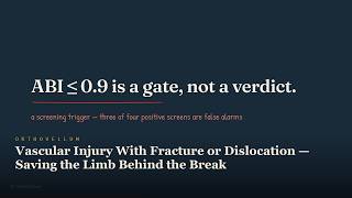
11:33
Vascular Injury With Fracture or Dislocation — Saving the Limb Behind the Break
Vascular Injury With Fracture or Dislocation is a limb‑threatening sequela that hides behind apparently normal pulses. Recognising hard signs, applying the 0.9 ABI/API gate, and using temporary shunts can save the limb during the critical reperfusion hour. This video breaks down the anatomy, assessment and management steps every clinician needs. ⏱ Chapters 0:00 Intro 0:21 A Tethered Artery Behind Every Dislocation 1:23 Posterior Elbow Dislocation — The Brachial… 1:57 Humeral Head Displaced Into The Axilla… 2:26 Tibial Plateau Fracture With A 4 Cm… 2:51 Recognise: Hard And Soft Signs 3:37 Absent Distal Runoff On Cta — An Actionable… 4:14 The 0.9 Gate 5:40 Vessel, Bone, Or Shunt? 7:25 Patent Popliteal Flow Through A Vein… 8:20 After The Clamp Comes Off 9:48 Deep Posterior Compartment Haematoma — The… 10:32 Recap & sources The video explains how fractures and dislocations can injure tethered arteries such as the popliteal, brachial and axillary vessels, why a normal pulse does not exclude injury, and how to differentiate hard from soft signs. It covers the 0.9 ABI/API screening threshold, the role of temporary shunts in damage‑control perfusion, and the urgency of reperfusion within the golden hour to prevent irreversible muscle loss and compartment syndrome. 🦴 IN THIS VIDEO YOU'LL LEARN • How tethering of the popliteal artery at the adductor hiatus and soleus arch makes it vulnerable in knee dislocation and tibial plateau fracture • Why the brachial artery is at risk in supracondylar fractures and elbow dislocations due to its anterior crossing of the joint • The mechanism of axillary artery injury in shoulder dislocation and proximal humeral fractures, especially in older patients with atherosclerotic vessels • How to identify hard signs of vascular injury (active bleeding, expanding/pulsatile haematoma, bruit, thrill, absent pulse, ischaemic limb) • How to recognise soft signs (history of bleeding, small stable haematoma, pulse asymmetry, neurological deficit, proximity wound) and when to order ABI/API or CTA • The clinical significance of an ABI or API value below 0.9 as a screening gate for further vascular imaging • When and how to apply a temporary intravascular shunt to restore flow while skeletal stabilisation or transfer is arranged • Why reperfusion after the golden hour increases the risk of reperfusion injury, compartment syndrome and the need for early fasciotomy • The importance of documenting neurovascular status before and after reduction, and repeating examinations during postoperative care 📌 KEY TAKEAWAYS ✔️ Hard signs mandate immediate vascular control; do not delay for imaging if the limb is threatened ✔️ A normal pulse is reassuring only when the whole examination, ABI/API and mechanism are reliable ✔️ The 0.9 ABI/API threshold guides screening: values below prompt CTA or duplex in stable patients ✔️ Temporary shunts are damage‑control tools, not definitive repair, buying time for skeletal stabilisation or transfer ✔️ Reperfusion within the first hour is critical to salvage muscle and avoid compartment syndrome ✔️ Serial neurovascular and compartment checks are essential after reduction, fixation and repair 👩⚕️ WHO THIS IS FOR This resource is designed for orthopaedic trainees, medical students, clinicians managing trauma, and interested patients who want to understand vascular injury associated with fractures and dislocations. 📚 RELATED TOPICS: Popliteal artery injury in knee dislocation · Brachial artery assessment after elbow trauma · Axillary artery risk in shoulder dislocation · ABI and ABI interpretation in extremity trauma · Temporary intravascular shunt techniques · Fasciotomy indications after reperfusion 🔗 More free orthopaedic teaching at Orthovellum.com — subscribe for weekly videos. 🎨 CREDITS • Original Orthovellum production. ⚠️ DISCLAIMER: Educational only — not individual medical advice. Always consult a qualified clinician for diagnosis and treatment. #vascularinjury #fracturedislocation #hardsigns #ABIscreening #temporaryshunt #reperfusionhour #poplitealartery #brachialartery #axillaryartery #kneedislocation #tibialplateaufracture #elbowdislocation #shoulderdislocation #compartmentsyndrome #fasciotomy
16 Aug 2026

7:52
Thumb Base Arthritis — The Price of the Human Grip — Quick Revision (exam review)
The 8-minute revision cut of the full OrthoVellum lecture: Thumb Base Arthritis — The Price of the Human Grip. Just the teaching core — every diagram and every rule, no intro, no recap. Watch the full lecture on the OrthoVellum channel for the complete build-up, and visit Orthovellum.com for the written topic with references. Educational only — not individual medical advice. Always consult a qualified clinician. #Orthopaedics #Orthopedics #MedEd #OrthoVellum #OrthopaedicSurgery
15 Aug 2026

10:01
Thumb Base Arthritis — The Price of the Human Grip
Thumb base arthritis, also known as carpometacarpal osteoarthritis, affects up to one in three women over fifty and is driven by failure of the anterior oblique (beak) ligament. The grind test reproduces pain by axial compression with rotation, while Eaton–Littler staging on a single radiograph guides treatment. This video explores the anatomy, diagnosis, staging and the evidence behind ligament reconstruction and tendon interposition (LRTI). ⏱ Chapters 0:00 Intro 0:56 A Saddle Built For Reach, Not Stability 1:54 The Restraints — Volar And Dorsal, Model… 2:49 Recognising It 3:33 Robert'S View — The True AP Of The Thumb… 4:03 Four Stages, One Radiograph 4:49 Staging Is The Treatment Map 5:49 What Actually Helps 7:01 Inside The Operation 7:41 The Operative Checklist 8:21 The Failure Mode — Subsidence And Lost… 8:53 Recap & sources The video details the saddle‑joint anatomy of the thumb carpometacarpal articulation, explains how attenuation of the beak ligament initiates dorsoradial subluxation and cartilage wear, demonstrates the grind test and its interpretation, walks through Eaton–Littler radiographic staging, compares conservative and surgical options by stage, and critically appraises the current evidence for ligament reconstruction and tendon interposition versus simple trapeziectomy. Understanding these points is essential for accurate diagnosis, appropriate staging, and informed treatment decisions for this common source of thumb pain. 🦴 IN THIS VIDEO YOU'LL LEARN • Identify the saddle joint anatomy of the thumb CMC articulation • Recognise how attenuation of the anterior oblique (beak) ligament initiates dorsoradial subluxation • Perform and interpret the grind test for thumb base arthritis • Apply Eaton–Littler staging using a single radiograph • Differentiate thumb CMC arthritis from De Quervain’s tenosynovitis and scaphotrapezial arthritis • Summarise conservative and surgical treatment algorithms by stage • Critically appraise the evidence for ligament reconstruction and tendon interposition (LRTI) versus simple trapeziectomy • Explain postoperative expectations and satisfaction rates after trapeziectomy with LRTI 📌 KEY TAKEAWAYS ✔️ The anterior oblique (beak) ligament is the primary stabiliser; its failure starts thumb CMC arthritis. ✔️ A positive grind test is pathognomonic for symptomatic thumb base arthritis. ✔️ Eaton–Littler stage III–IV disease usually warrants surgical intervention, most commonly trapeziectomy with LRTI. ✔️ Current evidence shows no clear superiority of adding LRTI to trapeziectomy, yet patient satisfaction remains over 90%. 👩⚕️ WHO THIS IS FOR This video is designed for orthopaedic trainees, medical students, hand therapists and clinicians managing thumb pain, as well as informed patients seeking to understand their condition. 📚 RELATED TOPICS: De Quervain's tenosynovitis · Scaphotrapezial-trapezoid (STT) arthritis · Thumb metacarpophalangeal joint hyperextension · Trapeziectomy techniques · Ligament reconstruction and tendon interposition (LRTI) outcomes · Basal thumb joint splinting 🔗 More free orthopaedic teaching at Orthovellum.com — subscribe for weekly videos. 🎨 CREDITS • Original Orthovellum production. ⚠️ DISCLAIMER: Educational only — not individual medical advice. Always consult a qualified clinician for diagnosis and treatment. #orthopaedics #handsurgery #thumbarthritis #cmcarthritis #grindtest #eatonlittler #lrtit #trapeziectomy #beakligament #thumbpain #handanatomy #osteoarthritis #jointsurgery #patienteducation #musculoskeletal
15 Aug 2026

8:23
Low Back Pain — When Should You Worry? — Quick Revision (exam review)
The 8-minute revision cut of the full OrthoVellum lecture: Low Back Pain — When Should You Worry?. Just the teaching core — every diagram and every rule, no intro, no recap. Watch the full lecture on the OrthoVellum channel for the complete build-up, and visit Orthovellum.com for the written topic with references. Educational only — not individual medical advice. Always consult a qualified clinician. #Orthopaedics #Orthopedics #MedEd #OrthoVellum #OrthopaedicSurgery
7 Aug 2026

11:39
Low Back Pain — When Should You Worry?
Low back pain affects up to 95% of people as non‑specific mechanical pain, but a small proportion hides serious pathology such as cancer, infection, fracture or cauda equina syndrome. The first clinical step is diagnostic triage – sorting every patient into non‑specific, radicular or red‑flag groups – because this decision drives imaging, treatment and urgency. Missing cauda equina can lead to permanent bladder, bowel and sexual dysfunction, so asking about saddle anaesthesia and bladder function is essential in every significant back‑or‑leg‑pain presentation. ⏱ Chapters 0:00 Intro 1:26 The Three-Way Fork 2:20 Many Pain Sources — Imaging Cannot Localise… 3:01 The Emergency You Cannot Miss 3:56 Red Flags — Tuna Fish 4:53 The Scan That Lies 5:49 When To Image 6:42 The Leg Tells You The Level 7:38 Traversing Root Vs Exiting Root 8:19 What Actually Helps 9:12 Yellow Flags Predict Chronicity, Not Disease 10:32 Recap & sources This video explains the diagnostic triage for low back pain, detailing how to distinguish the overwhelming majority of non‑specific mechanical pain from radicular pain and the small but critical fraction of serious specific pathology flagged by red flags. It covers the red‑flag mnemonics (TUNA FISH), the cauda equina emergency (SBBL), why routine imaging is discouraged, and the evidence‑based non‑operative management pathway that focuses on staying active, appropriate analgesia and addressing yellow flags. Understanding this framework prevents missed emergencies, avoids unnecessary investigations and guides effective, patient‑centred care. 🦴 IN THIS VIDEO YOU'LL LEARN • How to perform the three‑way triage of low back pain into non‑specific, radicular and serious pathology groups • Recognise the red‑flag features of malignancy, infection, fracture and inflammatory disease using the TUNA FISH mnemonic • Identify cauda equina syndrome by saddle anaesthesia, bladder dysfunction, bowel incontinence and bilateral leg signs (SBBL) • Understand why routine lumbar imaging is of low specificity and can worsen outcomes in non‑specific low back pain • Apply the ACTIVE management mnemonic to guide advice, exercise, analgesia and psychosocial assessment • Differentiate neurogenic urinary retention (CES‑R) from incomplete cauda equina (CES‑I) and their prognostic implications • Know when urgent MRI and emergency surgical decompression are indicated for suspected cauda equina • Use yellow‑flag screening tools such as STarT Back to target interventions that prevent chronic disability 📌 KEY TAKEAWAYS ✔️ Triage first: non‑specific vs radicular vs red‑flag directs every subsequent decision ✔️ Cauda equina is a surgical emergency – saddle anaesthesia plus bladder/bowel/sexual dysfunction demands urgent MRI and decompression ✔️ Routine imaging for ordinary low back pain is harmful; reserve scans for red flags, progressive neurology or surgical radicular pain ✔️ Stay active, use NSAIDs as first‑line analgesia and address yellow flags to prevent chronicity ✔️ Remember TUNA FISH for red flags and SBBL for cauda equina – they save lives and function ✔️ Effective management combines reassurance, exercise, biopsychosocial care and avoidance of routine opioids 👩⚕️ WHO THIS IS FOR This lecture is designed for orthopaedic trainees, medical students, junior doctors and allied health professionals who encounter low back pain in clinical practice. It is also valuable for interested patients seeking a clear, evidence‑based understanding of when back pain warrants further investigation. 📚 RELATED TOPICS: Radiculopathy and sciatica assessment · Spinal metastasis red flags · Vertebral osteoporotic fracture evaluation · Inflammatory back pain and axial spondyloarthritis · Psychosocial yellow flags and STarT Back tool · Lumbar disc herniation indications for surgery 🔗 More free orthopaedic teaching at Orthovellum.com — subscribe for weekly videos. 🎨 CREDITS • Original Orthovellum production. ⚠️ DISCLAIMER: Educational only — not individual medical advice. Always consult a qualified clinician for diagnosis and treatment. #lowbackpain #redflags #caudaequina #triage #nonspecificpain #radiculopathy #spinehealth #backpainmanagement #orthopaedics #physiotherapy #yellowflags #imagingguidelines
7 Aug 2026

6:33
Carpal Tunnel Explained — Why Your Hand Wakes You at Night — Quick Revision (exam review)
The 7-minute revision cut of the full OrthoVellum lecture: Carpal Tunnel Explained — Why Your Hand Wakes You at Night. Just the teaching core — every diagram and every rule, no intro, no recap. Watch the full lecture on the OrthoVellum channel for the complete build-up, and visit Orthovellum.com for the written topic with references. Educational only — not individual medical advice. Always consult a qualified clinician. #Orthopaedics #Orthopedics #MedEd #OrthoVellum #OrthopaedicSurgery
27 Jul 2026
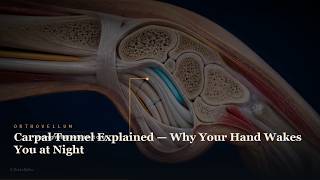
9:01
Carpal Tunnel Explained — Why Your Hand Wakes You at Night
Carpal Tunnel Syndrome is the most common compression neuropathy, caused by the median nerve being squeezed inside the wrist’s rigid tunnel. This explains why patients wake at night with burning, tingling numbness that eases after shaking the hand. Understanding the anatomy reveals why the little finger is spared and what truly relieves the pressure. ⏱ Chapters 0:00 Intro 0:59 A Tunnel That Cannot Expand 2:06 WHAT PRESSURE DOES 3:06 Why It Wakes You At Night 4:07 Which Fingers, And Why 5:15 Making The Diagnosis 6:25 What Actually Fixes It 7:52 Recap & sources This video breaks down the carpal tunnel’s bony and ligamentous structure, shows how increased pressure affects the median nerve, and links each anatomical detail to the classic symptoms — night‑time waking, shaking relief, and sparing of the little finger. By clarifying the mechanical basis of the condition, viewers gain a clear framework for recognising, explaining, and managing carpal tunnel syndrome in everyday practice. 🦴 IN THIS VIDEO YOU'LL LEARN • Identify the carpal bones that form the floor and walls of the tunnel • Describe the transverse carpal ligament (flexor retinaculum) as the roof • List the nine flexor tendons and their synovial sheaths that share the tunnel • Explain why the median nerve lies against the underside of the ligament • Understand why the tunnel cannot expand and how content swelling raises pressure • Link rising pressure to venous ischemia within the nerve and reversible symptoms • Describe the progression from ischemia to demyelination and axonal loss • Explain how wrist flexion or extension at night increases tunnel pressure • Recognise how nocturnal fluid shifts contribute to symptom onset • Clarify why the little finger (ulnar nerve territory) is spared • Identify clinical signs that reflect sensory loss in the median nerve distribution • Summarise why shaking the hand temporarily relieves symptoms • Differentiate reversible ischemic symptoms from permanent neuropathic changes • Outline the pathophysiological basis for night‑time worsening 📌 KEY TAKEAWAYS ✔️ The carpal tunnel is a rigid, non‑expandable space bounded by bone and ligament ✔️ Median nerve compression first causes ischemic symptoms that are reversible ✔️ Night‑time symptoms arise from wrist posture and fluid shifts while lying flat ✔️ The little finger is spared because the ulnar nerve runs outside the tunnel ✔️ Shaking the hand restores nerve blood flow, providing brief relief ✔️ Chronic pressure leads to demyelination and eventual axonal loss 👩⚕️ WHO THIS IS FOR Ideal for orthopaedic trainees, medical students, clinicians, and interested patients who want a clear, anatomy‑based understanding of carpal tunnel syndrome without exam‑specific focus. 📚 RELATED TOPICS: Ulnar nerve entrapment at the wrist (Guyon’s canal) · De Quervain’s tenosynovitis · Trigger finger (stenosing tenosynovitis) · Wrist osteoarthritis and its impact on tendon sheaths · Nerve gliding and tendon‑excursion exercises · Ergonomic workplace modifications for wrist health 🔗 More free orthopaedic teaching at Orthovellum.com — subscribe for weekly videos. 🎨 CREDITS • Original Orthovellum production. ⚠️ DISCLAIMER: Educational only — not individual medical advice. Always consult a qualified clinician for diagnosis and treatment. #carpaltunnelsyndrome #mediannerve #wristanatomy #nightnumbness #handshakerelief #nervecompression #orthopaedics #neurology #handpain #clinicalanatomy #patienteducation #musculoskeletalhealth #nerveischemia #demyelination #axonalloss
27 Jul 2026
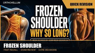
8:47
Frozen Shoulder Explained — Why It Hurts, and Why It Takes So Long — Quick Revision (exam review)
The 9-minute revision cut of the full OrthoVellum lecture: Frozen Shoulder Explained — Why It Hurts, and Why It Takes So Long. Just the teaching core — every diagram and every rule, no intro, no recap. Watch the full lecture on the OrthoVellum channel for the complete build-up, and visit Orthovellum.com for the written topic with references. Educational only — not individual medical advice. Always consult a qualified clinician. #Orthopaedics #Orthopedics #MedEd #OrthoVellum #OrthopaedicSurgery
27 Jul 2026

11:16
Frozen Shoulder Explained — Why It Hurts, and Why It Takes So Long
Frozen shoulder, also known as adhesive capsulitis, is a disorder where the shoulder joint capsule becomes inflamed, fibrotic and physically contracts, producing a true loss of motion that is identical whether the patient or the examiner moves the arm. The hallmark is a capsular restriction that feels like a wall, preventing everyday tasks such as reaching behind the back or fastening a seatbelt. Understanding what actually tightens inside the joint and how it differs from a rotator cuff tear is essential for correct diagnosis and management. ⏱ Chapters 0:00 Intro 1:07 What Is Actually Frozen 2:18 The Three Phases 3:35 Thawing — The Same Capsule, In Reverse 4:18 Treat The Phase You Are In 4:54 Telling It Apart 6:06 The Diagnosis Is Clinical — The X-Ray… 7:06 Who Gets It 8:10 What Actually Helps 9:08 When It Will Not Thaw 10:20 Recap & sources This video breaks down the pathophysiology of frozen shoulder, describing the inflammatory freezing phase, the fibrotic frozen phase and the gradual thawing phase, explains why recovery takes months to years, shows how to distinguish true capsular tightness from muscular weakness, and outlines evidence‑based interventions that can genuinely alter the natural history. 🦴 IN THIS VIDEO YOU'LL LEARN • How the shoulder capsule normally provides slack via the axillary recess • What histological changes occur during inflammation and fibrosis in adhesive capsulitis • Why the coracohumeral ligament and rotator interval tighten first • How to recognise the freezing, frozen and thawing phases clinically • Why passive and active range of motion are equally limited in true capsular contracture • How frozen shoulder differs from a rotator cuff tear on examination and imaging • Which factors (e.g., diabetes, immobilisation) influence the onset and duration • What interventions have been shown to shorten the thawing phase or improve outcomes 📌 KEY TAKEAWAYS ✔️ True capsular contracture limits motion regardless of who moves the arm ✔️ Night pain and progressive stiffness define the freezing phase ✔️ The axillary recess disappears as the capsule shrinks ✔️ Recovery is a slow remodelling process, not a quick fix ✔️ Early physiotherapy and intra‑articular steroid injection can improve outcomes ✔️ Distinguishing from rotator cuff tear avoids unnecessary surgery 👩⚕️ WHO THIS IS FOR This video is designed for orthopaedic trainees, medical students, clinicians managing shoulder disorders, and any patient or carer seeking a clear, evidence‑based explanation of frozen shoulder. 📚 RELATED TOPICS: rotator cuff pathology · shoulder impingement syndrome · calcific tendinitis · post‑traumatic shoulder stiffness · osteoarthritis of the glenohumeral joint · shoulder hydrodilatation techniques 🔗 More free orthopaedic teaching at Orthovellum.com — subscribe for weekly videos. 🎨 CREDITS • Original Orthovellum production. ⚠️ DISCLAIMER: Educational only — not individual medical advice. Always consult a qualified clinician for diagnosis and treatment. #frozenshoulder #adhesivescapsulitis #shoulderpain #shoulderstiffness #capsularcontracture #axillaryrecess #nightpain #rotatorcufftear #shoulderrehab #physiotherapy #corticosteroidinjection #orthopaedics
27 Jul 2026

9:44
Sciatica — Why Back Pain Shoots Down Your Leg — Quick Revision (exam review)
The 10-minute revision cut of the full OrthoVellum lecture: Sciatica — Why Back Pain Shoots Down Your Leg. Just the teaching core — every diagram and every rule, no intro, no recap. Watch the full lecture on the OrthoVellum channel for the complete build-up, and visit Orthovellum.com for the written topic with references. Educational only — not individual medical advice. Always consult a qualified clinician. #Orthopaedics #Orthopedics #MedEd #OrthoVellum #OrthopaedicSurgery
27 Jul 2026

12:42
Sciatica – Why Back Pain Shoots Down Your Leg – Disc Herniation & Nerve Root Pain
Sciatica is leg pain that follows the path of a lumbosacral nerve root, most often caused by a lumbar disc herniation pressing on that root. The sensation can feel electric, burning or like a cable being pulled, and it may be sharper far down the leg than at the spine itself. Understanding what the herniated disc actually does to the nerve root explains why the pain travels and how inflammation drives the symptoms. ⏱ Chapters 0:00 Intro 1:18 The Disc 2:27 How A Disc Herniates 3:24 Chemistry, Not Just Compression 4:13 Which Level, Which Root 5:23 Root By Root: Pain, Power, Reflex 6:18 Sensitive To Exclude, Specific To Confirm 7:11 The Red Flags 8:19 What Happens Without Surgery 9:23 The Scan Must Match The Leg 10:14 What Surgery Changes 11:34 Recap & sources This video breaks down the anatomy of a lumbar disc, shows how a herniation develops from bulge to sequestration, and explains why chemical irritation—not just mechanical compression—produces the classic burning sciatica. It maps which disc levels (L4‑L5, L5‑S1) generate specific pain patterns, highlights red‑flag signs that demand urgent review, and outlines what factors truly influence recovery and long‑term outcome. 🦴 IN THIS VIDEO YOU'LL LEARN • How a lumbar disc is structured and why its posterolateral corner is the weak point • The stepwise progression from disc bulge to protrusion, extrusion and sequestration • Why nucleus pulposus triggers an inflammatory response that sensitises the nerve root • Which lumbar levels (L2‑L3, L3‑L4, L4‑L5, L5‑S1) correspond to specific dermatomal pain distributions • How to differentiate true sciatic nerve root pain from referred pain or peripheral neuropathy • Key red‑flag symptoms and signs that suggest infection, tumour or cauda equina syndrome • What clinical examination manoeuvres (straight‑leg raise, crossed‑straight‑leg raise, femoral stretch test) reveal about nerve root involvement • Factors that predict natural resolution of disc herniations and when surgical intervention may be considered 📌 KEY TAKEAWAYS ✔️ Sciatica is a symptom, not a diagnosis; identify the underlying nerve‑root pathology ✔️ Disc herniation causes leg pain mainly through inflammatory mediators released by the nucleus pulposus ✔️ Posterolateral disc herniations at L4‑L5 and L5‑S1 produce the classic sciatica distribution ✔️ A positive straight‑leg raise test reproduces radicular pain and helps localise the affected root ✔️ Most lumbar disc herniations improve with time; surgery is reserved for progressive neurology or intolerable pain despite conservative care 👩⚕️ WHO THIS IS FOR This video is designed for orthopaedic trainees, medical students, clinicians managing spinal pain, and interested patients who want a clear, evidence‑based explanation of sciatica and disc herniation. 📚 RELATED TOPICS: lumbar spinal stenosis · cauda equina syndrome · piriformis syndrome · facet joint arthropathy · degenerative disc disease · epidural steroid injection 🔗 More free orthopaedic teaching at Orthovellum.com — subscribe for weekly videos. 🎨 CREDITS • Original Orthovellum production. ⚠️ DISCLAIMER: Educational only — not individual medical advice. Always consult a qualified clinician for diagnosis and treatment. #sciatica #discherniation #nerverootpain #lumbarspine #backpain #radiculopathy #straightlegraise #neurosurgery #orthopaediceducation #spineanatomy #inflammatorypain #treatmentoptions #patienteducation #backhealth
27 Jul 2026

9:18
Bone Remodelling — the BMU, the RANKL Axis, and Why Bone Is Never Finished — Quick Revision
The 9-minute revision cut of the full OrthoVellum lecture: Bone Remodelling — the BMU, the RANKL Axis, and Why Bone Is Never Finished. Just the teaching core — every diagram and every rule, no intro, no recap. Watch the full lecture on the OrthoVellum channel for the complete build-up, and visit Orthovellum.com for the written topic with references. Educational only — not individual medical advice. Always consult a qualified clinician. #Orthopaedics #Orthopedics #MedEd #OrthoVellum #OrthopaedicSurgery
27 Jul 2026
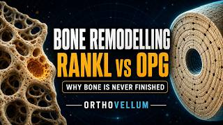
13:13
Bone Remodelling: BMU, RANKL Axis & Modelling vs Remodelling Explained
Bone remodelling is a continuous, lifelong process where the skeleton demolishes and rebuilds itself. Discover the Basic Multicellular Unit (BMU), the RANKL axis, and why bone is never finished. ⏱ Chapters 0:00 Intro — a crew is cutting bone right now 0:27 Why bone remodels at all: structure, calcium, load 1:17 Modelling is not remodelling 2:20 Osteoclasts — sealing zone, ruffled border, Howship's lacuna 3:15 Osteoblasts — the build crew and osteoid 4:03 The osteocyte — the mechanosensor 4:55 The BMU and its five phases 5:58 Coupling — the crew that follows 6:49 Fast to demolish, slow to rebuild 7:58 Cortical tunnels, trabecular trenches 9:02 The control system: RANKL, RANK and OPG 10:02 Oestrogen holds the brake on 10:57 Every drug is an intervention in this cycle 12:04 Recap and sources This video provides a comprehensive breakdown of bone remodelling physiology, explaining how the skeleton handles structural maintenance, metabolic calcium demands, and mechanical adaptation. We explore the vital cellular anatomy of the BMU, differentiate remodelling from modelling, and explain how coupling imbalances drive metabolic bone disease and modern pharmacological targets. 🦴 IN THIS VIDEO YOU'LL LEARN • The three reasons bone remodels: removing fatigue damage, calcium supply, and mechanical adaptation • The critical distinction between coupled bone remodelling and independent bone modelling • The exact cellular anatomy and function of the Basic Multicellular Unit (BMU) • How osteoclasts create Howship's lacunae using a sealing zone and ruffled border • How osteoblasts deposit unmineralised osteoid at one to two micrometres per day, as a sheet • The ten-to-fifteen-day mineralisation delay of the osteoid seam — and why that lag matters • How the RANK/RANKL/OPG axis acts as the master regulator of bone resorption • How osteocytes sense load and change what they secrete — loading suppresses sclerostin • The clinical consequences of trabecular perforation and irreversible microarchitectural damage • Why understanding modelling explains the potent anabolic effects of modern osteoporosis drugs 📌 KEY TAKEAWAYS ✔️ Remodelling replaces bone without changing its shape, while modelling changes bone size and architecture ✔️ Bone resorption and formation are normally tightly coupled within the BMU to prevent net bone loss ✔️ The RANKL to OPG ratio strictly dictates the rate of osteoclast formation and bone resorption ✔️ Trabecular bone remodels at a significantly higher rate than cortical bone, making it more vulnerable to early osteoporosis ✔️ Uncoupling resorption from formation is the fundamental physiological mechanism underlying osteoporosis 👩⚕️ WHO THIS IS FOR This video is designed for orthopaedic trainees, junior doctors, and medical students seeking a deep understanding of bone physiology. It is also highly valuable for curious patients interested in how the skeletal system works. 📚 RELATED TOPICS: Osteoporosis and Metabolic Bone Disease Pathology · RANKL Pathway Pharmacology and Denosumab · Bone Composition and Macroscopic Structure · Osteocyte Mechanotransduction and Wolff's Law · Wnt Signalling and Sclerostin Inhibition · Calcium Homeostasis and Parathyroid Hormone 🔗 More free orthopaedic teaching at Orthovellum.com — subscribe for weekly videos. 🎨 CREDITS • Original Orthovellum production. ⚠️ DISCLAIMER: Educational only — not individual medical advice. Always consult a qualified clinician for diagnosis and treatment. #orthopaedics #boneremodelling #bmubiology #rankl #osteoporosis #bonemetabolism #osteoclast #osteoblast #orthovellum #medicaleducation #bonescience #wolffslaw #osteocytes
27 Jul 2026
8:57
Surgical Approaches to the Hip — Posterior vs Direct Anterior vs Lateral — Quick Revision
The 9-minute revision cut of the full OrthoVellum lecture: Surgical Approaches to the Hip — Posterior vs Direct Anterior vs Lateral. Just the teaching core — every diagram and every rule, no intro, no recap. Watch the full lecture on the OrthoVellum channel for the complete build-up, and visit Orthovellum.com for the written topic with references. Educational only — not individual medical advice. Always consult a qualified clinician. #Orthopaedics #Orthopedics #MedEd #OrthoVellum #OrthopaedicSurgery
26 Jul 2026

20:40
Surgical Approaches to the Hip: Posterior vs Anterior vs Lateral Explained
Surgical approaches to the hip for total hip arthroplasty include the posterior, direct anterior, and lateral methods. Each window buys exposure by spending something—whether a muscle, a nerve, or a margin of stability. Understanding the anatomical trade-offs, dislocation directions, and nerve risks demystifies the debate over which approach is truly best. ⏱ Chapters 0:00 Intro 0:19 ADULT RECONSTRUCTION · THA 0:41 Every Window Spends Something 1:43 Dislocation Rate By Approach 2:36 Approaches Differ In The Complication They C… 2:55 The Hip In Layers 4:02 Three Windows Onto The Same Joint 4:42 Posterior — Exposure, Honestly Bought 5:54 Sciatic Nerve — Why It Is The Peroneal Divis… 7:09 Repair The Rotators And Capsule. Dislocation… 7:36 Direct Anterior — The Only True Plane 9:00 Is There A True Internervous Plane? 9:56 The Two Structures That Define The Anterior… 11:13 Lateral & Anterolateral 13:01 What The Evidence Shows 14:09 What Level I Evidence And Registries Show 15:27 Surgeon Volume Beats Approach Choice. 15:51 Choosing, In Practice 17:25 Position Decides Stability 18:46 The Lewinnek Zone Is Necessary — Not Suffici… 19:19 Recap & sources 20:23 S28 This video breaks down the posterior, direct anterior, anterolateral, and direct lateral approaches for total hip arthroplasty at a fellowship depth. We explore the anatomical layers, the specific internervous planes, and the exact nerves at risk in each surgical window. We also analyse the trade-off between hip stability, the abductor mechanism, and the surgeon's learning curve, supported by the latest AOANJRR registry data and Level I RCT meta-analyses. 🦴 IN THIS VIDEO YOU'LL LEARN • The three-way trade-off between hip stability, the abductor mechanism, and the surgical learning curve • How posterior capsule and short external rotator repair dramatically reduced historical dislocation rates • The specific internervous or intermuscular planes utilised by the posterior, anterior, and lateral approaches • Identifying and protecting the sciatic, lateral femoral cutaneous, femoral, and superior gluteal nerves • Why the direction of post-operative dislocation directly correlates with the direction the hip was opened • The safe zones for splitting the gluteus medius to prevent permanent Trendelenburg gait • How national registry data proves that revision rates are similar across all approaches when performed by experienced surgeons • The difference in surgical positioning and radiographic imaging requirements for each technique 📌 KEY TAKEAWAYS ✔️ No approach is free; each buys exposure by sacrificing a muscle, a nerve, or a margin of stability. ✔️ Soft tissue repair in the posterior approach reduces dislocation rates from 5% to under 2%. ✔️ The direct anterior approach is the only true internervous plane (superior gluteal vs femoral nerve), offering the lowest dislocation rate but a steep learning curve. ✔️ Protect the superior gluteal nerve by never splitting the gluteus medius more than 5cm proximal to the greater trochanter. ✔️ The approach determines the direction of dislocation: posterior dislocates in flexion/IR, anterior dislocates in extension/ER. 👩⚕️ WHO THIS IS FOR This video is designed for orthopaedic surgical trainees, junior doctors, and medical students seeking a fellowship-level understanding of hip arthroplasty. It is also highly valuable for practicing orthopaedic surgeons, healthcare professionals, and curious patients wanting to understand the surgical options and anatomical realities of total hip replacement. 📚 RELATED TOPICS: Total Hip Arthroplasty Component Positioning and Safe Zones · Sciatic Nerve Anatomy and Posterior Hip Dislocation Mechanisms · Abductor Mechanism Deficiency and Trendelenburg Gait · Smith-Petersen Interval and Hueter Direct Anterior Approach · AOANJRR Registry Data on Primary Hip Replacement Survival · Posterior Soft Tissue Repair Techniques in Hip Arthroplasty 🔗 More free orthopaedic teaching at Orthovellum.com — subscribe for weekly videos. 🎨 CREDITS • Original Orthovellum production. ⚠️ DISCLAIMER: Educational only — not individual medical advice. Always consult a qualified clinician for diagnosis and treatment. #orthopaedics #hipreplacement #hiparthroplasty #surgicalapproaches #orthopaedicsurgery #directanteriorapproach #posteriorapproach #hardingeapproach #hipanatomy #adultreconstruction #orthopedicsurgery #medicaleducation
26 Jul 2026

6:06
Nerve Injury & Regeneration: Seddon, Sunderland & Millimetre-a-Day Rule
Peripheral nerve injury classification depends on which anatomical layers are damaged, determining whether a nerve heals itself or requires surgical repair. Master the Seddon and Sunderland classifications alongside the biology of Wallerian degeneration and the crucial one millimetre per day axonal regeneration rule. ⏱ Chapters 0:00 Intro 0:13 BASIC SCIENCE · NERVE INJURY 0:32 The Anatomy That Decides Everything 1:25 Seddon Counts Outcomes. Sunderland Counts La… 1:41 Sunderland'S Five Degrees 2:45 Wallerian Degeneration — Demolition And Rebu… 3:42 Regeneration — A Millimetre A Day 4:35 Tinel'S Sign — The Marching Regeneration Fro… 5:07 Two Clocks: Schwann Bands Fade After 3–4 Mon… 5:33 Recap & sources This video provides a comprehensive, fellowship-level guide to nerve injury and regeneration, explaining the complex anatomy of peripheral nerves and the biological processes that dictate clinical outcomes. Covering everything from the initial cellular responses like Wallerian degeneration and chromatolysis to the formation of Schwann cell bands of Büngner, viewers will understand exactly how to classify nerve damage, predict recovery times, and determine the optimal windows for surgical nerve repair. 🦴 IN THIS VIDEO YOU'LL LEARN • The precise anatomical layers of a peripheral nerve from the axon out to the epineurium • How to accurately classify nerve injuries using both the Seddon and Sunderland grading systems • The step-by-step cellular biology of Wallerian degeneration following peripheral nerve transection • How chromatolysis reprogrammes the neuronal cell body from transmission mode to growth mode • The critical role of Schwann cells in clearing inhibitory debris and forming the bands of Büngner • Why the axonal growth cone advances at a rate of approximately one millimetre per day • How to clinically differentiate between neurapraxia, axonotmesis and neurotmesis • The importance of endoneurial tube preservation for guiding successful nerve regeneration • How to calculate expected recovery times and understand the optimal surgical windows for repair 📌 KEY TAKEAWAYS ✔️ First and second degree Sunderland injuries retain their endoneurial scaffold and heal spontaneously; third degree and deeper injuries fail without surgical intervention. ✔️ Wallerian degeneration is an active, organised demolition process beginning within 24 to 48 hours, essential for clearing inhibitory myelin debris. ✔️ Schwann cells completely change character to phagocytose debris and line up inside surviving endoneurial tubes to form the bands of Büngner. ✔️ Neuronal cell bodies undergo chromatolysis, moving the nucleus to the edge and peaking protein factory retooling at 7 to 14 days. ✔️ Regenerating axons rely on intact anatomical guide-rails; without them, they wander and form painful neuromas. ✔️ At a regeneration rate of one millimetre a day, proximal nerve injuries frequently exceed the 18 to 24 month window of motor endplate viability. 👩⚕️ WHO THIS IS FOR This video is designed for orthopaedic trainees, medical students, clinicians, and curious patients who want a deep, medically accurate understanding of peripheral nerve biology and the principles dictating surgical decision-making. 📚 RELATED TOPICS: Brachial plexus injury assessment and classification · Electrodiagnostic testing and EMG interpretation for nerve injuries · Surgical techniques for primary and secondary nerve repair · Carpal tunnel syndrome and compressive neuropathies · Management of painful neuromas · Peripheral nerve tumours and sheath lesions 🔗 More free orthopaedic teaching at Orthovellum.com — subscribe for weekly videos. 🎨 CREDITS • Original Orthovellum production. ⚠️ DISCLAIMER: Educational only — not individual medical advice. Always consult a qualified clinician for diagnosis and treatment. #orthopaedics #nerveinjury #neuroregeneration #sunderlandclassification #seddonclassification #walleriandegeneration #peripheralnerve #neurosurgery #handsurgery #orthopaedicsurgery #medicalbiology #anatomy #physiology #neuroapraxia #axonotmesis
21 Jul 2026

8:21
Salter-Harris Classification: Growth-Arrest Risk & Fixation Principles
Understanding the Salter-Harris classification of physeal fractures is essential for predicting growth-arrest risk in children. By tracking where the fracture line travels relative to the growth plate, orthopaedic surgeons can determine the threat to the germinal layer and commit to the correct operative plan. ⏱ Chapters 0:00 Intro 0:36 Foundations 1:04 Physeal architecture 1:55 SALTR — Fracture Exit Direction 2:31 Epidemiology 3:08 Recognising It 3:58 Radiograph · Type II 4:33 Radiograph · Type III 5:14 Transitional fractures 5:53 Managing It 6:35 Pitfalls 7:08 Operative Concepts 7:49 Recap & sources This video provides a comprehensive guide to the Salter-Harris classification system, exploring the anatomy of the physis and explaining why the fracture line's path dictates clinical outcomes. We cover the SALTR mnemonic (Types I-V), the importance of the Thurston-Holland fragment, and the Peterson modification, all while explaining how high-risk patterns and anatomical sites like the distal femur dictate the need for anatomic reduction and prolonged monitoring. 🦴 IN THIS VIDEO YOU'LL LEARN • How the fracture line's path through the physis, epiphysis, and metaphysis predicts the risk of growth arrest • The anatomical layers of the growth plate and why the hypertrophic zone is the weakest link • How to accurately apply the SALTR mnemonic to classify physeal injuries • Why the Thurston-Holland fragment is the radiographic hallmark of a Type II fracture • The management principles for intra-articular fractures requiring anatomic reduction • Why the distal femur carries a high risk of growth disturbance regardless of the fracture type • The role of CT and MRI in evaluating articular step-off and occult Type V crush injuries • How to safely apply physis-sparing fixation when treating unstable physeal fractures • How to monitor for manage late complications such as a physeal bar 📌 KEY TAKEAWAYS ✔️ The higher the Salter-Harris type number, the higher the risk of physeal growth disturbance. ✔️ Types III and IV are intra-articular fractures that demand anatomic open reduction to prevent post-traumatic arthritis. ✔️ Anatomical site matters as much as fracture type, with distal femoral physeal fractures causing growth disturbance in over half of cases. ✔️ Type V is a crush injury that is often diagnosed retrospectively after asymmetric growth appears. ✔️ Place fixation hardware parallel to the physis, as transphyseal threaded hardware can create an iatrogenic growth bar. ✔️ Monitor high-risk physeal fractures with serial radiographs for 12 to 18 months to detect early physeal bar formation. 👩⚕️ WHO THIS IS FOR This video is designed for orthopaedic trainees, medical students, and treating clinicians seeking a clear, systematic understanding of paediatric fracture management. Curious patients and caregivers looking to understand the prognosis and surgical decisions surrounding childhood growth plate injuries will also find it highly informative. 📚 RELATED TOPICS: Peterson classification of physeal fractures · Management of distal femoral physeal fractures · Langenskiöld procedure for physeal bar resection · Paediatric distal tibia and triplane fractures · Park-Harris growth arrest lines · Epiphysiodesis techniques for limb-length discrepancy 🔗 More free orthopaedic teaching at Orthovellum.com — subscribe for weekly videos. 🎨 CREDITS • Original Orthovellum production. ⚠️ DISCLAIMER: Educational only — not individual medical advice. Always consult a qualified clinician for diagnosis and treatment. #orthopaedics #paediatricorthopaedics #salterharris #fractureclassification #growthplate #physis #paediatricfractures #orthopedicsurgery #traumaorthopedics #medicaleducation #fracturefixation #bonGrowth #pediatricortho #orthoteaching
20 Jul 2026

14:15
ACL Injuries Explained: Why the Knee Gives Way & ACL Reconstruction
An ACL injury is a common yet complex mechanical failure that causes the knee to give way during pivoting and landing. Explore the precise mechanism of ACL tears, from the instant of injury to clinical examination, surgical reconstruction and return to sport. ⏱ Chapters 0:00 Intro 0:40 Core Answer 1:16 Anatomy 1:58 Non-contact mechanism 2:40 Load versus capacity 3:17 History before hardware 4:00 Examination · Lachman 4:39 Functional instability · pivot shift 5:19 Radiograph 6:02 MRI 6:44 Acute management 7:21 Operate or rehabilitate 8:12 Graft selection 8:55 Graft failure · age 9:36 Surgical timing 10:23 Reconstruction geometry 11:06 Failed reconstruction 11:55 Lateral augmentation 12:40 Return to sport 13:24 Recap This comprehensive Orthovellum lecture covers everything you need to know about anterior cruciate ligament injuries. We break down the biomechanics of the non-contact pivot mechanism, the diagnostic power of the Lachman and pivot shift tests and the characteristic MRI findings like bone bruises and Segond fractures. Learn the critical principles of ACL reconstruction, including graft selection, anatomic tunnel placement and the criteria for a safe return to sport. 🦴 IN THIS VIDEO YOU'LL LEARN • The functional anatomy of the ACL and its role as the primary restraint to anterior tibial translation • The classic non-contact injury mechanism involving deceleration, valgus collapse and internal rotation • How to perform and interpret the Lachman, anterior drawer and pivot shift clinical tests • The differential diagnosis for an acute haemarthrotic knee, including patellar dislocation and meniscal tears • Identifying key radiographic and MRI findings such as the Segond fracture and kissing contusion pattern • The rationale for selecting different graft types like bone-patellar tendon-bone and hamstring autografts • Why anatomic femoral tunnel placement is critical to avoid graft failure and recurrent instability • The importance of delaying surgical reconstruction to regain range of motion and prevent arthrofibrosis • The criteria and typical timeline for a safe return to sport after ACL reconstruction 📌 KEY TAKEAWAYS ✔️ A tense haemarthrosis within hours of a twisting injury is an ACL rupture until proven otherwise. ✔️ The pivot shift test is the most specific clinical sign of functional rotatory instability. ✔️ A Segond fracture on a radiograph is pathognomonic for an ACL tear. ✔️ Bone-patellar tendon-bone autograft remains the gold standard for young, high-demand athletes. ✔️ Wait at least 3 weeks post-injury before reconstruction to restore motion and reduce arthrofibrosis risk. ✔️ Allografts have a significantly higher failure rate in patients under 25 years old. 👩⚕️ WHO THIS IS FOR This video is designed for orthopaedic trainees, medical students and sports medicine clinicians seeking a rigorous understanding of ACL injuries, as well as curious patients looking to understand their diagnosis and surgical management. 📚 RELATED TOPICS: Anterolateral Ligament Complex and Lateral Extra-articular Tenodesis · Meniscal Tears: Repair vs Meniscectomy · Posterior Cruciate Ligament Injuries · Multi-ligamentous Knee Dislocation and Vascular Assessment · Patellar Instability and MPFL Reconstruction · Principles of Sports Physiotherapy and Neuromuscular Training 🔗 More free orthopaedic teaching at Orthovellum.com — subscribe for weekly videos. 🎨 CREDITS • Van Dyck P et al., Journal of the Belgian Society of Radiology 2016 — CC-BY 4.0 — https://pmc.ncbi.nlm.nih.gov/articles/PMC6100662/ • Adwan H et al., Cureus 2025 — CC-BY 4.0 — https://pmc.ncbi.nlm.nih.gov/articles/PMC12007960/ • DBCLS, CC BY 4.0 — sagittal knee section, recoloured to episode palette — CC-BY 4.0 — https://bioicons.com/ ⚠️ DISCLAIMER: Educational only — not individual medical advice. Always consult a qualified clinician for diagnosis and treatment. #orthopaedics #orthovellum #aclinjury #kneeinjury #sportsmedicine #aclreconstruction #orthopedicsurgery #acltear #knee #sportsinjury #kneesurgery #arthroscopy #aclrecovery #medicaleducation
11 Jul 2026

16:00
Avascular Necrosis of Hip: Early Diagnosis and Staging to Save the Native Joint
Avascular necrosis of the hip is a race against collapse — and timing is everything. A young patient with groin pain and a near-normal radiograph may already be heading toward femoral head failure. Identifying osteonecrosis on MRI before the crescent sign appears is the decision that determines whether you can preserve the native joint or commit the patient to decades of arthroplasty. This OrthoVellum lecture walks through femoral-head AVN from vascular anatomy and risk factors through Ficat, Steinberg, ARCO and Kerboul staging, real clinical imaging, and the treatment fork between core decompression, salvage, and total hip arthroplasty. ⏱ CHAPTERS 0:00 Intro 0:46 Chapter 1: Foundations 0:48 Catch AVN on MRI before subchondral collapse — once the crescent sign ap 1:58 One dominant vessel hugs the posterior neck — kink it, and the head dies 3:16 How the femoral head dies and collapses 4:59 Chapter 2: Recognising it 5:01 Insidious groin pain, painful internal rotation 6:23 Real MRI · bilateral femoral-head AVN · Kalekar et al., Cureus 2024 (CC 7:12 Real radiograph · bilateral femoral-head AVN · Bilge et al., JOSR (CC BY 7:54 Real radiograph · bilateral Stage III AVN · Kalekar et al., Cureus 2024 8:40 Chapter 3: Managing it 8:43 Is the head still spherical — and how old is the patient? 10:17 Inflection Points That Decide Joint Preservation 12:16 KEY NUMBERS 13:19 Take-home 14:44 Sources & disclaimer 🦴 KEY TAKEAWAYS • Catch AVN on MRI before subchondral collapse — joint preservation works in Ficat I–II. • The medial circumflex femoral artery and its posterosuperior retinacular branches leave the head vulnerable. • Image both hips: disease is bilateral in 50–80% within two years. • The crescent sign marks mechanical failure; after collapse, isolated decompression usually fails. • Stage with Ficat (when), Steinberg (how much), ARCO (where), and Kerboul angle (collapse risk). • Counsel young arthroplasty patients honestly about revision risk (~15–20% at 10 years). 🎨 CREDITS Original OrthoVellum educational production. Clinical images from attributed open-access sources (see video sources card). Narration: Kokoro TTS. Visual engine: OrthoVellum blend / HyperFrames lecture design. 🔗 More free orthopaedic teaching at Orthovellum.com — subscribe for weekly videos. ⚠️ Educational only — not individual medical advice. Always consult a qualified clinician for diagnosis and treatment. #avascularnecrosis #AVNhip #osteonecrosis #femoralhead #hippain #hippreservation #coredecompression #Ficatstaging #orthopaedics #orthopedics #hipsurgery #jointpreservation #medicaleducation #OrthoVellum
9 Jul 2026
9:44
Slipped Capital Femoral Epiphysis (SCFE): Diagnosis, Klein’s Line & Treatment
Slipped Capital Femoral Epiphysis (SCFE) explained clearly: anatomy, biomechanics, referred knee pain, radiographic diagnosis, Klein’s line / Trethowan sign, Loder stability, Southwick severity, complications, and safe in-situ fixation. This Orthovellum lecture is built for clinicians, medical students, orthopaedic trainees, and interested patients who want to understand why SCFE is easy to miss and why management decisions affect the femoral head blood supply. The core message: an adolescent with a limp or knee pain needs the hip examined and imaged. ⏱️ CHAPTERS: 0:00 s0 0:08 The Hip That Hides 0:22 The growth plate fails: the head stays in the socket, the neck slips o 0:45 The head sits on the physis — and its blood supply rides the posterosu 1:21 A vertical, weakened physis fails in shear — and the neck slips off th 1:58 A disease of the adolescent growth spurt 2:27 The atypical slip: think endocrine, and screen 3:01 One question predicts the outcome: can the child bear weight? 3:42 Grade the head–shaft angle — severity predicts impingement 4:14 Referred pain hides it; the Drehmann sign reveals it 4:53 s10 5:17 Klein’s line & the Trethowan sign 5:59 s12 6:25 Fix in situ — never force a reduction 7:31 s14 7:55 The complication that defines outcome 8:22 Know the complications — and the two cannot-miss traps 9:06 See the slip, pin the slip, save the hip 🦴 KEY TAKEAWAYS: • SCFE is a Salter–Harris type-I failure through the proximal femoral physis. • Pain is often referred to the thigh or knee; a normal knee does not exclude hip disease. • Klein’s line should clip the epiphysis; if it misses, think Trethowan sign. • Loder stability predicts avascular necrosis risk: stable slips are safer, unstable slips are emergencies. • Fix in situ with a central screw; do not force reduction of a chronic slip. • Counsel families about contralateral slip risk, especially in younger children or endocrine/renal causes. 🎨 Production: original OrthoVellum educational animation, radiographic teaching panels, and medical schematics. 🔗 More free orthopaedic teaching at Orthovellum.com — subscribe for weekly videos. ⚠️ Educational only — not individual medical advice. Always consult a qualified clinician for diagnosis and treatment. #orthopaedics #SCFE #paediatrics #medicaleducation #orthopaedicsurgery #hippain #Orthovellum
5 Jul 2026

20:37
Osteosarcoma: Diagnosis, MAP Chemotherapy & Limb Salvage Surgery Deep-Dive
Osteosarcoma is the most common primary malignant bone tumour, characterised by the direct formation of osteoid by malignant mesenchymal cells. In this deep-dive, we explore everything from its bimodal epidemiology and classic sunburst periosteal reactions to the MAP chemotherapy protocol and limb salvage surgery. ⏱ Chapters 0:00 Intro 0:16 Ch1 00 0:59 Ch1 01 1:39 Ch1 02 2:17 Ch2 Card 2:54 Ch2 01 3:43 Ch2 02 4:18 Ch2 03 5:02 Ch3 Card 5:41 Ch3 01 6:12 Ch3 02 6:55 Ch3 03 7:20 Ch4 Card 8:00 Ch4 01 8:30 Ch4 02 9:04 Ch4 03 9:38 Ch5 Card 10:22 Ch5 01 10:58 Ch5 02 11:43 Ch5 03 12:21 Ch6 Card 13:05 Ch6 01 13:36 Ch6 02 14:17 Ch6 03 14:51 Ch7 Card 15:32 Ch7 01 16:05 Ch7 02 16:40 Ch7 03 17:15 Ch8 Card 17:25 Ch8 00 18:06 Ch8 01 18:54 Ch8 02 19:40 Ch8 03 20:26 Closer This comprehensive lecture builds a complete clinical picture of osteosarcoma from first principles to bedside decision-making. We cover the tumour's distinct histological subtypes, pathognomonic radiographic findingsand the critically important Enneking staging system. Viewers will understand the rationale behind neoadjuvant chemotherapy, the principles of wide surgical resectionand the vital prognostic indicators that dictate patient survival. 🦴 IN THIS VIDEO YOU'LL LEARN • Define osteosarcoma histologically by the direct production of immature osteoid by malignant cells • Identify the bimodal age distribution and the anatomical predilection for the metaphysis of long bones • Recognise pathognomonic radiographic signs including the sunburst periosteal reaction and Codman triangle • Differentiate between conventional, telangiectatic, parostealand periosteal histological subtypes • Understand the MAP chemotherapy protocol (Methotrexate, Adriamycin, cisPlatin) and its systemic role • Apply the Enneking surgical staging system to dictate treatment pathways and surgical margins • Evaluate the role of limb salvage surgery and recognise when it is contraindicated • Analyse how histological necrosis greater than 90% correlates with patient prognosis • Formulate a differential diagnosis to distinguish osteosarcoma from Ewing sarcoma and aneurysmal bone cysts ⏱️ CHAPTERS 📌 KEY TAKEAWAYS ✔️ Osteosarcoma typically presents around the knee: distal femur (50%), proximal tibia (25%), proximal humerus (10%). ✔️ Sunburst and Codman triangle periosteal reactions indicate an aggressive high-grade bone lesion. ✔️ Conventional high-grade osteosarcoma requires neoadjuvant chemotherapy, wide surgical resectionand adjuvant chemotherapy. ✔️ Parosteal osteosarcoma is a distinct low-grade variant managed with wide resection alone, without chemotherapy. ✔️ Over 90% histological necrosis following neoadjuvant chemotherapy is the strongest indicator of a good prognosis. 👩⚕️ WHO THIS IS FOR This video is designed for orthopaedic trainees, junior doctorsand medical students seeking a rigorous understanding of primary bone tumours. Curious patients and caregivers looking for a comprehensive, clinically accurate overview of osteosarcoma treatment will also find it highly valuable. 📚 RELATED TOPICS: Ewing Sarcoma Clinical Presentation and Management · Enneking Surgical Staging of Musculoskeletal Tumours · Limb Salvage Surgery Techniques in Orthopaedic Oncology · Aneurysmal Bone Cyst vs Telangiectatic Osteosarcoma · Paget's Disease of Bone and Secondary Malignancy · Neoadjuvant and Adjuvant Chemotherapy Principles 🔗 More free orthopaedic teaching at Orthovellum.com — subscribe for weekly videos. 🎨 CREDITS • Original Orthovellum production. ⚠️ DISCLAIMER: Educational only — not individual medical advice. Always consult a qualified clinician for diagnosis and treatment. #orthopaedics #osteosarcoma #orthopaedicsurgery #bonetumour #medicaleducation #oncology #limbsalvage #bonesurgery #orthovellum #medicaleducation #pathology #femurtumour #cancertreatment
21 Jun 2026

22:11
Fracture Healing Explained: Primary vs Secondary Bone Healing & Biology
Fracture healing is a unique biological process where bone regenerates its original structure rather than forming a scar. Understanding the cellular biology, Perren's strain theoryand the Diamond Concept is essential for choosing the right orthopaedic fixation. In this comprehensive deep dive, we explore exactly how primary and secondary bone healing occur. ⏱ Chapters 0:00 Intro 0:10 Ch1 Card 0:58 Ch1 01 1:59 Ch1 02 2:49 Ch2 Card 3:26 Ch2 01 4:03 Ch2 02 4:37 Ch2 03 5:12 Ch3 Card 6:00 Ch3 01 6:46 Ch3 02 7:37 Ch3 03 8:21 Ch4 Card 9:02 Ch4 01 9:34 Ch4 02 10:09 Ch4 03 10:42 Ch4 04 11:15 Ch5 Card 12:01 Ch5 01 12:42 Ch5 02 13:25 Ch5 03 14:10 Ch6 Card 14:49 Ch6 01 15:27 Ch6 02 16:03 Ch6 03 16:35 Ch7 Card 17:15 Ch7 01 17:57 Ch7 02 18:33 Ch7 03 19:22 Ch8 Card 20:16 Ch8 01 21:10 Ch8 02 22:01 Closer This complete orthopaedic lecture covers the entire spectrum of fracture healing, from the basic science of bone matrix composition and cellular physiology to the biomechanics dictating clinical decision-making. We explore the three distinct healing pathways—primary, secondaryand distraction osteogenesis—alongside the four phases of indirect healing and the critical factors that influence bony union. Whether applying a compression plate for absolute stability or an intramedullary nail for relative stability, this video provides the foundational knowledge required to understand bone repair. 🦴 IN THIS VIDEO YOU'LL LEARN • The unique cellular physiology of osteoblasts, osteocytesand osteoclasts in bone regeneration • The difference between primary healing via cutting cones and secondary healing via enchondral ossification • How Perren's strain theory dictates whether granulation tissue, cartilageor bone can form in a fracture gap • The four sequential phases of secondary fracture healing: inflammation, soft callus, hard callusand remodelling • How the Diamond Concept (Cells, Scaffold, Signals, Mechanical Stability) defines the requirements for fracture union • Why absolute stability requires compression plating while relative stability utilises intramedullary nails and bridging fixation • The composition of the bone matrix, including hydroxyapatite, Type I collagenand key non-collagenous proteins • How blood supply changes from centrifugal to centripetal flow following medullary disruption • The key clinical and radiographic signs of fracture union, including the RUST score criteria ⏱️ CHAPTERS 📌 KEY TAKEAWAYS ✔️ Bone heals by true regeneration, restoring its original molecular and structural architecture without leaving scar tissue. ✔️ Primary healing requires absolute stability with less than 2% strain and leaves no callus, utilising Haversian cutting cones. ✔️ Secondary healing requires relative stability (2-10% strain) and progresses through a visible callus via enchondral ossification. ✔️ Soft callus is made of Type II collagen (cartilage), while hard callus is composed of Type I collagen (woven bone). ✔️ Nicotine, infectionand poor soft tissue envelopes are potent biological inhibitors that significantly increase non-union risk. 👩⚕️ WHO THIS IS FOR This video is designed for orthopaedic surgery trainees, junior doctorsand medical students seeking a rigorous understanding of bone physiology. It also serves as an excellent refresher for senior clinicians and provides highly detailed, accessible explanations for curious patients interested in orthopaedics. 📚 RELATED TOPICS: AO Principles of Fracture Fixation and Absolute vs Relative Stability · Perren's Strain Theory and Fracture Biomechanics · The Diamond Concept in Non-Union Management · Bone Grafting, BMPsand Osteoinductive Signals · Ilizarov Method and Distraction Osteogenesis Principles · RUST Score and Radiographic Assessment of Fracture Union 🔗 More free orthopaedic teaching at Orthovellum.com — subscribe for weekly videos. 🎨 CREDITS • Original Orthovellum production. ⚠️ DISCLAIMER: Educational only — not individual medical advice. Always consult a qualified clinician for diagnosis and treatment. #fracturehealing #orthopaedics #orthopedicsurgery #bonehealing #trauma #primaryhealing #secondaryhealing #bonescience #osteobiology #fracture #orthobullets #medicine
21 Jun 2026

4:56
Hip Dislocation Masterclass | Reduction, AVN, Sciatic Nerve, Pipkin & Thompson-Epstein
Hip dislocation is a true orthopaedic emergency where every hour counts. This masterclass breaks down posterior and anterior hip dislocations, the critical 6-hour rule for avascular necrosis, sciatic nerve injury patternsand the Pipkin and Thompson-Epstein classification systems that guide surgical management. ⏱ Chapters 0:00 Intro 0:23 Essentials 0:47 Classification 1:11 Pipkin 1:40 Mnemonic Said 2:00 Mnemonic Faber 2:22 Algorithm 2:46 Sciatic 3:16 Open 3:37 Numbers 3:59 Clinical reasoning — Neck 4:22 Clinical reasoning — Loose A comprehensive, clinically-focused walkthrough of traumatic hip dislocations covering mechanism of injury, the SAID and FABER clinical presentations, urgent closed reduction techniques including the Allis and Stimson methodsand the mandatory post-reduction CT scan. It explores associated injuries (femoral head, acetabular posterior wall, femoral neck fractures), the Thompson-Epstein and Pipkin classifications, sciatic nerve examination and prognosisand surgical decision-making including the Kocher-Langenbeck approach and the 40% posterior wall threshold. 🦴 IN THIS VIDEO YOU'LL LEARN • Recognise the mechanism and clinical signs of posterior versus anterior hip dislocations using the SAID and FABER mnemonics • Apply the 6-hour reduction rule and understand how delays drive avascular necrosis risk from 5% to over 40% • Perform and select between the Allis and Stimson closed reduction techniques safely • Interpret post-reduction CT scans to assess concentric reduction, incarcerated fragments, posterior wall and femoral head fractures • Use the Thompson-Epstein classification to stratify posterior dislocations and guide treatment • Apply the Pipkin classification to femoral head fractures and decide between excision, ORIF and arthroplasty • Examine, document and prognosticate sciatic and peroneal nerve injuries before and after reduction • Identify the 40% posterior wall threshold that mandates open reduction and internal fixation • Recognise the femoral neck fracture-dislocation pattern and avoid the trap of attempting closed reduction • Plan the Kocher-Langenbeck surgical approach with safe sciatic nerve handling and knee flexion ⏱️ CHAPTERS 📌 KEY TAKEAWAYS ✔️ Time is hip: reduce within 6 hours to keep AVN risk around 5%; beyond 12 hours it approaches 50% ✔️ Posterior dislocations (90%) present shortened, adducted and internally rotated (SAID); anterior (10%) present abducted and externally rotated (FABER) ✔️ CT after every reduction is mandatory — assess concentricity, incarcerated fragments, head and wall fractures ✔️ The peroneal division of the sciatic nerve is most vulnerable in posterior dislocations; document before and after, most recover by 2 years ✔️ Pipkin I is below the fovea (may be excised), Pipkin II is above (fix), and Pipkin III with a neck fracture carries very high AVN risk ✔️ A posterior wall fracture involving more than 40% of the articular surface is unstable and requires ORIF; a coexisting femoral neck fracture forbids closed reduction 👩⚕️ WHO THIS IS FOR An essential learning resource for orthopaedic trainees, foundation and core surgical doctors, medical students and emergency clinicians managing acute trauma, as well as curious patients and physiotherapists wanting a deep understanding of hip dislocation. 📚 RELATED TOPICS: Avascular necrosis of the femoral head · Femoral neck fractures · Acetabular fractures · Femoral head fractures (Pipkin classification) · Sciatic nerve injury and foot drop · Posterior wall acetabular fracture fixation · Kocher-Langenbeck surgical approach · Dashboard knee injury and PCL rupture · Polytrauma and ATLS principles · Traumatic arthritis of the hip 🔗 More free orthopaedic teaching at Orthovellum.com — subscribe for weekly videos. 🎨 CREDITS • Original Orthovellum production. ⚠️ DISCLAIMER: Educational only — not individual medical advice. Always consult a qualified clinician for diagnosis and treatment. #orthopaedics #orthopaedictrauma #hipdislocation #posteriorhipdislocation #anteriorhipdislocation #avascularnecrosis #sciaticnerve #pipkinclassification #thompsonepstein #allistechnique #stimsonstechnique #kocherlangenbeck #femoralheadfracture #orthopaedicmasterclass #orthovellum
21 Jun 2026

5:28
Gout Crystal Arthropathy Masterclass: Acute Management to Tophaceous Surgery
Master the diagnosis and orthopaedic management of gout crystal arthropathy, from acute podagra to chronic tophaceous disease. Learn to confidently identify negatively birefringent monosodium urate crystals and apply essential treatment algorithms. ⏱ Chapters 0:00 Intro 0:19 Essentials 0:45 Stages 1:08 Needle Mnemonic 1:27 Crystals Comparison 1:50 Imaging 2:14 Differential 2:34 Coins 2:54 Ult 3:20 Surgical Music 3:39 Management Algorithm 3:59 Keynumbers 4:17 Complications 4:37 Clinical reasoning — Podagra 4:58 Clinical reasoning — Tophus This in-depth masterclass covers the entire spectrum of gout and crystal-induced arthropathy. We explore the underlying pathophysiology of monosodium urate crystal deposition, break down the four clinical stages of the disease, and compare gout with pseudogout (CPPD). Viewers will learn the diagnostic gold standards, including joint aspiration and dual-energy CT imaging, alongside comprehensive acute medical management and the specific surgical indications for operating on chronic tophaceous deposits. 🦴 IN THIS VIDEO YOU'LL LEARN • How to identify needle-shaped, negatively birefringent monosodium urate crystals under polarised light • The four clinical stages of gout from asymptomatic hyperuricaemia to chronic tophaceous arthropathy • Distinguishing gout from pseudogout based on crystal characteristics and chondrocalcinosis • Interpreting key imaging findings such as the double-contour sign on ultrasound and punched-out erosions on radiographs • Applying the COINS acronym for acute gout flare management including colchicine and NSAIDs • The critical importance of not initiating urate-lowering therapy like allopurinol during an acute attack • Understanding the NLRP3 inflammasome pathway and its role in crystal-induced inflammation • Recognising when surgical intervention is appropriate for chronic tophaceous gout using the MUSIC acronym • The pathological structure of a tophus and how urate-lowering therapy can reverse crystal deposition 📌 KEY TAKEAWAYS ✔️ Joint aspiration showing negatively birefringent, needle-shaped crystals remains the undisputed diagnostic gold standard. ✔️ Never start urate-lowering therapy during an acute gout attack; treat the flare with anti-inflammatories first. ✔️ A high synovial white cell count does not exclude coexisting crystals, so always rule out septic arthritis. ✔️ Dual-energy CT reliably colour-codes urate deposits, but early acute gout may yield false negatives. ✔️ Surgery for tophi is strictly reserved for mechanical obstruction, ulceration, infection, nerve impingement, or cosmetic concerns. 👩⚕️ WHO THIS IS FOR This video is designed for orthopaedic trainees, junior doctors, and medical students seeking a high-yield understanding of metabolic bone disease. It also serves as a detailed resource for clinicians managing acute monoarthritis and patients wanting a deeper look into the orthopaedic management of gout. 📚 RELATED TOPICS: Pseudogout and Calcium Pyrophosphate Deposition Disease (CPPD) · Septic Arthritis vs Acute Gouty Arthritis Differential Diagnosis · NLRP3 Inflammasome and Autoinflammatory Bone Conditions · Pharmacology of Urate-Lowering Therapy: Allopurinol and Febuxostat · Surgical Debridement Techniques for Tophaceous Gout 🔗 More free orthopaedic teaching at Orthovellum.com — subscribe for weekly videos. 🎨 CREDITS • Original Orthovellum production. ⚠️ DISCLAIMER: Educational only — not individual medical advice. Always consult a qualified clinician for diagnosis and treatment. #orthopaedics #gout #crystalarthropathy #podagra #rheumatology #monosodiumurate #pseudogout #orthopedicsurgery #jointaspiration #medicaleducation #tophaceousgout #acroarthritis #MSUcrystals
20 Jun 2026
6:11
Paget's Disease of Bone Masterclass: Orthopaedic Management & Complications
Paget's disease of bone is a chronic disorder of disorganised bone remodelling that poses unique challenges in orthopaedic surgery. In this masterclass, we cover everything from its classic biochemical hallmarks and radiological phases to managing pathological fractures and preventing massive operative bleeding. ⏱ Chapters 0:00 Intro 0:23 Essentials 0:57 Paget Mnemonic 1:24 Disease Phases 1:57 Biochemistry 2:26 Radiographic Signs 2:54 Fast Mnemonic 3:20 Surgical Algorithm 3:51 Pharmacology 4:23 Complications Numbers 4:54 Clinical reasoning — Incidental 5:28 Clinical reasoning — Sarcoma This comprehensive orthopaedic masterclass delves into the pathophysiology, diagnosisand surgical management of Paget's disease. Viewers will explore the three distinct radiological phases, from lytic flame-shaped lesions to burnt-out sclerotic boneand learn how to differentiate this condition from metastatic disease. We also detail critical operative strategies for handling highly vascular, mechanically compromised pagetic bone and the medical management of bone turnover using bisphosphonates. 🦴 IN THIS VIDEO YOU'LL LEARN • How to identify the hallmark biochemistry of markedly elevated alkaline phosphatase with normal calcium metabolism • Recognising the three radiological phases: lytic, mixedand sclerotic • Identifying pathognomonic X-ray signs like the flame-shaped lytic front, brim signand picture-frame vertebra • Using the FAST mnemonic to anticipate orthopaedic complications like fractures, arthritisand sarcomatous transformation • Surgical strategies to mitigate severe intraoperative haemorrhage in highly vascular pagetic bone • The importance of using long implants to bypass abnormal bone tissue during fracture fixation • Understanding the role of potent bisphosphonates like zoledronic acid as first-line medical therapy • Differentiating bone expansion in Paget's disease from metastatic bone lesions • Recognising the clinical presentation and devastating prognosis of sarcomatous transformation ⏱️ CHAPTERS 📌 KEY TAKEAWAYS ✔️ Markedly raised alkaline phosphatase with normal calcium and phosphate is the biochemical signature of Paget's disease. ✔️ Physical bone expansion is the single best radiographic discriminator from metastatic disease. ✔️ Despite appearing dense and sclerotic on imaging, woven pagetic bone is mechanically weak and prone to transverse stress fractures. ✔️ Administer bisphosphonates months before elective surgery to significantly reduce intraoperative haemorrhage. ✔️ Sarcomatous transformation to osteosarcoma is rare but devastating, classically presenting with a sudden increase in pain and a soft tissue mass. ✔️ A single 5mg intravenous infusion of zoledronic acid normalises alkaline phosphatase in around 90% of patients. 👩⚕️ WHO THIS IS FOR This video is designed for orthopaedic trainees, medical studentsand clinicians seeking a deep understanding of metabolic bone disease. Curious patients looking to understand the orthopaedic implications of Paget's disease will also find this masterclass highly informative. 📚 RELATED TOPICS: Metabolic bone diseases and osteoporosis · Osteosarcoma and sarcomatous transformation · Bisphosphonates mechanism of action in orthopaedics · Pathological fractures and bone lesions · Bone tumour radiology and lytic lesions · Hyperparathyroidism vs malignancy biochemical profiles 🔗 More free orthopaedic teaching at Orthovellum.com — subscribe for weekly videos. 🎨 CREDITS • Original Orthovellum production. ⚠️ DISCLAIMER: Educational only — not individual medical advice. Always consult a qualified clinician for diagnosis and treatment. #PagetsDisease #Orthopaedics #MetabolicBoneDisease #BonePathology #OrthopaedicSurgery #BoneRemodelling #Osteosarcoma #Bisphosphonates #Orthovellum #MedicalEducation #Radiology #PathologicalFracture
20 Jun 2026

6:32
Thoracolumbar Fractures Masterclass: TLICS, AO Spine & Surgical Management
Thoracolumbar fractures are the most common spinal injuries, typically occurring at the T10-L2 junction. Master the TLICS classification, AO Spine trauma principlesand posterior ligamentous complex assessment to confidently manage burst fractures and spinal trauma. ⏱ Chapters 0:00 Intro 0:22 Essentials 1:02 Denis 1:29 Plc Mnemonic 1:57 Classification 2:27 Tlics 3:01 Imaging Signs 3:33 Algorithm 4:11 Surgical Technique 4:46 Numbers 5:14 Clinical reasoning — 1 5:49 Clinical reasoning — 2 This comprehensive orthopaedic masterclass covers the anatomy, biomechanicsand classification systems of thoracolumbar fractures. We explore the Denis three-column concept, the Thoracolumbar Injury Classification and Severity (TLICS) scoreand the AO Spine classification, providing a structured framework for surgical decision-making, neurological assessmentand determining optimal conservative or operative management. 🦴 IN THIS VIDEO YOU'LL LEARN • The anatomical and biomechanical significance of the thoracolumbar junction (T10-L2) • How to apply the Denis three-column concept to assess spinal stability • The four key structures of the posterior ligamentous complex (PLC) • How to calculate and interpret the TLICS score for treatment decisions • The differences between AO Spine Type A, Band C injuries • How to identify the four cardinal MRI signs of posterior ligamentous complex disruption • Differentiating conus medullaris injuries from cauda equina syndromes • The indications for non-operative bracing versus posterior pedicle screw stabilisation • The principles of urgent spinal decompression for incomplete neurological deficits ⏱️ CHAPTERS 📌 KEY TAKEAWAYS ✔️ The thoracolumbar junction (T12-L1) is highly vulnerable as a transition zone between the rigid thoracic spine and mobile lumbar spine. ✔️ The posterior ligamentous complex (PLC) is the primary determinant of mechanical stability and the posterior tension band resisting flexion. ✔️ A TLICS score of 4 or more indicates surgical management, driven heavily by PLC status and incomplete neurological deficit. ✔️ MRI is essential in borderline cases to check for STIR or T2 hyperintensity and interspinous widening, which confirm PLC disruption. ✔️ Urgent surgical decompression within 24 hours is the standard principle for incomplete cord or cauda equina injuries. 👩⚕️ WHO THIS IS FOR This video is designed for orthopaedic trainees, medical students, junior doctorsand physiotherapists seeking a deep understanding of spinal trauma. Curious patients looking to understand thoracolumbar fracture management and surgical decision-making will also find this masterclass highly informative. 📚 RELATED TOPICS: Cervical Spine Trauma and Clearance · Cauda Equina Syndrome Assessment · Spinal Cord Injury and STASCIS Principles · Posterior Lumbar Interbody Fusion (PLIF) · Diffuse Idiopathic Skeletal Hyperostosis (DISH) · Osteoporotic Vertebral Compression Fractures 🔗 More free orthopaedic teaching at Orthovellum.com — subscribe for weekly videos. 🎨 CREDITS • Original Orthovellum production. ⚠️ DISCLAIMER: Educational only — not individual medical advice. Always consult a qualified clinician for diagnosis and treatment. #orthopaedics #spinesurgery #thoracolumbar #spinalfracture #orthopaedictrauma #tlics #spine #neurosurgery #spinaltrauma #medicaleducation #burstfracture #orthovellum
20 Jun 2026

5:26
Multiple Myeloma Orthopaedic Masterclass: Lytic Lesions, CRAB Criteria & Mirels Score
Multiple myeloma is the most common primary malignancy of bone and a critical topic for orthopaedic surgeons. Understand the pathognomonic purely lytic lesions of multiple myeloma, the CRAB diagnostic criteriaand how to calculate impending fracture risk using the Mirels score. ⏱ Chapters 0:00 Intro 0:24 Essentials 0:57 Crab 1:28 Crab Mnemonic 1:44 Imaging 2:14 Mirels 2:41 Staging Algorithm 3:10 Management 3:48 Medical 4:17 Clinical reasoning — Fracture 4:49 Clinical reasoning — Cord This in-depth masterclass covers everything orthopaedic trainees and clinicians need to know about multiple myeloma. We explore the pathophysiology of plasma cell neoplasia, why DKK1 and sclerostin create punched-out lesions with absolutely no sclerotic responseand how end-organ damage defines the disease. Learn exactly how to apply the Mirels scoring system for prophylactic fixation, choose the right intramedullary constructsand coordinate skeletal protection with haematology. 🦴 IN THIS VIDEO YOU'LL LEARN • Identify multiple myeloma as the most common primary malignancy of bone with purely lytic punched-out lesions • Recognise the CRAB criteria for end-organ damage including hypercalcaemia, renal failure, anaemiaand bone disease • Understand why bone scans are virtually useless and why whole-body MRI is the most sensitive imaging modality • Calculate impending pathological fracture risk using the four parameters of the Mirels scoring system • Determine the surgical threshold for prophylactic fixation versus non-operative medical management • Select appropriate orthopaedic implants like intramedullary nails augmented with PMMA cement for long bones • Manage oncological emergencies such as spinal cord compression with high-dose dexamethasone and urgent decompression • Avoid early construct failure by bypassing progressive skip lesions and avoiding isolated plates or short nails • Understand the role of mandatory bisphosphonate therapy in reducing skeletal-related events by forty percent ⏱️ CHAPTERS 📌 KEY TAKEAWAYS ✔️ Myeloma lesions are purely osteolytic with absolutely no sclerotic or blastic healing response. ✔️ Any patient presenting with elevated calcium, renal insufficiency, anaemiaor a single lytic bone lesion must be evaluated for underlying myeloma. ✔️ A Mirels score of 9 or higher mandates prophylactic fixationand over 50 percent cortical destruction in a long bone strongly prompts fixation regardless of total score. ✔️ Always use intramedullary nails to protect the entire bone and avoid isolated plates or short nails that will fail inevitably at progressive skip lesions. ✔️ Ensure dental clearance prior to starting bisphosphonates to mitigate the risk of osteonecrosis of the jaw. 👩⚕️ WHO THIS IS FOR This masterclass is designed for orthopaedic trainees, medical studentsand practising clinicians seeking a comprehensive understanding of plasma cell neoplasia and skeletal protection. Curious patients looking to understand the orthopaedic management of lytic bone lesions will also find this overview highly valuable. 📚 RELATED TOPICS: Mirels Score for Impending Pathological Fracture · Metastatic Bone Disease and Pathological Fractures · MGUS and Smoldering Myeloma Differential Diagnosis · Bisphosphonate Therapy and Osteonecrosis of the Jaw · Surgical Management of Spinal Cord Compression · Plasma Cell Dyscrasias and Paraproteinaemias 🔗 More free orthopaedic teaching at Orthovellum.com — subscribe for weekly videos. 🎨 CREDITS • Original Orthovellum production. ⚠️ DISCLAIMER: Educational only — not individual medical advice. Always consult a qualified clinician for diagnosis and treatment. #MultipleMyeloma #Orthopaedics #OrthopedicSurgery #BoneTumour #LyticLesions #CRABCriteria #MirelsScore #PathologicalFracture #PlasmaCellNeoplasm #OrthoVellum #BonePathology #SkeletalProtection #Haematology #OrthopaedicOncology
20 Jun 2026

6:58
Osteochondroma Masterclass: Most Common Benign Bone Tumour Explained
Osteochondroma is the most common benign bone tumour, representing 35% of all benign osseous lesions. In this Orthovellum masterclass, we cover everything from the pathological origins of this cartilage-capped exostosis to the critical red flags for malignant transformation. ⏱ Chapters 0:00 Intro 0:18 Essentials 0:56 Pathophysiology 1:32 Classification 2:05 Caps 2:36 Imaging 3:12 Imaging Rules 3:53 Pncc 4:25 Management 5:11 Complications 5:46 Clinical reasoning — Adolescent 6:16 Clinical reasoning — Adult This comprehensive video guide breaks down the pathophysiology, geneticsand imaging of osteochondroma. We explore the crucial distinction between sporadic solitary lesions and hereditary multiple exostoses (HME), demonstrating how to use the CAPS and PNCC mnemonics to confidently identify malignant transformation and determine when surgical excision is indicated. 🦴 IN THIS VIDEO YOU'LL LEARN • The pathognomonic imaging feature of cortical and medullary continuity with the parent bone • How aberrant cartilage herniates through the perichondral ring of the growth plate • The difference between solitary osteochondromas and hereditary multiple exostoses (EXT1/EXT2) • Morphological differences between pedunculated and sessile lesions • How to apply the CAPS mnemonic to identify red flags for malignant transformation • Why the two-centimetre rule for cartilage cap thickness is vital for adult patients • The clinical indications for surgical excision using the PNCC mnemonic • How to distinguish benign enchondral ossification in children from low-grade chondrosarcoma • The appropriate use of plain radiographs versus MRI in diagnosing and monitoring lesions ⏱️ CHAPTERS 📌 KEY TAKEAWAYS ✔️ An osteochondroma must stop growing at skeletal maturity; continued growth signals potential malignant transformation. ✔️ The risk of secondary chondrosarcoma is 1% in solitary lesions but rises to 5-25% in hereditary multiple exostoses (HME). ✔️ A cartilage cap greater than 2cm on MRI in a skeletally mature adult is highly suspicious for malignancy. ✔️ Sessile lesions on flat bones like the scapula or pelvis carry a higher malignancy risk due to thicker cartilage caps. ✔️ Surgical excision is indicated for mechanical pain, neurovascular compromise, cosmesisor concern for malignancy. 👩⚕️ WHO THIS IS FOR This masterclass is designed for orthopaedic trainees, medical studentsand clinicians seeking a high-yield understanding of bone tumours. Curious patients looking to understand the pathology and management of osteochondromas will also find this comprehensive breakdown highly valuable. 📚 RELATED TOPICS: Secondary chondrosarcoma and malignant transformation · Hereditary Multiple Exostoses (HME) and EXT1/EXT2 genetics · Benign vs malignant bone tumour imaging features · Enchondral ossification and growth plate pathology · Differential diagnosis of cartilage-capped bone lesions · Surgical excision techniques for exostosis 🔗 More free orthopaedic teaching at Orthovellum.com — subscribe for weekly videos. 🎨 CREDITS • Original Orthovellum production. ⚠️ DISCLAIMER: Educational only — not individual medical advice. Always consult a qualified clinician for diagnosis and treatment. #orthopaedics #osteochondroma #bonetumour #orthopedicsurgery #chondrosarcoma #hereditarymultipleexostoses #medicaleducation #orthosurgery #pathology #bonedisease #radiology #HME #skeletalpathology #orthoteaching #cartilagecap
20 Jun 2026

6:35
Osteoid Osteoma Masterclass: Imaging, Nidus, RFA Treatment & More
Osteoid osteoma is a classic benign bone-forming tumour that every orthopaedic clinician must confidently recognise. In this in-depth masterclass, we explore its unique pathophysiology, classic nocturnal pain patternand modern minimally invasive management. ⏱ Chapters 0:00 Intro 0:24 Essentials 1:05 Classification 1:37 Nidus 2:02 Vs Osteoblastoma 2:38 Imaging 3:17 Sclerosis Mnemonic 3:47 Management 4:17 Rfa Numbers 4:44 Prognosis 5:17 Clinical reasoning — Classic 5:50 Clinical reasoning — Atypical This video provides a comprehensive breakdown of osteoid osteoma, a benign osteoblastic tumour defined by a central nidus under two centimetres. We cover everything from anatomical classification and the prostaglandin-mediated pain pathway to the CT-guided imaging hallmarks and radiofrequency ablation techniques that define modern orthopaedic practice. 🦴 IN THIS VIDEO YOU'LL LEARN • The pathophysiology of nocturnal pain and why NSAIDs provide rapid relief • The strict two-centimetre size threshold that differentiates osteoid osteoma from osteoblastoma • Anatomical classifications including cortical, cancellous, subperiostealand intra-articular lesions • Why CT is the gold standard imaging modality for identifying the lucent nidus • How MRI findings can be misleadingly aggressive due to extensive surrounding oedema • The classic double-density sign seen on bone scans • First-line radiofrequency ablation techniques and achieving successful day-case treatment • Specific clinical scenarios where surgical excision is mandated over thermal ablation ⏱️ CHAPTERS 📌 KEY TAKEAWAYS ✔️ Nidus size is the sole differentiator between osteoid osteoma and osteoblastoma, as their histology is identical. ✔️ Classic presentation is night pain in young males that is dramatically relieved by NSAIDs within twenty to thirty minutes. ✔️ CT is the undisputed gold standard for visualising the nidus and planning intervention. ✔️ CT-guided radiofrequency ablation is the first-line treatment, achieving over ninety percent success rates. ✔️ Never use ablation near an open physis due to the severe risk of growth plate injury. 👩⚕️ WHO THIS IS FOR This masterclass is essential for orthopaedic trainees, medical studentsand clinicians seeking a deep understanding of benign bone tumours. Curious patients looking to understand the diagnosis and treatment of their condition will also find this overview highly informative. 📚 RELATED TOPICS: Osteoblastoma diagnosis and management · Benign bone tumours of the appendicular skeleton · CT-guided radiofrequency ablation in orthopaedics · Intra-articular hip synovitis differential diagnosis · Painful scoliosis in adolescents 🔗 More free orthopaedic teaching at Orthovellum.com — subscribe for weekly videos. 🎨 CREDITS • Original Orthovellum production. ⚠️ DISCLAIMER: Educational only — not individual medical advice. Always consult a qualified clinician for diagnosis and treatment. #orthopaedics #osteoidosteoma #bonetumour #orthopaedicsurgery #medicaleducation #radiofrequencyablation #rheumatology #radiology #bonedisease #orthosurgery #medstudent #doctor #surgeon #tumour
20 Jun 2026

5:59
Ewing Sarcoma Masterclass: Diagnosis, Staging & VDC-IE Treatment
Ewing sarcoma is the second most common primary bone malignancy in children and adolescents, requiring urgent multimodal treatment. This in-depth orthopaedic masterclass covers everything from the diagnostic EWS-FLI1 translocation to complex surgical decision-making and the VDC-IE chemotherapy protocol. ⏱ Chapters 0:00 Intro 0:17 Essentials 0:45 Ewing Mnemonic 1:17 Pathophysiology 1:59 Imaging Signs 2:34 Enneking Staging 3:05 Management 3:53 Chemo Mnemonic 4:26 Prognosis Numbers 4:55 Clinical reasoning — Mimic 5:25 Clinical reasoning — Margins This video provides a comprehensive deep dive into Ewing sarcoma, a highly malignant small round cell tumour. We explore the underlying molecular biology, including the t(11;22) translocationand review classic radiographic findings such as onion-skin periosteal reactions. The masterclass breaks down the Enneking staging system, details the VDC-IE neoadjuvant chemotherapy regimenand outlines the critical principles of wide surgical excision and limb salvage. Whether you are learning about musculoskeletal oncology for the first time or consolidating your clinical knowledge, this video explains why early recognition and strict multimodal management are vital for improving patient survival. 🦴 IN THIS VIDEO YOU'LL LEARN • How to identify the clinical presentation of Ewing sarcoma and why it frequently mimics osteomyelitis • The molecular significance of the t(11;22) EWS-FLI1 translocation and its role as a diagnostic hallmark • Recognising classic radiographic features like permeative lytic destruction, onion-skin periosteal reactionand Codman triangle • Interpreting histological findings including small round blue cells, glycogen-rich PAS positive stainingand diffuse CD99 membrane positivity • Applying the Enneking surgical staging system to stratify localised versus metastatic bone tumours • Understanding the five-agent VDC-IE chemotherapy protocol and its specific drug toxicities • The critical importance of neoadjuvant chemotherapy before achieving local control with wide surgical excision • How tumour necrosis percentage, tumour volumeand metastatic status dictate patient prognosis and survival rates • The role of radiation therapy at 55.8 Gray for unresectable pelvic tumours and positive surgical margins ⏱️ CHAPTERS 📌 KEY TAKEAWAYS ✔️ Ewing sarcoma classically presents in the diaphysis of long bones in patients aged 10-20 years old. ✔️ Fever and elevated inflammatory markers can mimic infection, making tissue biopsy essential for diagnosis. ✔️ The EWS-FLI1 fusion oncogene from the t(11;22) translocation is the diagnostic gold standard. ✔️ Treatment strictly follows a multimodal paradigm: neoadjuvant chemotherapy followed by wide surgical excision. ✔️ Operating before administering chemotherapy is a critical error that significantly worsens patient survival. ✔️ Over 90% tumour necrosis post-chemotherapy predicts a better prognosis, while metastatic disease drops survival to 30%. 👩⚕️ WHO THIS IS FOR This masterclass is designed for orthopaedic trainees, junior doctorsand medical students seeking a comprehensive understanding of bone malignancies. It is also highly relevant for practising clinicians and curious patients wanting to understand the complex surgical and oncological management of Ewing sarcoma. 📚 RELATED TOPICS: Osteosarcoma diagnosis and surgical management · Small round blue cell tumours of bone · Enneking surgical staging system for musculoskeletal tumours · Limb salvage surgery in paediatric orthopaedics · Neoadjuvant and adjuvant chemotherapy protocols in sarcoma · Onion-skin and sunburst periosteal reactions 🔗 More free orthopaedic teaching at Orthovellum.com — subscribe for weekly videos. 🎨 CREDITS • Original Orthovellum production. ⚠️ DISCLAIMER: Educational only — not individual medical advice. Always consult a qualified clinician for diagnosis and treatment. #orthopaedics #ewingsarcoma #bonetumour #orthopaedicsurgery #orthopedics #pediatriconcology #musculoskeletaloncology #pathology #osteosarcoma #surgeoneducation #medicalstudent #orthoteaching #saratoma
20 Jun 2026
6:25
Chondrosarcoma Masterclass: Subtypes, Diagnosis, Grading & Surgical Management
Chondrosarcoma is a malignant cartilage-forming tumour and the second most common primary malignancy of bone. Because its avascular matrix makes it profoundly resistant to chemotherapy and radiotherapy, surgery remains the only curative treatment. ⏱ Chapters 0:00 Intro 0:28 Essentials 1:00 Subtypes 1:37 Ccpmd 2:02 Radiographic 2:37 Pest 2:59 Grading 3:27 Enneking 4:00 Management 4:37 Prognosis Numbers 5:11 Clinical reasoning — Pelvis 5:41 Clinical reasoning — Dedifferentiated This comprehensive orthopaedic oncology masterclass breaks down everything you need to know about chondrosarcoma, from its unique pathophysiology and cartilage matrix biology to complex surgical decision-making. We explore the five distinct tumour subtypes, histological grading, the Enneking staging systemand the critical clinical challenge of distinguishing low-grade chondrosarcoma from a benign enchondroma. 🦴 IN THIS VIDEO YOU'LL LEARN • Why chondrosarcoma is resistant to chemotherapy and radiotherapy due to its avascular cartilage matrix • How to distinguish a benign enchondroma from a low-grade chondrosarcoma using the PEST criteria • The five distinct subtypes of chondrosarcoma using the CCPM-D mnemonic (Central, Clear cell, Periosteal, Mesenchymal, Dedifferentiated) • How histological grading (Grades I to III) directly dictates prognosis and ten-year survival rates • The anatomical distribution of these tumours and why they favour the pelvis and proximal femur • How the Enneking staging system integrates tumour grade and anatomical compartmentalisation • The devastating prognosis and biphasic pathology of dedifferentiated chondrosarcoma • The clinical importance of the HEY1-NCOA2 fusion in diagnosing mesenchymal chondrosarcoma • The essential role of wide surgical resection as the primary curative management option ⏱️ CHAPTERS 📌 KEY TAKEAWAYS ✔️ Surgical resection is the ONLY curative treatment; chondrosarcoma is notoriously resistant to both chemo and radiotherapy. ✔️ Histological grade is the single most important prognostic factor, with survival dropping from 90% (Grade I) to 29% (Grade III). ✔️ New pain, endosteal scalloping beyond two-thirds, size over 5cmand positive tracer uptake strongly suggest malignant transformation. ✔️ Dedifferentiated chondrosarcoma is a deadly biphasic tumour requiring aggressive high-grade sarcoma protocols. ✔️ Pelvic chondrosarcomas carry a worse prognosis due to late presentation and the difficulty of achieving wide surgical margins. 👩⚕️ WHO THIS IS FOR This masterclass is designed for orthopaedic trainees, medical students, junior doctorsand practising clinicians seeking a deep understanding of bone and soft tissue tumours. Curious patients looking to understand the science behind cartilage-forming bone malignancies will also find it highly informative. 📚 RELATED TOPICS: Enchondroma vs chondrosarcoma imaging distinctions · Enneking staging system for musculoskeletal tumours · Dedifferentiated chondrosarcoma pathology · Osteosarcoma diagnosis and management · Primary malignant bone tumours overview · Core needle biopsy techniques in orthopaedic oncology 🔗 More free orthopaedic teaching at Orthovellum.com — subscribe for weekly videos. 🎨 CREDITS • Original Orthovellum production. ⚠️ DISCLAIMER: Educational only — not individual medical advice. Always consult a qualified clinician for diagnosis and treatment. #chondrosarcoma #orthopaedics #orthopaediconcology #bonetumour #malignantbonetumour #cartilagetumour #orthopaedicsurgery #medicine #surgery #musculoskeletal #oncology #pathology #enchondroma
20 Jun 2026

4:37
Morton's Neuroma Masterclass: Diagnosis, Mulder's Click & Surgical Neurectomy
Master the clinical diagnosis and management of Morton's neuroma, the common compressive neuropathy of the interdigital nerve. Learn how to confidently identify the pathognomonic Mulder's click and navigate both conservative and surgical treatments. ⏱ Chapters 0:00 Intro 0:19 Essentials 0:46 Classification 1:13 Mulders 1:34 Imaging 1:58 Algorithm 2:23 Surgical 2:45 Numbers 3:09 Differentials 3:39 Clinical reasoning — Classic 4:02 Clinical reasoning — Recurrent This in-depth Orthovellum masterclass covers everything you need to know about Morton's neuroma, from its underlying pathophysiology of perineural fibrosis to stepwise clinical management. We explore why the third web space is most commonly affected, how to differentiate this neuropathy from other causes of metatarsalgiaand the critical clinical pearls for ultrasound imaging and dorsal neurectomy. 🦴 IN THIS VIDEO YOU'LL LEARN • Understand the pathophysiology of Morton's neuroma as perineural fibrosis rather than a true neoplasm • Identify why the third web space is the most commonly affected location due to dual plantar nerve contributions • Correctly perform and interpret Mulder's click test for interdigital nerve compression • Differentiate Morton's neuroma from other forefoot pathologies like metatarsalgia, MTP synovitisand Freiberg's disease • Implement conservative management strategies including footwear modification and metatarsal padding • Determine the indications and expected outcomes for corticosteroid injections • Explain the dorsal surgical approach for neurectomy and why it is preferred over the plantar approach • Counsel patients on expected postoperative permanent numbness between the affected toes • Recognise and manage the commonest surgical complication: stump neuroma ⏱️ CHAPTERS 📌 KEY TAKEAWAYS ✔️ Morton's neuroma is not a true nerve tumour; it is perineural fibrosis from chronic compression beneath the transverse metatarsal ligament. ✔️ The third web space is most commonly affected (65% of cases) because the nerve there receives contributions from both medial and lateral plantar nerves. ✔️ Mulder's click is the highly specific (95%) pathognomonic sign, but Morton's neuroma is ultimately a clinical diagnosis. ✔️ Permanent numbness between the affected toes after surgical neurectomy is an expected outcome, not a complication. ✔️ A stump neuroma is the commonest actual surgical complication, occurring in 10-15% of cases. 👩⚕️ WHO THIS IS FOR This masterclass is designed for orthopaedic trainees, junior doctorsand medical students seeking a comprehensive understanding of forefoot pathology. Clinicians managing foot and ankle conditions, as well as curious patients looking for detailed, medically accurate information on Morton's neuroma, will also find it highly valuable. 📚 RELATED TOPICS: Interdigital Neuroma Differential Diagnoses · Metatarsalgia and Plantar Plate Injuries · Freiberg's Disease and Metatarsal Avascular Necrosis · Corticosteroid Injections in Foot and Ankle Surgery · Dorsal vs Plantar Approaches in Forefoot Surgery 🔗 More free orthopaedic teaching at Orthovellum.com — subscribe for weekly videos. 🎨 CREDITS • Original Orthovellum production. ⚠️ DISCLAIMER: Educational only — not individual medical advice. Always consult a qualified clinician for diagnosis and treatment. #mortonsneuroma #orthopaedics #footandankle #muldersclick #interdigitalneuroma #metatarsalgia #orthovellum #medicalstudent #forefootpain #neurectomy #podiatry
20 Jun 2026

6:42
Hallux Rigidus Masterclass: Carp Classification, Cheilectomy & Arthrodesis
Master the clinical assessment and surgical management of hallux rigidus, the most common arthritic condition of the foot. We explore the Carp classification, grading systemsand the critical decision-making process between joint-preserving cheilectomy and first metatarsophalangeal joint arthrodesis. ⏱ Chapters 0:00 Intro 0:21 Essentials 1:02 Carp 1:48 Imaging 2:33 Dorsal 3:03 Vdn 3:29 Staging 4:05 Algorithm 4:48 Numbers 5:32 Clinical reasoning — Grading 6:09 Clinical reasoning — Fusion This in-depth Orthovellum masterclass provides a comprehensive breakdown of first metatarsophalangeal joint arthritis. Viewers will understand the biomechanical demands of the joint, learn how to interpret weight-bearing radiographs for grading severityand explore the management algorithm. The video covers the Carp and Hattrup-Johnson classification systems in detail, alongside the specific indications for cheilectomy versus arthrodesis, providing a complete framework for treating hallux rigidus. 🦴 IN THIS VIDEO YOU'LL LEARN • Understand the biomechanics of the first MTP joint and normal dorsiflexion requirements for gait • Identify the radiographic findings and clinical signs used to grade hallux rigidus • Differentiate between primary idiopathic and secondary causes of first MTP arthritis • Apply the Carp classification (Grades 0-4) to guide your orthopaedic surgical decision-making • Use the Hattrup-Johnson system as a simpler alternative for research and clinical communication • Identify the specific indications for a joint-preserving cheilectomy using the DORSAL mnemonic • Determine the optimal fusion position for first MTP arthrodesis using the VDN rule • Recognise when to escalate from cheilectomy to arthrodesis based on plantar cartilage preservation ⏱️ CHAPTERS 📌 KEY TAKEAWAYS ✔️ Hallux rigidus grading directly dictates treatment: Carp Grades 1-2 are typically managed with cheilectomy, while Grades 3-4 require arthrodesis. ✔️ A cheilectomy is contraindicated if cartilage loss extends beyond the dorsal third of the joint; the plantar two-thirds must be preserved. ✔️ Ideal first MTP arthrodesis fusion position requires 10-15 degrees of valgus, 15-20 degrees of dorsiflexion relative to the groundand neutral rotation. ✔️ The grind test (compression and rotation of the hallux) is highly specific for reproducing pain from intra-articular arthritis. ✔️ First MTP arthrodesis provides definitive management for advanced disease with a 90-95% fusion rate and high patient satisfaction. 👩⚕️ WHO THIS IS FOR This masterclass is essential for orthopaedic trainees, medical studentsand clinicians seeking a comprehensive understanding of foot and ankle pathology. Curious patients suffering from big toe joint stiffness and pain will also find valuable insights into their condition. 📚 RELATED TOPICS: First Metatarsophalangeal Joint Arthrodesis Surgical Technique · Cheilectomy and Dorsal Impingement Syndrome · Hallux Valgus Correction and Intermetatarsal Angles · Sesamoiditis and Turf Toe Pathomechanics · Osteochondritis Dissecans of the Metatarsal Head · Inflammatory Arthropathy and Gout in the Foot 🔗 More free orthopaedic teaching at Orthovellum.com — subscribe for weekly videos. 🎨 CREDITS • Original Orthovellum production. ⚠️ DISCLAIMER: Educational only — not individual medical advice. Always consult a qualified clinician for diagnosis and treatment. #halluxrigidus #orthopaedics #footandankle #carpclassification #cheilectomy #arthrodesis #mtpjoint #orthopaedicsurgery #footsurgery #halluxlimitus #medicalstudent #orthoteaching
20 Jun 2026
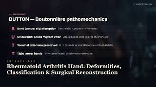
6:04
Rheumatoid Arthritis Hand: Deformities, Classification & Surgical Reconstruction
Rheumatoid arthritis affects the hand in 90% of patients, producing the classic deformities every clinician must recognise — ulnar drift, boutonnière, swan-neckand the Nalebuff thumb patterns. This masterclass walks you through the pathomechanics, mnemonicsand a logical proximal-to-distal surgical algorithm that corrects deforming forces from the wrist down to the fingertips. ⏱ Chapters 0:00 Intro 0:24 Essentials 0:58 Classification 1:30 Finger Deformities 2:09 Deformity Mechanics 2:40 Swan Neck 3:14 Tendon Ruptures 3:45 Algorithm 4:21 Complications 4:56 Clinical reasoning — Vj 5:27 Clinical reasoning — Mcp A comprehensive, clinically-focused masterclass on rheumatoid arthritis of the hand covering synovial pathophysiology, the 2010 ACR/EULAR diagnostic framework, characteristic deformities of the thumb, MCP joints and fingers, tendon attrition syndromes (Vaughan-Jackson and Mannerfelt), and the staged surgical reconstruction pathway from synovectomy through MCP arthroplasty to PIP arthrodesis. Designed to build a clear, structured approach for anyone managing inflammatory hand disease. 🦴 IN THIS VIDEO YOU'LL LEARN • The pathophysiology of rheumatoid pannus, synovial hypertrophy and the two-year DMARD window for preventing erosive damage • How to differentiate boutonnière deformity (PIP flexion, DIP extension) from swan-neck deformity (PIP hyperextension, DIP flexion) using the BUTTON and SWAN mnemonics • The Nalebuff classification of rheumatoid thumb deformities from Type I boutonnière through to Type VI arthritis mutilans • Staging and treatment thresholds for boutonnière and swan-neck finger deformities based on passive correction and angle of lag • Why MCP ulnar drift occurs — radial collateral ligament destruction, extensor subluxation and intrinsic pull • Vaughan-Jackson extensor tendon rupture sequence and Mannerfelt flexor pollicis longus attrition, plus the role of the Darrach procedure in caput ulnae syndrome • The proximal-to-distal surgical algorithm: medical optimisation, wrist correction first, then MCP arthroplasty with soft tissue rebalancing • The role of serology — rheumatoid factor sensitivity versus anti-CCP specificity — and key demographics including the 3:1 female predominance and HLA-DR4 association ⏱️ CHAPTERS 📌 KEY TAKEAWAYS ✔️ 90% of rheumatoid patients develop hand involvement — early DMARD therapy within 2 years is the single biggest determinant of outcome ✔️ Boutonnière arises from central slip disruption with volar lateral band migration; swan-neck from volar plate laxity, intrinsic tightness, FDS attenuation or flexor nodules ✔️ Fixed boutonnière deformity over 50° or chronic for 6–12 months is best treated with PIP arthrodesis in functional position, not arthroplasty ✔️ Vaughan-Jackson ruptures follow a predictable sequence (EDM → ring EDC → middle EDC) over a dorsally prominent ulnar head — address caput ulnae early to prevent further ruptures ✔️ Always correct the wrist before the fingers; extensor tendons must pass over a stable foundation or recurrent deformity is inevitable ✔️ Stop methotrexate two weeks before and after surgery to minimise infection riskand only achieve low disease activity before proceeding to reconstruction 👩⚕️ WHO THIS IS FOR Ideal for orthopaedic and hand surgery trainees, rheumatology clinicians, physiotherapists, medical studentsand informed patients seeking a deep understanding of how rheumatoid arthritis affects the hand and how it is reconstructed. 📚 RELATED TOPICS: Boutonnière Deformity Pathomechanics and Repair · Swan-Neck Deformity: Causes and Surgical Options · Vaughan-Jackson Syndrome and Extensor Tendon Rupture · Mannerfelt Lesion and Flexor Pollicis Longus Attrition · MCP Joint Arthroplasty in Rheumatoid Arthritis · Caput Ulnae Syndrome and the Darrach Procedure 🔗 More free orthopaedic teaching at Orthovellum.com — subscribe for weekly videos. 🎨 CREDITS • Original Orthovellum production. ⚠️ DISCLAIMER: Educational only — not individual medical advice. Always consult a qualified clinician for diagnosis and treatment. #orthopaedics #handsurgery #rheumatoidarthritis #rheumatology #handdeformity #boutonniere #swanneck #ulnardrift #DMARDs #MCParthroplasty #PIPfusion #Darrachprocedure #Nalebuffclassification #medicaleducation #orthopedics
19 Jun 2026

6:23
Lateral Epicondylitis (Tennis Elbow) Masterclass | ECRB Tendinosis, Cozen Test, PRP Treatment
Lateral epicondylitis — commonly called tennis elbow — is a degenerative tendinosis of the extensor carpi radialis brevis (ECRB) origin, not a true inflammation. This masterclass unpacks the pathogenesis, the Nirschl classification, the key clinical tests including Cozen and Maudsleyand the evidence-based management pathway from eccentric exercises through to surgical release. ⏱ Chapters 0:00 Intro 0:20 Essentials 0:57 Classification 1:29 Anatomy 2:01 Clinical Signs 2:42 Cozen Mnemonic 3:08 Management Mnemonic 3:39 Algorithm 4:20 Surgical Technique 4:54 Key Numbers 5:20 Clinical reasoning — 1 5:49 Clinical reasoning — 2 This comprehensive OrthoVellum masterclass covers lateral epicondylitis in depth, from the angiofibroblastic degeneration of the ECRB origin to conservative care, injection therapyand operative release. Learners gain a clear grasp of epidemiology (1–3% incidence, peak age 35–50), the common extensor origin anatomy, the COZEN and TENNIS mnemonics, the differential with radial tunnel syndromeand why platelet-rich plasma outperforms corticosteroid for durable outcomes. It is essential viewing for anyone managing elbow pain in clinic. 🦴 IN THIS VIDEO YOU'LL LEARN • Explain why lateral epicondylitis is a degenerative tendinosis rather than an inflammatory tendinitis • Identify the ECRB origin as the primary site of pathology and describe the biomechanics of overload • Apply the Nirschl classification to stage tendinosis severity and guide treatment decisions • Perform and interpret Cozen's test, Mill's testand Maudsley's test and differentiate tennis elbow from radial tunnel syndrome • Construct a structured conservative management plan using the TENNIS mnemonic and Tyler twist eccentric protocol • Compare the evidence for platelet-rich plasma versus corticosteroid injection in lateral epicondylitis • List the indications, techniquesand risks of surgical debridement of the ECRB origin • Recognise when imaging (USS or MRI) is appropriate and how to interpret ECRB tendon changes • Describe the risk factors, natural historyand occupational associations of tennis elbow ⏱️ CHAPTERS 📌 KEY TAKEAWAYS ✔️ Lateral epicondylitis is angiofibroblastic tendinosis of the ECRB, not inflammation — histology lacks prostaglandins ✔️ 80–90% of patients respond to conservative management; surgery is reserved for those failing 6–12 months ✔️ PRP is superior to corticosteroid for long-term relief; steroids only provide 4–8 weeks of benefit and increase recurrence ✔️ Maximum tenderness is 1 cm distal and anterior to the lateral epicondyle; PIN syndrome pain sits 4 cm distal ✔️ The underside of the ECRB origin is the Nirschl lesion — grey, friable, avascular degenerative tissue 👩⚕️ WHO THIS IS FOR This masterclass is designed for orthopaedic and sports medicine trainees, physiotherapists, general practitionersand allied health clinicians managing elbow pain, as well as informed patients seeking a deep understanding of their condition. 📚 RELATED TOPICS: Medial Epicondylitis (Golfer's Elbow) · Radial Tunnel Syndrome and PIN Entrapment · Common Extensor Origin Anatomy · Eccentric Exercise Protocols in Tendinopathy · Elbow Arthroscopy Portals and Indications · Corticosteroid Versus PRP Injections in Tendinopathy 🔗 More free orthopaedic teaching at Orthovellum.com — subscribe for weekly videos. 🎨 CREDITS • Original Orthovellum production. ⚠️ DISCLAIMER: Educational only — not individual medical advice. Always consult a qualified clinician for diagnosis and treatment. #orthopaedics #orthopedic #sportsmedicine #tenniselbow #lateralepicondylitis #ECRB #tendinosis #elbowpain #NirschlClassification #CozenTest #plateletrichplasma #elbowsurgery #medicaleducation #tendonpathology
19 Jun 2026

8:34
Mallet Finger Masterclass: Splinting Protocol, Surgery & Doyle Classification
Master the diagnosis and management of mallet finger, the classic Zone I extensor tendon injury. Learn the critical 6-8 week splinting protocol, the Doyle classificationand the surgical indications every clinician needs to know. ⏱ Chapters 0:00 Intro 0:23 Essentials 1:20 Classification 2:05 Anatomy 2:47 Swan Neck 3:22 Surgical Indications 3:55 Imaging 4:40 Differential 5:17 Algorithm 6:09 Ishiguro 6:48 Outcomes 7:25 Clinical reasoning — Chronic 7:56 Clinical reasoning — Bony This OrthoVellum masterclass provides a comprehensive deep dive into mallet finger injuries. We cover the underlying anatomy of the extensor apparatus, mechanism of injuryand the Doyle classification system. The video heavily emphasises clinical decision-making, specifically how to differentiate stable injuries that require strict continuous extension splinting from complex cases demanding surgical intervention like Ishiguro extension block pinning. 🦴 IN THIS VIDEO YOU'LL LEARN • How to classify mallet finger injuries using the Doyle classification system • The strict 6 to 8 week continuous extension splinting protocol and patient education • Identifying surgical indications using the LOSS mnemonic • The anatomical mechanism of swan neck deformity secondary to chronic mallet finger • Recognising and managing the paediatric Seymour fracture • How to perform and understand the principles of Ishiguro extension block pinning • Differentiating tendinous mallet from differentials like jersey finger and sagittal band rupture • Evaluating radiographs to determine articular fragment size and volar subluxation ⏱️ CHAPTERS 📌 KEY TAKEAWAYS ✔️ Splint first: almost all closed mallet injuries, including bony mallets, are treated with continuous extension splinting. ✔️ A single episode of DIPJ flexion during the splinting period completely resets the 6-8 week healing clock. ✔️ Volar subluxation of the distal phalanx or a large fracture fragment indicates an unstable joint requiring surgery. ✔️ Splint fit is crucial; dorsal skin necrosis is the most common complication of tight splints. ✔️ A residual extensor lag of less than 10 degrees is an excellent, highly functional outcome. 👩⚕️ WHO THIS IS FOR This video is designed for orthopaedic trainees, medical students, junior doctorsand hand therapists seeking a comprehensive understanding of hand trauma. Curious patients looking to understand their mallet finger diagnosis and treatment may also find it highly informative. 📚 RELATED TOPICS: Zone 1 Extensor Tendon Injuries · Swan Neck Deformity Pathophysiology · Paediatric Phalangeal Fractures · Jersey Finger (FDP Avulsion) · Hand Therapy and Splinting Protocols · Dorsal Hand Anatomy and Extensor Apparatus 🔗 More free orthopaedic teaching at Orthovellum.com — subscribe for weekly videos. 🎨 CREDITS • Original Orthovellum production. ⚠️ DISCLAIMER: Educational only — not individual medical advice. Always consult a qualified clinician for diagnosis and treatment. #orthopaedics #malletfinger #handsurgery #orthovellum #extensortendon #handtrauma #doyleclassification #medicaleducation #splinting #fracturefixation #anatomy #clinicals #bonfracture #surgeon #medicaleducation
19 Jun 2026

7:20
De Quervain Tenosynovitis Masterclass: Anatomy, Finkelstein Test & Treatment
De Quervain tenosynovitis is a stenosing condition of the first dorsal compartment causing significant radial wrist pain. In this Orthovellum masterclass, we explore the underlying myxoid degeneration, critical anatomical variations like the EPB septumand evidence-based management strategies. ⏱ Chapters 0:00 Intro 0:22 Essentials 0:56 Anatomy 1:33 Dorsal Compartments 2:06 Classification 2:36 Staging 3:14 Tests 3:55 Imaging 4:31 Algorithm 5:06 Surgical Technique 5:39 Complications 6:13 Clinical reasoning — Failure 6:40 Clinical reasoning — Injection This video provides a comprehensive overview of de Quervain tenosynovitis, moving beyond the historical misconception of primary inflammation to explain the true mechanical and degenerative pathophysiology. We cover everything from the classic presentation in postpartum women to the Shima anatomical classification, clinical severity stagingand the complete management algorithm from splinting and corticosteroid injections to surgical release. 🦴 IN THIS VIDEO YOU'LL LEARN • Understand the pathophysiology of myxoid degeneration and fibrocartilaginous thickening in the first dorsal compartment • Identify the contents of all six dorsal extensor compartments with a focus on the abductor pollicis longus and extensor pollicis brevis • Recognise the classic patient demographics, including the high prevalence in postpartum and lactating women • Perform the Finkelstein (Eichhoff) manoeuvre and the more specific WHAT test for clinical diagnosis • Differentiate de Quervain tenosynovitis from intersection syndrome, Wartenberg syndromeand basal thumb osteoarthritis • Apply the Shima cadaveric classification to identify the separate EPB subcompartment present in 30 to 50 percent of patients • Determine appropriate treatment pathways based on clinical severity staging from mild to severe disease • Execute the surgical release of the first dorsal compartment while protecting the superficial radial nerve • Prevent surgical failure by ensuring complete release of all subcompartments and septa ⏱️ CHAPTERS 📌 KEY TAKEAWAYS ✔️ De Quervain tenosynovitis is a degenerative stenosing tenovaginitis, not a primary inflammatory synovitis. ✔️ The Finkelstein test (fist over thumb with ulnar deviation) is pathognomonic and sufficient for clinical diagnosis. ✔️ Corticosteroid injection into the tendon sheath is the first-line therapy for moderate disease with a 70 to 80 percent success rate. ✔️ A separate septum creating a hidden subcompartment for the extensor pollicis brevis is present in up to 50 percent of patients. ✔️ Failing to identify and release a separate EPB subcompartment is the most common cause of failed surgical management. ✔️ The superficial radial nerve crosses the surgical field in roughly 60 percent of cases and must be carefully protected to prevent neuroma. 👩⚕️ WHO THIS IS FOR This masterclass is designed for orthopaedic trainees, junior doctorsand medical students seeking a high-yield understanding of hand and wrist pathology. It is also highly valuable for physiotherapists, general practitionersand curious patients wanting a detailed breakdown of radial wrist pain and its management. 📚 RELATED TOPICS: Intersection Syndrome of the Wrist · Wartenberg Syndrome (Superficial Radial Nerve Entrapment) · Basal Thumb Osteoarthritis · Dorsal Extensor Compartments of the Wrist · Corticosteroid Injections in Hand Surgery · Stenosing Tenosynovitis Pathophysiology 🔗 More free orthopaedic teaching at Orthovellum.com — subscribe for weekly videos. 🎨 CREDITS • Original Orthovellum production. ⚠️ DISCLAIMER: Educational only — not individual medical advice. Always consult a qualified clinician for diagnosis and treatment. #dequervain #tenosynovitis #orthopaedics #handsurgery #finkelsteintest #radialwristpain #orthovellum #anatomy #medicaleducation #extensorcompartments #wristpain
19 Jun 2026

5:45
Scaphoid Fractures Masterclass: Blood Supply, Herbert Classification & SNAC Wrist
Scaphoid fractures are the most common carpal bone injury, carrying a notorious risk of avascular necrosis and nonunion. Master the retrograde blood supply, Herbert classificationand acute management decisions in this comprehensive orthopaedic masterclass. ⏱ Chapters 0:00 Intro 0:19 Essentials 0:46 Blood Supply 1:19 Herbert 1:49 Location Risk 2:14 Panda 2:44 Snac 3:11 Occult Algorithm 3:38 Management 4:14 Numbers 4:40 Clinical reasoning — Occult 5:09 Clinical reasoning — Displaced This video provides an in-depth breakdown of scaphoid fractures, covering everything from unique carpal anatomy and the dorsal vascular ridge to the predictable pattern of SNAC wrist arthritic collapse. We explore the Herbert classification system, surgical indications using the PANDA mnemonic, occult fracture imaging trapsand the differences between conservative casting and operative fixation using a headless compression screw. 🦴 IN THIS VIDEO YOU'LL LEARN • The clinical significance of the scaphoid's retrograde blood supply from the dorsal carpal branch of the radial artery • How to apply the Herbert classification to differentiate stable and unstable acute fractures • Why 10-20% of scaphoid fractures are radiographically occult on initial X-rays • The five PANDA indications for operative surgical fixation • How fracture location dictates the risk of avascular necrosis and nonunion • The modern imaging gold standard of immediate MRI for clinically suspected but X-ray negative injuries • The predictable four-stage progression of SNAC (Scaphoid Nonunion Advanced Collapse) wrist • How to identify carpal instability and DISI patterns on lateral wrist radiographs • The principles of bone grafting and vascularised grafts for established scaphoid nonunion ⏱️ CHAPTERS 📌 KEY TAKEAWAYS ✔️ Proximal pole fractures have the highest risk of avascular necrosis due to their complete reliance on retrograde intraosseous blood flow. ✔️ Anatomic snuffbox tenderness with normal initial radiographs still warrants either immobilisation or an immediate MRI. ✔️ Herbert Type A fractures are stable and managed conservatively, whilst Type B fractures are inherently unstable and require surgical fixation. ✔️ Untreated nonunion predictably leads to SNAC wrist, progressing from the radial styloid to pancarpal arthritis. ✔️ Displacement greater than 1mm or a scapholunate angle over 60 degrees mandates operative intervention. 👩⚕️ WHO THIS IS FOR This masterclass is designed for orthopaedic trainees, junior doctorsand medical students seeking a comprehensive understanding of carpal injuries. It also serves as a highly detailed resource for curious patients wanting to learn more about the science behind scaphoid fracture management. 📚 RELATED TOPICS: Avascular Necrosis of the Proximal Pole · Scaphoid Nonunion Advanced Collapse (SNAC Wrist) · Herbert Screw Fixation Technique · Vascularised Bone Grafting for Scaphoid Nonunion · Perilunate and Trans-scaphoid Dislocations · Carpal Instability and DISI Patterns 🔗 More free orthopaedic teaching at Orthovellum.com — subscribe for weekly videos. 🎨 CREDITS • Original Orthovellum production. ⚠️ DISCLAIMER: Educational only — not individual medical advice. Always consult a qualified clinician for diagnosis and treatment. #orthopaedics #scaphoidfracture #orthovellum #handtrauma #carpalfracture #avascularnecrosis #herbertclassification #orthopedicsurgery #fooshinjury #snacwrist #bonesurgery #medicaleducation #orthoteaching
19 Jun 2026

6:04
Subtrochanteric Fractures Explained: Why the Nail Entry Point Decides the Outcome
Subtrochanteric fractures occur in the highest stress zone of the femur and present a unique surgical challenge due to the classic FEAR deformity. Master the biomechanics, Russell-Taylor classificationand the critical principle of reducing the fracture before reaming. ⏱ Chapters 0:00 Intro 0:21 Essentials 0:58 Fear 1:23 Russell Taylor 1:51 Seinsheimer 2:19 Biomechanics 2:52 Reduction 3:19 Algorithm 3:54 Atypical 4:27 Numbers 4:58 Clinical reasoning — Reduction 5:22 Clinical reasoning — Pearls This comprehensive orthopaedic masterclass provides an in-depth look at subtrochanteric femur fractures, exploring why they occur within five centimetres of the lesser trochanter and why powerful muscle forces cause the proximal fragment to displace. We cover the biomechanical reasons why intramedullary nails vastly outperform plates, how to utilise the Russell-Taylor and Seinsheimer classification systems to guide your surgical approachand the crucial steps for achieving and maintaining reduction using the BLOCKING mnemonic. 🦴 IN THIS VIDEO YOU'LL LEARN • Identify the unique anatomical and biomechanical properties of the subtrochanteric zone • Recognise the classic FEAR deformity (Flexed, Externally rotated, Abducted) caused by unopposed muscle pulls • Apply the Russell-Taylor classification to determine the correct nail entry point based on piriformis fossa integrity • Differentiate fracture stability and fragment patterns using the Seinsheimer classification • Understand why long cephalomedullary nails are the gold standard fixation compared to plates • Implement the critical surgical rule of confirming reduction on AP and lateral imaging before reaming • Utilise the BLOCKING mnemonic for surgical adjuncts including ball-spike pushers, cerclage wiresand blocking screws • Identify atypical bisphosphonate-associated fractures using the LATERAL BEAK criteria • Recognise the importance of accepting slight valgus alignment and preventing varus malunion ⏱️ CHAPTERS 📌 KEY TAKEAWAYS ✔️ The nail follows the reamer: always achieve and confirm reduction before reaming to avoid locking in a malreduction. ✔️ Match the leg position to the proximal fragment by flexing, abductingand slightly externally rotating to overcome the FEAR deformity. ✔️ Always use a long cephalomedullary nail to share load, reduce the lever armand protect the entire femur from stress risers. ✔️ If the piriformis fossa is fractured (Russell-Taylor Type II), you must switch to a trochanteric entry nail. ✔️ Accept slight valgus and NEVER accept varus; use blocking (Poller) screws proactively in the concavity to guide nail trajectory. ✔️ Be vigilant for atypical fractures in patients with over five years of bisphosphonate use, looking for lateral cortex beaking. 👩⚕️ WHO THIS IS FOR This masterclass is designed for orthopaedic surgical trainees, medical students, junior doctorsand clinicians seeking a comprehensive understanding of lower limb trauma. Curious patients with a background in human biology will also find this detailed breakdown highly informative. 📚 RELATED TOPICS: Atypical Bisphosphonate Femur Fractures · Russell-Taylor Classification System · Cephalomedullary Nailing Technique · Blocking Screws (Poller Screws) in Intramedullary Nailing · Proximal Femoral Anatomy and Muscular Attachments · Femoral Shaft Stress Fractures 🔗 More free orthopaedic teaching at Orthovellum.com — subscribe for weekly videos. 🎨 CREDITS • Original Orthovellum production. ⚠️ DISCLAIMER: Educational only — not individual medical advice. Always consult a qualified clinician for diagnosis and treatment. #orthopaedics #orthopedicsurgery #subtrochantericfracture #femurfracture #traumaorthopaedics #cephalomedullarynail #FEARdeformity #intramedullarynail #fracturefixation #orthoteaching #atypicalfracture #bonetrauma
19 Jun 2026

4:56
Pilon Fractures Explained: Distal Tibia Patterns, CT Planning & Fixation
Pilon fractures are complex intra-articular injuries of the distal tibial plafond where the soft tissue envelope dictates absolutely all timing decisions. Master the staged management of these high-energy injuries using the Span, Scan, Plan protocol to minimise wound complications and optimise articular reduction. ⏱ Chapters 0:00 Intro 0:19 Essentials 0:48 Classification 1:06 Ao 1:29 Span 1:53 Columns 2:14 Imaging 2:40 Algorithm 3:09 Fixation 3:36 Numbers 3:57 Clinical reasoning — Soft 4:24 Clinical reasoning — Pilon This comprehensive masterclass covers the diagnosis, classificationand definitive management of tibial plafond fractures. We explore the Ruedi-Allgower and AO/OTA classification systems, the three-column articular mapping conceptand the critical staged surgical protocol required to safely navigate the anteromedial watershed zone. From initial spanning external fixation through to definitive ORIF, you will learn the precise surgical approaches and fixation strategies necessary to manage these notoriously unforgiving orthopaedic injuries. 🦴 IN THIS VIDEO YOU'LL LEARN • How to classify pilon fractures using the Ruedi-Allgower and AO/OTA (43-C) systems • The importance of the anteromedial soft tissue envelope and how it dictates surgical timing • Applying the Span, Scan, Plan philosophy for staged orthopaedic management • Interpreting CT scans using the MAP framework of anteromedial, anterolateraland posterior columns • Identifying key anatomical fracture fragments including the Tillaux-Chaput and Volkmann fragments • The criteria for proceeding to definitive fixation, including the wrinkle sign and fracture blister management • Choosing the correct surgical approach based on the articular column requiring reconstruction • Understanding the back-to-front and lateral-to-medial sequence for articular block reconstruction • Managing severe open pilon fractures (Grade IIIB) and knowing when to consider primary arthrodesis ⏱️ CHAPTERS 📌 KEY TAKEAWAYS ✔️ Soft tissue is king: never plate through swollen or blistered skin ✔️ Syndesmosis is often intact in pilon fractures because the primary mechanism is axial loading, not rotation ✔️ A minimum 7cm skin bridge must be maintained between surgical incisions ✔️ Haemorrhagic blisters must be left intact until epithelialisation, while clear blisters can be incised carefully ✔️ Over 50% of patients develop post-traumatic arthritis, directly correlating with the quality of articular reduction 👩⚕️ WHO THIS IS FOR This masterclass is designed for orthopaedic trainees, junior doctorsand medical students seeking a deep understanding of lower limb trauma. Clinicians managing acute orthopaedic presentations and curious patients looking for detailed, high-level information on distal tibial injuries will also find it highly valuable. 📚 RELATED TOPICS: Rotational Ankle Fractures and Syndesmotic Injury · Tibial Plateau Fractures: Principles of Bicondylar Reconstruction · External Fixation Principles in Orthopaedic Trauma · Compartment Syndrome of the Leg and Foot · Post-Traumatic Osteoarthritis of the Ankle 🔗 More free orthopaedic teaching at Orthovellum.com — subscribe for weekly videos. 🎨 CREDITS • Original Orthovellum production. ⚠️ DISCLAIMER: Educational only — not individual medical advice. Always consult a qualified clinician for diagnosis and treatment. #orthopaedics #pilonfracture #orthopaedicsurgery #traumasurgery #anklefracture #orthopedics #tibialplafond #fracturefixation #orif #bonesurgery #medicaleducation #orthopedictrauma
19 Jun 2026

8:08
Acute Compartment Syndrome Masterclass: Diagnosis, 6 P's & Fasciotomy
Acute compartment syndrome is a true orthopaedic emergency where early recognition of raised compartment pressure and timely fasciotomy make all the difference. In this in-depth masterclass, we cover everything from the critical six-hour window to the subtle early signs of ischaemia. ⏱ Chapters 0:00 Intro 0:25 Essentials 1:08 Classification 1:51 Anatomy 2:25 Ps 3:01 Adsl 3:37 Cast 4:12 Delta 4:54 Algorithm 5:41 Technique 6:28 Numbers 6:59 Clinical reasoning — Delta 7:30 Clinical reasoning — Pearls This comprehensive video explores the pathophysiology, clinical diagnosisand surgical management of acute compartment syndrome across all major anatomical regions. We delve into the critical pressure thresholds, the progressive 'six P's' clinical signsand the regional fasciotomy techniques required to prevent irreversible muscle necrosis and devastating contractures. 🦴 IN THIS VIDEO YOU'LL LEARN • How to diagnose acute compartment syndrome clinically without delaying fasciotomy for pressure measurement • The critical pressure thresholds for fasciotomy, including absolute pressure over 30mmHg and delta P under 30 • Why tibial shaft fractures are the most common cause and how ischaemia-reperfusion injury contributes • The six P's of compartment syndrome and why pain on passive stretch is the most sensitive early sign • How to identify and release the deep posterior compartment of the leg to avoid incomplete fasciotomy • The regional anatomy of the leg, forearm, thigh, handand foot compartments • Why a palpable pulse does not rule out compartment syndrome due to its microvascular nature • How to stage severity from impending to established or necrotic compartment syndrome • The devastating consequences of delayed diagnosis, including Volkmann's ischaemic contracture ⏱️ CHAPTERS 📌 KEY TAKEAWAYS ✔️ Acute compartment syndrome is a clinical diagnosis; never delay fasciotomy for pressure readings if suspicion is high. ✔️ Fasciotomy within six hours offers the best outcomes; irreversible muscle necrosis begins after eight hours. ✔️ Pain out of proportion to the injury and pain on passive stretch are the earliest and most reliable indicators. ✔️ The deep posterior compartment is the most commonly missed structure during a leg fasciotomy. ✔️ Pulses remain palpable until extremely late because compartment syndrome is a microvascular problem. ✔️ Always remove tight casts or dressings immediately if compartment syndrome is suspected. 👩⚕️ WHO THIS IS FOR This masterclass is essential viewing for orthopaedic trainees, junior doctorsand medical students. It also provides valuable insights for clinicians managing acute trauma and curious patients wanting to understand this limb-threatening surgical emergency. 📚 RELATED TOPICS: Fasciotomy surgical techniques and incisions · Volkmann's ischaemic contracture pathophysiology · Tibial shaft fracture complications and management · Chronic exertional compartment syndrome · Crush syndrome and ischaemia-reperfusion injury 🔗 More free orthopaedic teaching at Orthovellum.com — subscribe for weekly videos. 🎨 CREDITS • Original Orthovellum production. ⚠️ DISCLAIMER: Educational only — not individual medical advice. Always consult a qualified clinician for diagnosis and treatment. #compartmentSyndrome #orthopaedics #fasciotomy #trauma #orthopaedicsurgery #orthopaedicemergency #acutecompartmentsyndrome #surgicaleducation #medicalstudent #tibialfracture #clinicalsigns
19 Jun 2026

6:25
Talus Fractures Masterclass: Hawkins Classification, AVN & Blood Supply
Talus fractures are high-energy, unforgiving injuries where a precarious blood supply dictates outcomes. Master the Hawkins classification, talar blood supplyand urgent reduction protocols to prevent avascular necrosis. ⏱ Chapters 0:00 Intro 0:18 Essentials 0:58 Blood Supply 1:35 Classification 2:16 Imaging 3:02 Algorithm 3:42 Approach 4:28 Numbers 5:06 Clinical reasoning — Avascular 5:47 Clinical reasoning — Signs This in-depth OrthoVellum masterclass covers everything you need to know about managing talus fractures. We explore the unique ligamentous blood supply, the progressive AVN risks outlined in the Hawkins classificationand critical imaging findings like the Canale view and Hawkins sign. The video also breaks down surgical approaches, including the anteromedial workhorse incisionand provides clear protocols for emergency reduction and ORIF timing. 🦴 IN THIS VIDEO YOU'LL LEARN • How to apply the Hawkins classification to stratify avascular necrosis risk • The three main arteries supplying the talus and how displaced fractures disrupt them • Why urgent reduction within 6 to 12 hours is critical for skin and neurovascular safety • How to correctly perform and interpret the Canale view for talar neck fractures • The clinical significance of Hawkins sign at 6 to 8 weeks post-injury • Why the absence of Hawkins sign does not confirm avascular necrosis • When to use non-operative management versus ORIF with screws • How to execute the anteromedial approach and dual-incision strategy for fixation • The mechanisms of high-energy dorsiflexion injuries and open fracture management ⏱️ CHAPTERS 📌 KEY TAKEAWAYS ✔️ AVN risk is dictated by initial fracture displacement and the number of dislocated joints. ✔️ The artery of the tarsal canal is the dominant blood supply to the talar body. ✔️ Hawkins sign indicates revascularisation and makes AVN unlikely, but its absence requires further follow-up. ✔️ Hawkins III and IV fractures are surgical emergencies requiring immediate reduction. ✔️ Varus malunion is the most common deformity following talar neck fractures. 👩⚕️ WHO THIS IS FOR This masterclass is designed for orthopaedic trainees, medical students, junior doctorsand clinicians seeking a comprehensive refresher on foot and ankle trauma, as well as informed patients curious about complex orthopaedic injuries. 📚 RELATED TOPICS: Talar body fractures and the Sneppen classification · Snowboarder's fracture (lateral process of the talus) · Subtalar dislocation reduction techniques · Post-traumatic ankle arthritis and arthrodesis · Osteochondral lesions of the talar dome 🔗 More free orthopaedic teaching at Orthovellum.com — subscribe for weekly videos. 🎨 CREDITS • Original Orthovellum production. ⚠️ DISCLAIMER: Educational only — not individual medical advice. Always consult a qualified clinician for diagnosis and treatment. #orthopaedics #talusfracture #footandankle #hawkinsclassification #avascularnecrosis #orthopaedicsurgery #talarneckfracture #trauma #hawkinssign #footsurgery #orthoteaching #medicaleducation
19 Jun 2026

5:43
Patella Fractures Masterclass: Tension Band Fixation & Extensor Mechanism
Patella fractures account for 1% of all skeletal injuries and are critical to knee extension biomechanics. Master the tension band principle, AO classificationand surgical decision-making for displaced patellar fractures. ⏱ Chapters 0:00 Intro 0:24 Essentials 0:57 Classification 1:27 Ao 1:57 Gaps 2:27 Lever 2:58 Biomechanics 3:28 Algorithm 4:02 Numbers 4:33 Clinical reasoning — Extension 5:02 Clinical reasoning — Implant This Orthovellum masterclass provides a comprehensive deep dive into the assessment, classificationand management of patella fractures. We explore the biomechanics of the extensor mechanism, the GAPS mnemonic for operative indicationsand how tension band wiring converts destructive tensile forces into healing articular compression. 🦴 IN THIS VIDEO YOU'LL LEARN • How to classify patella fractures using both descriptive patterns and the AO/OTA (bone 34) system • The biomechanical function of the patella using the LEVER mnemonic to understand extensor power • How to apply the GAPS mnemonic to identify crucial operative indications for patellar fractures • The mechanical principles of tension band wiring and how it converts tensile forces into dynamic compression • Why testing active knee extension before administering analgesia is a critical clinical step • The differences between managing transverse fractures, vertical splitsand highly comminuted patterns • Decision-making frameworks for choosing between conservative cylinder casting and ORIF • The clinical importance of the medial and lateral retinacular tissues during fracture assessment • Indications and techniques for partial patellectomy versus hardware fixation in severe injuries ⏱️ CHAPTERS 📌 KEY TAKEAWAYS ✔️ Operative fixation is indicated if fracture displacement exceeds 2-3mm or articular step-off exceeds 2mm ✔️ Always assess active straight leg raise BEFORE giving analgesia to avoid masking a disrupted extensor mechanism ✔️ Tension band fixation works by converting anterior tensile forces into beneficial articular compression during knee flexion ✔️ Around 50% of patients require hardware removal after K-wire fixation due to soft tissue prominence ✔️ Vertical patella fractures are often inherently stable and can typically be managed conservatively ✔️ Intact retinacular expansions can sometimes allow a false-negative straight leg raise in a fractured patella 👩⚕️ WHO THIS IS FOR This masterclass is designed for orthopaedic surgical trainees, junior doctorsand medical students seeking a deep understanding of lower limb trauma. Curious patients looking to understand the biomechanics and surgical rationale behind kneecap fractures will also find it highly informative. 📚 RELATED TOPICS: Quadriceps and patellar tendon ruptures · Tension band principles in olecranon fractures · Bipartite patella differentiation · Total patellectomy consequences and rehabilitation · Patellofemoral joint biomechanics and contact forces · Cannulated screw fixation techniques in orthopaedics 🔗 More free orthopaedic teaching at Orthovellum.com — subscribe for weekly videos. 🎨 CREDITS • Original Orthovellum production. ⚠️ DISCLAIMER: Educational only — not individual medical advice. Always consult a qualified clinician for diagnosis and treatment. #orthopaedics #patellafracture #kneefracture #extensormechanism #tensionband #orthovellum #traumasurgery #kneebiomechanics #orthopedicsurgery #AOclassification #fracturefixation #medicaleducation #kneeinjury
19 Jun 2026
6:13
Acute Compartment Syndrome Explained: The 5 Ps, Pressure Thresholds & the Fasciotomy Decision
Both-bone forearm fractures demand anatomic reduction to restore the critical mechanical axis of rotation. Master the principles of ORIF plating, radial bow restorationand compartment syndrome vigilance in this orthopaedic masterclass. ⏱ Chapters 0:00 Intro 0:26 Essentials 1:10 Classification 1:50 Pattern 2:19 Ao 2:51 Mnemonic 3:31 Algorithm 4:17 Numbers 4:50 Clinical reasoning — 1 5:37 Clinical reasoning — 2 This video provides a comprehensive deep dive into the evaluation, classificationand surgical management of both-bone forearm fractures in adults. We cover why anatomical reduction is non-negotiable for pronation and supination, how to utilise the AO/OTA classification and FOREARM mnemonic to guide your fixation strategyand the critical importance of identifying high-risk complications like compartment syndrome and radioulnar synostosis. 🦴 IN THIS VIDEO YOU'LL LEARN • How to apply the FOREARM mnemonic for orthogonal plating and fracture fixation principles • The surgical indications and biomechanical rationale for using 3.5mm LC-DCP or locking plates • Techniques to meticulously restore the magnitude and location of the natural radial bow • How fracture geometry dictates the use of compression, lag screwor bridge plating constructs • Selecting the correct surgical approach based on fracture location along the radial and ulnar shafts • Recognising and surgically managing compartment syndrome in high-energy forearm trauma • Identifying fracture-dislocation patterns including Galeazzi and Monteggia variants • Understanding the AO/OTA alphanumeric classification system for forearm injuries ⏱️ CHAPTERS 📌 KEY TAKEAWAYS ✔️ Anatomic reduction is mandatory; rotational malalignment greater than 10 degrees significantly impairs forearm function. ✔️ Achieve a minimum of six cortices (three screws) on each side of the fracture for both the radius and ulna. ✔️ Maximum radial bow is located at the proximal-middle junction and must be restored to optimise supination. ✔️ Use orthogonal plating 90 degrees apart to minimise the risk of radioulnar synostosis. ✔️ Compartment syndrome is a clinical diagnosis carrying a 2-5% risk in these high-energy injuries. 👩⚕️ WHO THIS IS FOR This masterclass is designed for orthopaedic surgical trainees, junior doctorsand medical students. Curious patients looking for an in-depth understanding of adult forearm fracture surgery will also find this educational. 📚 RELATED TOPICS: AO Principles of Internal Fixation · Forearm Anatomy and Biomechanics · Monteggia and Galeazzi Fracture-Dislocations · Compartment Syndrome of the Forearm · Radioulnar Synostosis Prevention · Surgical Approaches to the Forearm 🔗 More free orthopaedic teaching at Orthovellum.com — subscribe for weekly videos. 🎨 CREDITS • Original Orthovellum production. ⚠️ DISCLAIMER: Educational only — not individual medical advice. Always consult a qualified clinician for diagnosis and treatment. #orthopaedics #orthovellum #bothbonefracture #forearmfracture #orif #traumasurgery #compartmentsyndrome #orthopaedicsurgery #aoortho #fracturefixation #radialbow #bonetrauma #surgicalmasterclass
19 Jun 2026

8:29
Humeral Shaft Fractures Explained: Functional Bracing, Plating & the Radial Nerve
Master the clinical assessment and management of humeral shaft fractures with this comprehensive orthopaedic masterclass. Learn exactly how to evaluate radial nerve function using the WEST mnemonic and confidently navigate the notoriously tricky Holstein-Lewis fracture pattern. ⏱ Chapters 0:00 Intro 0:24 Essentials 1:04 Ao 1:47 Deformity 2:25 West 3:00 Holstein 3:40 Alignment 4:16 Stopbrace 5:02 Algorithm 5:52 Implants 6:39 Numbers 7:18 Clinical reasoning — Primary 7:49 Clinical reasoning — Secondary This video delivers an in-depth, keyword-rich guide to humeral diaphyseal fractures, covering everything from core epidemiology and AO/OTA classification to muscular deforming forces and acceptable alignment parameters. Whether you are deciding between functional bracing and operative fixationor determining when to explore a radial nerve palsy, this masterclass provides the critical orthopaedic knowledge you need for optimal patient outcomes. 🦴 IN THIS VIDEO YOU'LL LEARN • How to confidently classify humeral shaft fractures using the AO/OTA 12-A, Band C system • The muscular deforming forces acting on the proximal and distal fragments at different fracture levels • How to systematically examine the radial nerve using the WEST mnemonic before and after manipulation • The specific clinical features and high nerve risk associated with the Holstein-Lewis distal third spiral fracture • The acceptable alignment limits for non-operative management, including the 20-30-3-15 rule • The indications for surgical stabilisation using the STOP BRACE criteria • Why functional bracing remains the gold standard, achieving union in over ninety percent of compliant patients • The critical difference in management between a primary radial nerve palsy and a secondary palsy ⏱️ CHAPTERS 📌 KEY TAKEAWAYS ✔️ Functional bracing achieves over 90% union rates; the humerus is remarkably forgiving due to shoulder and elbow compensation. ✔️ Always document radial nerve function (wrist and finger extension) before AND after any fracture manipulation. ✔️ A secondary radial nerve palsy developing after manipulation requires urgent exploration, whereas a primary palsy is observed for 3-4 months. ✔️ The Holstein-Lewis fracture (distal third lateral spiral) carries a 22-32% risk of radial nerve palsy and strongly warrants consideration for ORIF. ✔️ Tolerable angulation is generous: up to 20° sagittal, 30° coronal, 3cm shorteningand 15° rotation. 👩⚕️ WHO THIS IS FOR This masterclass is essential viewing for orthopaedic trainees, junior doctorsand medical students seeking a high-yield understanding of upper limb trauma. Interested patients looking to understand their humeral fracture and the rationale behind bracing or surgery will also find it highly informative. 📚 RELATED TOPICS: Radial nerve palsy and posterior interosseous nerve anatomy · Principles of functional bracing in orthopaedic trauma · Open reduction internal fixation (ORIF) techniques for the humerus · Intramedullary nailing vs plating for humeral diaphyseal fractures · Floating elbow injuries and polytrauma management · Upper limb peripheral nerve examination 🔗 More free orthopaedic teaching at Orthovellum.com — subscribe for weekly videos. 🎨 CREDITS • Original Orthovellum production. ⚠️ DISCLAIMER: Educational only — not individual medical advice. Always consult a qualified clinician for diagnosis and treatment. #orthopaedics #orthopedicsurgery #humeralshaftfracture #trauma #radialnerve #functionalbracing #holsteinlewis #orthoteaching #medicaleducation #fracturefixation #anatomy #bonetracture #upperlimb
19 Jun 2026

6:22
Femoral Shaft Fractures Masterclass: Intramedullary Nailing, Rotation & Polytrauma
Learn the definitive approach to femoral shaft fractures in this orthopaedic masterclass. Master intramedullary nailing techniques, including antegrade versus retrograde entry points, Winquist-Hansen classificationand damage control orthopaedics in polytrauma. ⏱ Chapters 0:00 Intro 0:24 Essentials 0:59 Anatomy 1:32 Winquist 1:59 Retrograde 2:30 Entry 3:03 Rotation 3:33 Algorithm 4:06 Neckscreen 4:37 Numbers 5:06 Clinical reasoning — 1 5:35 Clinical reasoning — 2 This comprehensive video provides an in-depth look at the evaluation and surgical management of femoral shaft fractures. We cover critical anatomical deforming forces, the Winquist-Hansen classification for guiding locking strategyand essential clinical decision-making for the physiologically unstable polytrauma patient. Gain confidence in choosing the correct entry point, avoiding avascular necrosisand preventing the common pitfall of malrotation. 🦴 IN THIS VIDEO YOU'LL LEARN • How to apply the Winquist-Hansen classification to determine static versus dynamic locking strategies • The decision-making process for antegrade versus retrograde intramedullary nailing • How to identify and manage the five percent of patients with an ipsilateral femoral neck fracture • The principles of damage control orthopaedics versus early total care in polytrauma • Intraoperative techniques using the CORTICES mnemonic to assess and prevent malrotation • Why the trochanteric entry point is now preferred over the piriformis fossa • The predictable muscle deforming forces acting on proximal and distal third fractures • How to anticipate and manage the one to one and a half litres of hidden blood loss in closed fractures ⏱️ CHAPTERS 📌 KEY TAKEAWAYS ✔️ Reamed, locked intramedullary nailing within twenty-four hours is the gold standard for stable patients. ✔️ If the patient is physiologically unstable, apply a spanning external fixator for damage control orthopaedics. ✔️ Always fix the ipsilateral femoral neck fracture before nailing the shaft. ✔️ External rotation malreduction is the most common technical error; always compare clinically with the contralateral leg. ✔️ Avoid the piriformis entry point in children and use caution in young adults to prevent capital femoral avascular necrosis. 👩⚕️ WHO THIS IS FOR This masterclass is designed for orthopaedic surgical trainees, junior doctorsand medical students building their trauma knowledge. Curious patients looking for a detailed, high-level understanding of their femur fracture surgery will also find it highly informative. 📚 RELATED TOPICS: Subtrochanteric femur fractures and deforming forces · Damage control orthopaedics and the lethal triad · Floating knee injuries and ipsilateral tibial shaft fractures · Femoral neck fracture fixation techniques · Intramedullary nail biomechanics and static versus dynamic locking 🔗 More free orthopaedic teaching at Orthovellum.com — subscribe for weekly videos. 🎨 CREDITS • Original Orthovellum production. ⚠️ DISCLAIMER: Educational only — not individual medical advice. Always consult a qualified clinician for diagnosis and treatment. #orthopaedics #femoralshaftfracture #trauma #intramedullarynailing #orthopaedicsurgery #femurfracture #damagecontrol #polytrauma #winquisthansen #orthoteaching #medicalstudent #fracturefixation
19 Jun 2026

6:49
Tibial Shaft Fractures Masterclass: Classification, Nailing & Compartment Syndrome
Tibial shaft fractures are the most common long bone fracture in orthopaedic practice, presenting unique challenges due to their high risk of open injuries and compartment syndrome. This comprehensive masterclass covers everything from the Gustilo-Anderson classification to intramedullary nailing and acceptable alignment. ⏱ Chapters 0:00 Intro 0:28 Essentials 1:03 Gustilo 1:37 Ao 2:07 Alignment 2:39 Compartments 3:07 Fiveps 3:35 Abx 3:59 Algorithm 4:38 Fasciotomy 5:11 Numbers 5:42 Clinical reasoning — Compartment 6:11 Clinical reasoning — Fixation Master the complete management of tibial shaft fractures with this in-depth orthopaedic review. We explore the critical anatomy of the tibial shaft and leg compartments, fracture classifications (AO/OTA and Gustilo-Anderson), and the crucial clinical assessment protocols for identifying compartment syndrome early. The video also details the orthopaedic management paradigm, covering antibiotic selection, the 5-5-10-10 alignment ruleand surgical decision-making for intramedullary nailing versus external fixation. 🦴 IN THIS VIDEO YOU'LL LEARN • How to classify tibial shaft fractures using the AO/OTA 42-series and Gustilo-Anderson systems • The four fascial compartments of the leg using the ALPD mnemonic • How to recognise the early and late clinical signs of compartment syndrome using the 5 Ps • The 5-5-10-10 rule for acceptable fracture reduction and alignment • Selecting the correct antibiotic prophylaxis for open fractures using the GAP mnemonic • Why intramedullary nailing is the gold standard fixation for tibial shaft fractures • The critical time windows for administering IV antibiotics and performing surgical debridement • How to manage severe Gustilo Type IIIB and IIIC open fractures requiring flap coverage or vascular repair • The anatomical reasons why the tibia has the highest open fracture rate of any long bone ⏱️ CHAPTERS 📌 KEY TAKEAWAYS ✔️ The tibia has the highest open fracture rate of any long bone (25%) due to its subcutaneous anteromedial border. ✔️ The anterior compartment is typically the first to fail in compartment syndrome; pain out of proportion is the earliest clinical sign. ✔️ A reamed locked intramedullary nail is the treatment of choice for most tibial shaft fractures. ✔️ Use the GAP mnemonic for antibiotics: Cephalosporin, Add aminoglycoside for Type III, Penicillin for farm contamination. ✔️ Automatic Gustilo Type III upgrades include high-energy mechanism, vascular injury, farm contaminationor segmental fractures. 👩⚕️ WHO THIS IS FOR This masterclass is designed for orthopaedic trainees, medical studentsand practising clinicians seeking a comprehensive understanding of lower limb trauma. Curious patients looking for an in-depth, educational perspective on tibial fracture management will also find it highly informative. 📚 RELATED TOPICS: Compartment Syndrome of the Lower Leg · Gustilo-Anderson Open Fracture Classification · Intramedullary Nailing Techniques · Tibial Plateau Fractures · AO/OTA Fracture Classification System · Open Fracture Management Protocols 🔗 More free orthopaedic teaching at Orthovellum.com — subscribe for weekly videos. 🎨 CREDITS • Original Orthovellum production. ⚠️ DISCLAIMER: Educational only — not individual medical advice. Always consult a qualified clinician for diagnosis and treatment. #orthopaedics #tibialfracture #trauma #orthopaedicsurgery #compartmentsyndrome #fracturefixation #gustiloanderson #intramedullarynail #medicaleducation #bonesurgery #tibialshaft #openfracture
19 Jun 2026

8:28
Metastatic Bone Disease Masterclass: BPLTK Primaries, Mirels Score & Surgery
Metastatic bone disease is the most common malignancy affecting the skeleton, far exceeding primary bone tumours. In this comprehensive orthopaedic masterclass, we cover everything from the classic BPLTK primary tumours and radiographic patterns to critical surgical decision-making using the Mirels score. ⏱ Chapters 0:00 Intro 0:25 Essentials 1:13 Classification 1:54 Bpltk 2:24 Radiographic 3:05 Mirels 3:39 Mirels Algorithm 4:21 Management 5:22 Embolization 5:53 Cord Compression 6:48 Clinical reasoning — Proximal-Femur 7:36 Clinical reasoning — Unknown-Primary This video provides an in-depth, clinically focused review of metastatic bone disease for orthopaedic learners. We explore the underlying pathophysiology, including Batson's venous plexus and the vicious cycle of osteoclast and osteoblast activation. You will learn how to identify lytic, blasticand mixed lesions, interpret the Mirels scoring system for impending pathological fracturesand understand the critical pre-operative requirement of embolisation for highly vascular kidney and thyroid metastases. The overarching goal of management is palliationand we detail how to achieve durable fixation that allows immediate weight-bearing for patients. 🦴 IN THIS VIDEO YOU'LL LEARN • Identify the five most common primary tumours that metastasise to bone using the BPLTK mnemonic • Differentiate between lytic, blasticand mixed radiographic appearances and their underlying cellular biology • Calculate and interpret the Mirels scoring system to assess impending pathological fracture risk • Understand the indications and thresholds for prophylactic orthopaedic fixation • Recognise the critical importance of pre-operative embolisation for renal and thyroid bone metastases • Explain the anatomical and physiological mechanisms of bone spread, including Batson's valveless venous plexus • Identify the clinical red flags and emergency management of metastatic spinal cord compression • Appreciate the palliative surgical goals of metastatic bone disease management ⏱️ CHAPTERS 📌 KEY TAKEAWAYS ✔️ Metastatic bone disease is 25-30 times more common than primary bone tumours. ✔️ Kidney and thyroid metastases are highly vascular and require embolisation 24-48 hours before surgery. ✔️ Prostate classically causes blastic lesions, while kidney, thyroidand lung cause lytic destruction. ✔️ A Mirels score of 9 or higher indicates a high fracture risk and warrants prophylactic fixation. ✔️ Orthopaedic constructs for metastatic disease must be durable and allow immediate weight-bearing. 👩⚕️ WHO THIS IS FOR This masterclass is highly relevant for orthopaedic trainees, junior doctorsand medical students seeking a comprehensive understanding of skeletal metastases. Interested patients and caregivers looking to understand the orthopaedic management of secondary bone tumours will also find this educational. 📚 RELATED TOPICS: Mirels Scoring System for Pathological Fractures · Primary Bone Tumours vs Secondary Bone Metastases · Orthopaedic Management of Spinal Cord Compression · Batson's Venous Plexus and Tumour Spread · Pre-operative Embolisation in Orthopaedic Oncology · The Vicious Cycle of Bone Destruction 🔗 More free orthopaedic teaching at Orthovellum.com — subscribe for weekly videos. 🎨 CREDITS • Original Orthovellum production. ⚠️ DISCLAIMER: Educational only — not individual medical advice. Always consult a qualified clinician for diagnosis and treatment. #orthopaedics #metastaticbonedisease #orthopedicsurgery #mirelsscore #bonetumour #pathologicalfracture #orthopaedicsurgery #medicaleducation #orthooncology #skeletalmetastases #bonemetastasis #orthopaedicteaching
18 Jun 2026
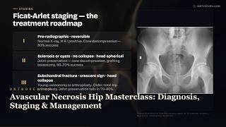
6:31
Avascular Necrosis Hip Masterclass: Diagnosis, Staging & Management
Avascular necrosis (AVN) of the hip is a devastating cause of progressive femoral head osteonecrosis in young adults. Learn the critical staging, pathophysiologyand surgical management strategies to determine when to preserve the joint versus when to replace it. ⏱ Chapters 0:00 Intro 0:29 Essentials 1:01 Aseptic 1:38 Medal 2:12 Ficat 2:42 Steinberg 3:09 Algorithm 3:43 Drill 4:11 Revision 4:42 Numbers 5:15 Clinical reasoning — Manage 5:46 Clinical reasoning — Pearls This OrthoVellum masterclass provides a comprehensive deep dive into the evaluation and management of avascular necrosis of the femoral head. Covering everything from the tenuous vascular anatomy and ASEPTIC risk factors to Ficat-Arlet and Steinberg staging systems, this video outlines the exact clinical thresholds for joint preservation versus total hip arthroplasty. 🦴 IN THIS VIDEO YOU'LL LEARN • How to identify the critical radiographic crescent sign indicating imminent femoral head collapse • Using the ASEPTIC mnemonic to categorise the diverse causes of osteonecrosis • The vascular anatomy of the femoral head and why retinacular vessel disruption is catastrophic • Applying the Ficat-Arlet and Steinberg classification systems to guide orthopaedic treatment • Indications and biomechanical principles of core decompression for pre-collapse AVN • Determining when to perform a valgus osteotomy versus a total hip arthroplasty • Why lesion size drastically affects the prognosis of joint-preserving surgery • The unique orthopaedic considerations and peri-operative protocols for sickle cell disease patients ⏱️ CHAPTERS 📌 KEY TAKEAWAYS ✔️ Always image the contralateral hip as 50-80% of AVN cases are bilateral within two years. ✔️ The crescent sign on X-ray signals a subchondral fracture, marking the failure point for joint preservation. ✔️ Core decompression works by reducing intraosseous pressure from over 30mmHg to under 10mmHg, but only works before head collapse. ✔️ Medial circumflex femoral artery provides 70-80% of blood supply; its disruption in femoral neck fractures causes a 30% AVN rate. ✔️ Total hip arthroplasty in AVN patients has higher failure and revision rates than in primary osteoarthritis. 👩⚕️ WHO THIS IS FOR This masterclass is essential for orthopaedic trainees, junior doctorsand medical students seeking a comprehensive understanding of adult reconstruction. Curious patients suffering from hip pain or osteonecrosis will also find valuable insights into their condition and surgical options. 📚 RELATED TOPICS: Femoral Neck Fractures and Fixation · Total Hip Arthroplasty in Young Adults · Core Decompression Surgical Technique · Vascularised Fibular Grafting for AVN · Steinberg and ARCO Classification of Osteonecrosis 🔗 More free orthopaedic teaching at Orthovellum.com — subscribe for weekly videos. 🎨 CREDITS • Original Orthovellum production. ⚠️ DISCLAIMER: Educational only — not individual medical advice. Always consult a qualified clinician for diagnosis and treatment. #orthopaedics #avascularnecrosis #osteonecrosis #hippain #femoralhead #jointpreservation #adultreconstruction #orthovellum #coredecompression #totalhiparthroplasty #orthopedicsurgery #crescentsign #femoralneckfracture #medicalstudent #orthopedics
18 Jun 2026

8:14
Femoroacetabular Impingement Masterclass: CAM, Pincer, Alpha Angle & FADIR
Femoroacetabular impingement (FAI) is a major cause of anterior hip pain and early osteoarthritis in young, active patients. In this Orthovellum masterclass, we break down everything you need to know about CAM and pincer morphology, alpha angle measurementand clinical management. ⏱ Chapters 0:00 Intro 0:23 Essentials 1:03 Pathophysiology 1:39 Classification 2:17 Cam Mnemonic 2:41 Pincer Mnemonic 3:13 Clinical Signs 3:48 Fadir Mnemonic 4:18 Imaging 4:56 Algorithm 5:39 Scope Mnemonic 6:12 Numbers 6:48 Clinical reasoning — 1 7:30 Clinical reasoning — 2 This video provides a comprehensive, structured deep dive into the mechanical pathologies, morphological classificationsand surgical decision-making for femoroacetabular impingement. Viewers will understand the distinct outside-in and inside-out damage mechanisms of CAM and pincer lesions, learn how to confidently interpret diagnostic imaging like alpha angles and crossover signsand master the clinical bedside examination using the FADIR test. 🦴 IN THIS VIDEO YOU'LL LEARN • Define femoroacetabular impingement and its role in driving early hip osteoarthritis • Differentiate CAM morphology from pincer morphology based on mechanical pathophysiology • Identify the diagnostic threshold for an alpha angle greater than 55 degrees on lateral imaging • Recognise the radiographic signs of acetabular overcoverage, including the crossover sign and os acetabuli • Understand why 85% of symptomatic FAI cases present with a mixed morphology requiring combined surgical approaches • Perform the FADIR test accurately and understand its exceptional 94-99% clinical sensitivity • Explain the difference between outside-in anterosuperior cartilage delamination and inside-out posteroinferior contre-coup tears • Determine appropriate patient selection for hip arthroscopy versus conservative management ⏱️ CHAPTERS 📌 KEY TAKEAWAYS ✔️ Femoroacetabular impingement requires a triad for diagnosis: characteristic symptoms, clinical signsand confirming imaging. ✔️ CAM lesions drive outside-in shear forces causing anterosuperior labral damage, while pincer lesions cause inside-out posteroinferior contre-coup tears. ✔️ An alpha angle exceeding 55 degrees is the critical radiographic diagnostic threshold for CAM morphology. ✔️ The FADIR test (flexion, adduction, internal rotation) is the gold standard clinical provocative manoeuvre with 94-99% sensitivity. ✔️ Mixed morphology is the rule rather than the exception, occurring in 85% of symptomatic FAI cases. 👩⚕️ WHO THIS IS FOR This masterclass is an essential resource for orthopaedic trainees, medical studentsand practising clinicians seeking a structured understanding of hip pathology, as well as inquisitive patients wanting to deeply understand their FAI diagnosis and surgical options. 📚 RELATED TOPICS: Hip arthroscopy surgical techniques and indications · Acetabular labral tears and chondral damage repair · Developmental dysplasia of the hip (DDH) · Slipped capital femoral epiphysis (SCFE) and Perthes disease · Early onset hip osteoarthritis and joint preservation · The Warwick agreement on femoroacetabular impingement syndrome 🔗 More free orthopaedic teaching at Orthovellum.com — subscribe for weekly videos. 🎨 CREDITS • Original Orthovellum production. ⚠️ DISCLAIMER: Educational only — not individual medical advice. Always consult a qualified clinician for diagnosis and treatment. #orthopaedics #femoroacetabularimpingement #fai #hiparthroscopy #hipsurgery #orthopedicsurgery #sportsmedicine #camlesion #pincerimpingement #hippain #alphaangle #fadirstest #medicalstudent #orthoteaching
18 Jun 2026

7:49
Patellofemoral Instability Masterclass: MPFL, Dysplasia & Surgical Management
Master the complexities of patellofemoral instability in this comprehensive orthopaedic masterclass. We explore critical anatomical risk factors using the PATELLA mnemonic, acute dislocation managementand the 'a-la-carte' surgical approach to patellar stabilisation. ⏱ Chapters 0:00 Intro 0:26 Essentials 1:11 Risk Factors 2:00 Imaging Signs 2:39 Dejour 3:18 Recurrence 3:52 Flap 4:19 Algorithm 5:14 Safe 5:43 Numbers 6:30 Clinical Reasoning 1 7:05 Clinical Reasoning 2 Patellofemoral instability is a common yet complex condition requiring a deep understanding of knee anatomy and biomechanics. This video provides an in-depth analysis of acute patellar dislocations, the natural history of recurrenceand the comprehensive radiological evaluation required to diagnose trochlear dysplasia and lateral malalignment. We cover everything from initial conservative management and the FLAP indications for early surgery, to MPFL reconstruction, tibial tubercle osteotomyand trochleoplasty. 🦴 IN THIS VIDEO YOU'LL LEARN • How to identify anatomical risk factors for instability using the PATELLA mnemonic • The role of the medial patellofemoral ligament (MPFL) as the primary restraint to lateral translation • How to apply the fifty-eighty rule for dislocation recurrence risk • When to operate on first-time dislocators using the FLAP surgical indications • How to evaluate trochlear dysplasia using the Dejour classification system • The clinical significance of the TT-TG distance and an Insall-Salvati ratio greater than 1.2 • How to recognise the pathognomonic kissing contusion pattern on MRI • The rationale behind the 'a-la-carte' surgical approach for patellar stabilisation • The critical importance of the Schoettle point for MPFL femoral tunnel placement ⏱️ CHAPTERS 📌 KEY TAKEAWAYS ✔️ The MPFL provides 50-60% of the restraint to lateral patellar translation in the first 30 degrees of flexion. ✔️ First-time dislocators without loose bodies are managed conservatively but carry a 50% recurrence risk. ✔️ A TT-TG distance greater than 20mm indicates pathological lateral offset warranting a tibial tubercle osteotomy. ✔️ The crossing sign on a lateral radiograph is the most sensitive indicator of trochlear dysplasia. ✔️ Surgical indications after a first dislocation include osteochondral fragments, large MPFL avulsionsand high-risk anatomical abnormalities. 👩⚕️ WHO THIS IS FOR This masterclass is designed for orthopaedic trainees, medical studentsand sports medicine clinicians seeking a comprehensive understanding of patellar instability. Curious patients looking to understand the surgical rationale for their knee condition will also find it highly informative. 📚 RELATED TOPICS: MPFL Reconstruction Surgical Technique · Tibial Tubercle Osteotomy (TTO) Indications · Sulcus Deepening Trochleoplasty · Assessing Patellar Height: Insall-Salvati vs Caton-Deschamps · Management of Osteochondral Defects in the Knee · Clinical Assessment of the Unstable Knee 🔗 More free orthopaedic teaching at Orthovellum.com — subscribe for weekly videos. 🎨 CREDITS • Original Orthovellum production. ⚠️ DISCLAIMER: Educational only — not individual medical advice. Always consult a qualified clinician for diagnosis and treatment. #orthopaedics #patellofemoralinstability #patelladislocation #kneesurgery #mpflreconstruction #sportsmedicine #trochleardysplasia #orthopedicsurgery #kneeinstability #mpfl #dejourclassification #orthovellum
18 Jun 2026

5:46
Olecranon Fractures Masterclass: Mayo Classification, TBW & Plate Fixation
Olecranon fractures disrupt the critical interplay between the extensor mechanism and the ulnohumeral articulation. Master the management of olecranon fractures, from assessing active elbow extension to applying the Mayo classification and the 2-30-50 rule. ⏱ Chapters 0:00 Intro 0:17 Essentials 0:44 Classification 1:14 Imaging 1:51 Algorithm 2:30 Tbw 3:06 Plate 3:39 Numbers 4:05 Complications 4:34 Clinical reasoning — Simple 5:04 Clinical reasoning — Complex This OrthoVellum masterclass provides an in-depth exploration of olecranon fractures, a common injury representing roughly ten percent of all elbow trauma. We cover the essential anatomy, biomechanics, clinical assessmentand the Mayo classification framework to guide your surgical decision-making between conservative management, tension band wiring (TBW), and plate fixation. 🦴 IN THIS VIDEO YOU'LL LEARN • How to assess the extensor mechanism and identify when a patient cannot extend against gravity • Applying the Mayo classification system based on fracture displacement, comminutionand elbow stability • Interpreting the true lateral elbow radiograph for anterior ulnar subluxation and terrible triad injuries • The principles and technique of tension band wiring using the TBW mnemonic • Specific indications for choosing plate fixation over tension band wiring using the PLATE mnemonic • Conservative management protocols and criteria for Mayo Type I injuries • Recognising complex trans-olecranon fracture-dislocation patterns • Applying the 2-30-50 rule for articular step-off, obliquityand symptomatic hardware removal thresholds • Considering fragment excision with triceps advancement in elderly, low-demand patients ⏱️ CHAPTERS 📌 KEY TAKEAWAYS ✔️ Inability to extend the elbow against gravity indicates complete extensor mechanism disruption and demands surgical intervention. ✔️ A fracture displacement or articular step-off greater than 2mm is the primary threshold for surgical fixation. ✔️ Tension band wiring is reserved for simple transverse or short oblique fractures; comminution or obliquity over 30 degrees mandates plating. ✔️ Always scrutinise the lateral radiograph for associated coronoid and radial head fractures indicating a terrible triad pattern. ✔️ Counsel patients pre-operatively that approximately 50% of tension band wiring cases require symptomatic hardware removal, compared to 22% for plates. 👩⚕️ WHO THIS IS FOR This video is designed for orthopaedic trainees, medical studentsand clinicians seeking a comprehensive understanding of elbow trauma. Curious patients looking for detailed, highly technical information on olecranon fracture management will also find it highly valuable. 📚 RELATED TOPICS: Tension Band Wiring Biomechanics and Principles · Trans-olecranon Fracture-Dislocations · The Terrible Triad of the Elbow · Monteggia Fracture Patterns · Proximal Ulna Anatomy and the Coronoid Process · Elbow Instability Assessment 🔗 More free orthopaedic teaching at Orthovellum.com — subscribe for weekly videos. 🎨 CREDITS • Original Orthovellum production. ⚠️ DISCLAIMER: Educational only — not individual medical advice. Always consult a qualified clinician for diagnosis and treatment. #orthopaedics #orthovellum #olecranonfracture #elbowtrauma #tensionbandwiring #orthopaedicsurgery #fracturefixation #mayoclassification #traumasurgery #orthopedics #elbowsurgery #medicaleducation #bonesurgery #extensormechanism
17 Jun 2026
7:09
Cubital Tunnel Syndrome Masterclass: Ulnar Nerve Compression at the Elbow
Explore the complete clinical picture of cubital tunnel syndrome, the second most common entrapment neuropathy of the upper limb. This orthopaedic masterclass covers ulnar nerve compression at the elbow, from pathophysiology to surgical decompression. ⏱ Chapters 0:00 Intro 0:20 Essentials 1:00 Classification 1:40 Anatomy 2:23 Compression Sites 2:57 Clinical Signs 3:44 Hand Muscles 4:24 Algorithm 5:17 Complications 5:56 Clinical reasoning — Manage 6:33 Clinical reasoning — Pearls This video provides an in-depth breakdown of cubital tunnel syndrome, detailing the anatomy of the fibro-osseous tunnel and the dynamic effects of elbow flexion on intra-neural pressure. We explore the McGowan classification system to guide management decisions, demonstrate essential clinical signs like Froment's and Wartenberg'sand compare conservative treatments against surgical interventions like in-situ decompression and anterior transposition. 🦴 IN THIS VIDEO YOU'LL LEARN • How to identify the anatomical boundaries of the cubital tunnel including Osborne's ligament and the arcade of Struthers • The dynamic pathophysiology of ulnar nerve compression and how elbow flexion increases intra-neural pressure • How to accurately grade cubital tunnel syndrome severity using the McGowan classification system • How to elicit and interpret classic motor signs of ulnar palsy including Froment's sign and Wartenberg's sign • Understanding the ulnar paradox and why clawing is paradoxically worse in low ulnar nerve lesions • The four potential sites of ulnar nerve compression around the elbow using the FOAM mnemonic • Identifying all hand muscles receiving ulnar innervation using the HALF PAD mnemonic • Differentiating between surgical indications and recognising when urgent decompression is required • Comparing surgical techniques including in-situ decompression versus anterior transposition ⏱️ CHAPTERS 📌 KEY TAKEAWAYS ✔️ Elbow flexion narrows the cubital tunnel by fifty-five percent and increases intra-neural pressure sixfold. ✔️ Froment's sign indicates adductor pollicis weakness and signals definitive motor involvement. ✔️ Conservative management is only appropriate for McGowan Grade I disease with entirely normal motor function. ✔️ Visible intrinsic muscle atrophy constitutes Grade III disease and requires urgent surgical decompression. ✔️ Motor recovery may be incomplete if surgery is delayed after the onset of muscle atrophy. 👩⚕️ WHO THIS IS FOR This masterclass is designed for orthopaedic trainees, medical students, junior doctorsand physiotherapists, as well as curious patients wanting a comprehensive understanding of their diagnosis. 📚 RELATED TOPICS: Ulnar nerve anatomy and entrapment sites · Froment's sign and Wartenberg's sign pathophysiology · McGowan grading system for nerve compression · Ulnar paradox and claw hand deformity · In-situ decompression vs anterior transposition 🔗 More free orthopaedic teaching at Orthovellum.com — subscribe for weekly videos. 🎨 CREDITS • Original Orthovellum production. ⚠️ DISCLAIMER: Educational only — not individual medical advice. Always consult a qualified clinician for diagnosis and treatment. #orthopaedics #cubitaltunnelsyndrome #ulnarnerve #handurgery #entrapmentneuropathy #orthovellum #neuropathy #elbowanatomy #medicalEducation #Fromentssign #WartenbergsSign
17 Jun 2026

6:31
Frozen Shoulder Explained: The Stages, Why It Hurts & What Actually Speeds Recovery
Adhesive capsulitis, commonly known as frozen shoulder, is a clinical diagnosis characterised by a painful, progressive global loss of both active and passive shoulder motion. In this Orthovellum masterclass, we explore the underlying capsular inflammation that progresses to fibrosis, explaining why loss of passive external rotation is the hallmark sign. ⏱ Chapters 0:00 Intro 0:14 Essentials 0:48 Pathology 1:20 Staging 1:50 Diabetes 2:30 Imaging 3:03 Algorithm 3:37 Pains 4:05 Numbers 4:35 Surgicaltechnique 5:09 Clinical reasoning — Diagnose 5:46 Clinical reasoning — Resistant This comprehensive video covers everything you need to know about adhesive capsulitis, from the initial synovial inflammation and angiogenesis to the dense, disorganised collagen deposition that causes a contracted, low-volume joint. We delve into the three classic clinical stages—freezing, frozenand thawing—and outline the escalating treatment ladder, emphasising why early intra-articular corticosteroid injections are highly effective for pain, while gentle stretching physiotherapy is crucial for recovering stiffness. Understanding this condition is vital, as it is strongly linked to diabetes mellitus and, despite being self-limiting, can take one to three years to fully resolve. 🦴 IN THIS VIDEO YOU'LL LEARN • How to clinically diagnose adhesive capsulitis using the hallmark sign of loss of passive external rotation • The underlying pathophysiology cascade from synovial inflammation to myofibroblast-driven capsular fibrosis • The three classic clinical stages of frozen shoulder: freezing, frozenand thawing • Why diabetes mellitus is the strongest risk factor and how it predicts a more resistant disease course • How to classify frozen shoulder by cause into primary (idiopathic) and secondary (intrinsic, extrinsic, systemic) categories • The role of imaging in excluding secondary causes like osteoarthritis or a locked posterior dislocation rather than making the diagnosis • How to apply an escalating treatment ladder matched to the specific stage of the disease • Why forced stretching is counterproductive and how needle hydrodilatation fits into the management pathway • The surgical indications for manipulation under anaesthesia or arthroscopic capsular release in refractory cases ⏱️ CHAPTERS 📌 KEY TAKEAWAYS ✔️ Frozen shoulder is a strictly clinical diagnosis; radiographs are usually normal and used primarily to exclude other pathologies. ✔️ Loss of passive external rotation with the arm at the side is the signature discriminating clinical sign. ✔️ Diabetes mellitus is the strongest risk factor, often resulting in bilateral, treatment-resistant disease. ✔️ An intra-articular corticosteroid injection provides the greatest benefit during the painful, inflammatory freezing stage. ✔️ Most cases are self-limiting over a one to three-year period, but recovery is significantly prolonged. ✔️ Forced stretching is counterproductive against an actively contracting, fibrotic capsule. 👩⚕️ WHO THIS IS FOR This masterclass is designed for orthopaedic trainees, medical studentsand clinicians seeking a comprehensive understanding of shoulder pathology. Curious patients suffering from shoulder stiffness will also find valuable insights into the condition's natural history and treatment. 📚 RELATED TOPICS: Rotator interval anatomy and coracohumeral ligament function · Glenohumeral osteoarthritis versus adhesive capsulitis · Myofibroblast pathology in Dupuytren disease · Technique for shoulder intra-articular corticosteroid injections · Arthroscopic capsular release surgical technique · Secondary stiff shoulder following trauma or immobilisation 🔗 More free orthopaedic teaching at Orthovellum.com — subscribe for weekly videos. 🎨 CREDITS • Original Orthovellum production. ⚠️ DISCLAIMER: Educational only — not individual medical advice. Always consult a qualified clinician for diagnosis and treatment. #orthopaedics #adhesivecapsulitis #frozenshoulder #shoulderpain #orthovellum #shoulderinstability #medicalstudent #orthopedicsurgery #shoulderstiffness #diabetes #sportsmedicine #capsularrelease
17 Jun 2026

6:52
Anterior Shoulder Instability Masterclass: Bankart vs Latarjet & Bone Loss
Anterior shoulder instability is the most common major joint dislocation, yet deciding between an arthroscopic Bankart repair and a Latarjet procedure remains a complex clinical challenge. This comprehensive orthopaedic masterclass breaks down glenoid bone loss, the on-track off-track conceptand essential pathoanatomy to guide your surgical decision-making. ⏱ Chapters 0:00 Intro 0:24 Essentials 0:59 Classification 1:24 Lesions 2:04 Bone 2:35 Staging 3:04 Isis 3:31 Imaging 4:02 Algorithm 4:34 Bsc 5:00 Numbers 5:37 Clinical reasoning — Young 6:10 Clinical reasoning — Recurrent This video provides a complete, structured framework for understanding traumatic anterior glenohumeral dislocation and subluxation. We explore the critical anatomy of the static and dynamic stabilisers, delve into key lesions like the Bankart tear and Hill-Sachs fractureand explain how to quantify glenoid bone loss using 3D CT reconstruction. By covering decision tools like the BONE mnemonic and the Instability Severity Index Score (ISIS), this masterclass equips you with the knowledge to confidently navigate the management algorithm for shoulder instability. 🦴 IN THIS VIDEO YOU'LL LEARN • How to accurately quantify glenoid bone loss using the best-fit circle method on 3D CT reconstructions • The critical surgical thresholds deciding whether to perform an arthroscopic Bankart repair or a Latarjet procedure • The difference between an on-track and off-track Hill-Sachs lesion using the glenoid track concept • How to reliably identify essential pathoanatomy including Bankart, bony Bankart, ALPSA, HAGLand GLAD lesions • How to calculate and interpret the Instability Severity Index Score (ISIS) to predict recurrence risk • The significance of age as a predictor of recurrence, including the 67% recurrence rate in patients under 20 • The specific radiograph views needed to detect instability, including West Point and Stryker notch views • The BONE mnemonic for remembering the key indicators favouring a Latarjet procedure • The triple effect of the Latarjet procedure: bone block augmentation, conjoint tendon sling effectand capsular repair ⏱️ CHAPTERS 📌 KEY TAKEAWAYS ✔️ Glenoid bone loss greater than 25% absolutely requires a Latarjet or formal bone graft; a simple Bankart repair will fail. ✔️ Age is the strongest predictor of recurrence: patients under 20 have a 67% recurrence rate after a first-time dislocation. ✔️ An off-track Hill-Sachs lesion engages the anterior glenoid rim and requires a Latarjet or remplissage procedure. ✔️ An ISIS score greater than 6 predicts a high recurrence risk with isolated Bankart repair, pushing the indication towards a Latarjet. ✔️ A bony Bankart lesion carries a higher recurrence risk after arthroscopic repair compared to a purely soft tissue labral tear. 👩⚕️ WHO THIS IS FOR This masterclass is designed for orthopaedic trainees, medical studentsand sports medicine clinicians seeking a high-yield, structured understanding of shoulder instability. Curious patients suffering from shoulder dislocations may also find the detailed explanations of surgical decisions highly informative. 📚 RELATED TOPICS: Arthroscopic Bankart Repair Technique and Outcomes · Latarjet Procedure: Surgical Technique and Complications · Remplissage for Engaging Hill-Sachs Lesions · Management of First-Time Shoulder Dislocations in Young Athletes · Static and Dynamic Stabilisers of the Glenohumeral Joint · Glenoid Bone Loss Measurement and the Best-Fit Circle Method 🔗 More free orthopaedic teaching at Orthovellum.com — subscribe for weekly videos. 🎨 CREDITS • Original Orthovellum production. ⚠️ DISCLAIMER: Educational only — not individual medical advice. Always consult a qualified clinician for diagnosis and treatment. #orthopaedics #shoulderinstability #anteriordislocation #bankartrepair #latarjet #hillsachslesion #sportsmedicine #shouldersurgery #orthopedicsurgery #glenoidboneloss #shoulderdislocation #orthoteaching #medicalstudent #surgeoneducation
17 Jun 2026

4:11
Clubfoot Treatment Explained: The Ponseti Technique, Step by Step
Congenital talipes equinovarus, commonly known as clubfoot, is a rigid deformity present at birth that requires structured, sequential management. In this Orthovellum masterclass, we explore the Ponseti method, the CAVE deformity sequenceand the critical role of foot abduction bracing in preventing relapse. ⏱ Chapters 0:00 Intro 0:28 Essentials 1:00 Cave 1:21 Pirani 1:43 Classification 2:07 Algorithm 2:33 Numbers 2:54 Relapse 3:16 Clinical reasoning — Newborn 3:39 Clinical reasoning — Relapse This video provides a comprehensive breakdown of clubfoot pathoanatomy, classificationand evidence-based treatment. We demonstrate why the Ponseti protocol of serial casting, percutaneous Achilles tenotomyand bracing achieves over ninety percent correction ratesand detail how to objectively monitor deformity resolution using the Pirani scoring system. 🦴 IN THIS VIDEO YOU'LL LEARN • How to identify the four components of the CAVE deformity: cavus, adductus, varusand equinus • The pathoanatomy of clubfoot including the smaller, medially deviated talus and contracted soft tissues • How to calculate and interpret the Pirani score using the PELHMM mnemonic • The step-by-step Ponseti casting sequence and why correcting in the wrong order causes rocker-bottom deformity • The indications and timing for performing a percutaneous Achilles tenotomy • The foot abduction bracing protocol required to maintain long-term correction • How to classify clubfeet as idiopathic, neuromuscularor syndromic and understand their differing prognoses • The essential role of family education and bracing compliance in preventing the thirty to fifty percent relapse rate • The clinical approach to managing recurrent deformity and when to consider posteromedial release ⏱️ CHAPTERS 📌 KEY TAKEAWAYS ✔️ Correct the CAVE components strictly in sequence: cavus, adductus, varusand finally equinus. ✔️ A Pirani score greater than four predicts the need for a percutaneous Achilles tenotomy in over eighty percent of cases. ✔️ Never force dorsiflexion before correcting adductus and varus to avoid creating a midfoot break. ✔️ Foot abduction brace compliance is the single most important factor in preventing relapse. ✔️ Always screen for associated neuromuscular conditions like spina bifida and arthrogryposis. ✔️ Manage relapse first by repeating the Ponseti casting protocol before considering extensive surgery. 👩⚕️ WHO THIS IS FOR This masterclass is designed for orthopaedic trainees, medical studentsand clinicians seeking a comprehensive understanding of paediatric foot deformities, as well as curious patients and families looking to understand the gold-standard management of clubfoot. 📚 RELATED TOPICS: Developmental Dysplasia of the Hip (DDH) · Arthrogryposis Multiplex Congenita · Congenital Vertical Talus · Percutaneous Achilles Tenotomy Technique · Posteromedial Release for Resistant Clubfoot · Neuromuscular Foot Deformities in Cerebral Palsy 🔗 More free orthopaedic teaching at Orthovellum.com — subscribe for weekly videos. 🎨 CREDITS • Original Orthovellum production. ⚠️ DISCLAIMER: Educational only — not individual medical advice. Always consult a qualified clinician for diagnosis and treatment. #orthopaedics #clubfoot #paediatricorthopaedics #ponseti #congenitaltalipes #orthopaedicsurgery #medicalstudent #doctor #piraniscore #orthoteaching #paediatrics #footandankle
17 Jun 2026

6:01
Legg-Calvé-Perthes Disease Masterclass: Classification, Containment & Surgery
Master the orthopaedic principles of Legg-Calvé-Perthes disease, including idiopathic avascular necrosis of the femoral head in children. Learn the critical Herring lateral pillar classification and containment principles. ⏱ Chapters 0:00 Intro 0:26 Essentials 0:56 Nfrr 1:31 Gleam 2:01 Herring 2:40 Late 3:09 Algorithm 4:08 Stulberg 4:37 Numbers 4:59 Clinical reasoning — Manage 5:25 Clinical reasoning — Pearls This in-depth masterclass covers the complete pathology, epidemiologyand management of Legg-Calvé-Perthes disease. We explore the four radiological stages of the condition's natural history, the most reliable classification systemsand the surgical decision-making framework required to preserve the developing hip and prevent early osteoarthritis. 🦴 IN THIS VIDEO YOU'LL LEARN • The epidemiology, associationsand underlying vascular pathology of idiopathic avascular necrosis in the paediatric hip • Waldenström's four radiographic stages: Necrosis, Fragmentation, Reossificationand Remodelling (NFRR) • The Herring lateral pillar classification system and how it dictates modern treatment and prognosis • Catterall's head-at-risk signs using the GLEAM mnemonic to identify hips requiring proactive intervention • The LATE mnemonic for recalling poor prognostic factors, including the critical importance of skeletal age • The biological rationale and clinical application of the containment principle during the healing process • Age-dependent surgical decision-making, including when to observe versus when to operate • Surgical containment and salvage options such as varus femoral osteotomy and Salter pelvic osteotomy ⏱️ CHAPTERS 📌 KEY TAKEAWAYS ✔️ Skeletal age at onset is the most important prognostic factor; children under six have tremendous remodelling potential. ✔️ Herring lateral pillar classification is the modern gold standard, assessed during the maximal fragmentation stage. ✔️ The primary goal of treatment is to maintain a contained femoral head within the acetabulum to ensure a spherical mature hip. ✔️ The natural history of Perthes is self-limiting over two to four years, making time-dependent observation key in younger patients. ✔️ Surgical containment is typically indicated for children aged six to eight with Herring B/C or C classifications. 👩⚕️ WHO THIS IS FOR This masterclass is designed for orthopaedic surgical trainees, junior doctorsand medical students seeking a comprehensive understanding of paediatric hip disorders. Curious patients and caregivers looking for an in-depth, medically accurate overview of Legg-Calvé-Perthes disease management will also find it highly valuable. 📚 RELATED TOPICS: Developmental dysplasia of the hip (DDH) · Slipped capital femoral epiphysis (SCFE) · Paediatric avascular necrosis management · Salter innominate osteotomy technique · Femoral varus derotation osteotomy · Catterall classification of Perthes disease 🔗 More free orthopaedic teaching at Orthovellum.com — subscribe for weekly videos. 🎨 CREDITS • Original Orthovellum production. ⚠️ DISCLAIMER: Educational only — not individual medical advice. Always consult a qualified clinician for diagnosis and treatment. #PerthesDisease #Orthopaedics #PaediatricOrthopaedics #OrthoVellum #AvascularNecrosis #HerringClassification #PaediatricSurgery #HipSurgery #OrthopaedicSurgery #MedicalEducation #CatterallClassification
17 Jun 2026

5:28
Spinal Cord Injury Masterclass: ASIA Scale, Shock & Early Surgery
Master the acute management of spinal cord injury with this comprehensive orthopaedic masterclass. Learn the critical differences between neurogenic and spinal shock, ASIA classificationand why maintaining a mean arterial pressure of 85-90 mmHg is vital for preventing secondary cord injury. ⏱ Chapters 0:00 Intro 0:18 Essentials 0:49 Shock 1:22 Asia 1:45 MRI Signs 2:14 Secondary Injury 2:39 Algorithm 3:17 Map 3:36 Numbers 4:04 Clinical reasoning — Manage 4:43 Clinical reasoning — Pearls This video provides an in-depth, keyword-rich overview of acute spinal cord injury, covering everything from initial ATLS resuscitation and precise neurological classification to surgical timing and prognostic indicators. We explore the pathophysiology of primary versus secondary injury, demystify the controversial use of methylprednisoloneand explain the evidence behind early decompression surgery within 24 hours. 🦴 IN THIS VIDEO YOU'LL LEARN • How to accurately classify spinal cord injuries using the ASIA Impairment Scale from A to E • The crucial difference between neurogenic shock and spinal shockand how they are managed • Why maintaining a mean arterial pressure of 85 to 90 mmHg for up to 7 days prevents secondary ischaemic injury • How to properly assess for sacral sparing at S4-S5 to distinguish complete from incomplete injuries • The role of sagittal T2-weighted MRI in evaluating cord compression, oedemaand haemorrhage • Why current clinical guidelines recommend against the routine use of high-dose methylprednisolone • The clinical significance of the bulbocavernosus reflex in resolving spinal shock • How to differentiate central cord, anterior cordand Brown-Sequard incomplete syndromes • The prognostic implications of MRI signal changes and ASIA grades for patient ambulation potential ⏱️ CHAPTERS 📌 KEY TAKEAWAYS ✔️ Preventing secondary injury is the primary therapeutic target: strictly avoid hypoxia and hypotension. ✔️ Early surgery within 24 hours for incomplete injuries with compression significantly improves neurological outcomes. ✔️ Neurogenic shock causes hypotension and bradycardia above T6, requiring vasopressor support like norepinephrine. ✔️ Always check for sacral sparing: perianal sensation and voluntary anal contraction dictate the prognosis. ✔️ A normal CT scan does not exclude cord injury; MRI is mandatory if neurological deficits are present. 👩⚕️ WHO THIS IS FOR This masterclass is essential viewing for orthopaedic trainees, medical studentsand emergency or trauma clinicians seeking a comprehensive understanding of acute spinal trauma. Interested patients and caregivers looking to understand the mechanics and medical management of spinal cord injuries will also find this clinical breakdown highly informative. 📚 RELATED TOPICS: Central Cord Syndrome Management · Cauda Equina Syndrome vs Conus Medullaris Syndrome · ATLS Principles in Polytrauma · Cervical Spine Fractures and Dislocations · SCIWORA in Paediatric and Adult Populations · Vasopressor Choice in Neurogenic Shock 🔗 More free orthopaedic teaching at Orthovellum.com — subscribe for weekly videos. 🎨 CREDITS • Original Orthovellum production. ⚠️ DISCLAIMER: Educational only — not individual medical advice. Always consult a qualified clinician for diagnosis and treatment. #spinalcordinjury #orthopaedics #trauma #ASIAscale #neurogenicshock #spinalshock #neurosurgery #spinesurgery #medicaleducation #traumasurgery #ASIAclassification #orthopaedictrauma
17 Jun 2026

6:16
Cervical Myelopathy Masterclass: Diagnosis, Nurick Grade & Surgical Approach
Cervical myelopathy is the most common cause of spinal cord dysfunction in adults over fifty-five, driven by chronic cervical cord compression. Master the clinical signs, MRI signal changesand surgical decision-making to halt progressive neurological decline. ⏱ Chapters 0:00 Intro 0:19 Essentials 0:58 Classification 1:38 Nurick 2:06 Mnemonics 2:33 Clinical Signs 3:05 Imaging Signs 3:33 Algorithm 4:10 Approach Selection 4:44 Numbers 5:14 Clinical reasoning — Manage 5:42 Clinical reasoning — Pearls This comprehensive orthopaedic masterclass covers everything you need to know about degenerative cervical myelopathy. We explore the underlying pathophysiology of spinal cord compression, break down the Nurick classification systemand detail how to differentiate upper motor neuron lesions from lower motor nerve root issues. From interpreting T2 hyperintensity on MRI to deciding between an anterior cervical discectomy fusion (ACDF) and a posterior laminoplasty, this video provides a structured framework for managing this progressive condition. 🦴 IN THIS VIDEO YOU'LL LEARN • How to identify progressive upper motor neuron signs such as hyperreflexia, Hoffman signand the inverted radial reflex • The pathophysiological mechanisms of cervical cord compression including direct mechanical compression, ischaemiaand repetitive microtrauma • How to apply the Nurick classification to grade functional impairment and guide surgical timing • Differentiating cervical myelopathy from radiculopathy and distinguishing upper motor neuron versus lower motor neuron lesions • Interpreting MRI signal changes, including T2 hyperintensity, T1 hypointensityand the prognostic implications of myelomalacia and the snake-eye sign • Using the VITAMINS mnemonic to structure your differential diagnosis and rule out structural mimics • Selecting the appropriate surgical approach based on anatomical compression patterns, such as ACDF for anterior pathology and posterior decompression for multilevel stenosis • Understanding why early surgical intervention at Nurick grades 1 and 2 yields significantly better clinical outcomes than delayed surgery ⏱️ CHAPTERS 📌 KEY TAKEAWAYS ✔️ Cervical myelopathy is a progressive disease; 80% of patients will deteriorate without surgical decompression. ✔️ T2 hyperintensity on MRI indicates cord oedema or gliosis and predicts a poorer functional recovery. ✔️ Surgical decompression halts progression and improves symptoms in roughly 70-80% of patients. ✔️ Anterior compression at one or two levels favours ACDF, while multilevel stenosis (more than three levels) typically requires a posterior approach. ✔️ Do not wait for severe disability: early intervention before chair-bound status is critical for preserving neurological function. 👩⚕️ WHO THIS IS FOR This masterclass is designed for orthopaedic trainees, medical students, junior doctorsand clinicians seeking a deep understanding of spinal cord compression, as well as curious patients wanting to learn about the diagnosis and surgical management of degenerative cervical myelopathy. 📚 RELATED TOPICS: Ossification of the posterior longitudinal ligament (OPLL) · Anterior Cervical Discectomy and Fusion (ACDF) surgical techniques · Cervical laminoplasty vs laminectomy outcomes · Differentiating myelopathy from amyotrophic lateral sclerosis (ALS) · Degenerative cervical radiculopathy and nerve root compression · The Pavlov ratio and congenital cervical stenosis 🔗 More free orthopaedic teaching at Orthovellum.com — subscribe for weekly videos. 🎨 CREDITS • Original Orthovellum production. ⚠️ DISCLAIMER: Educational only — not individual medical advice. Always consult a qualified clinician for diagnosis and treatment. #cervicalmyelopathy #orthopaedics #spinesurgery #spinalcordcompression #spondylosis #neurosurgery #medicine #medicalstudent #orthopaedicsurgery #spinestenosis #clinicalneurology #mri #surgery #doctor
17 Jun 2026
5:51
Intertrochanteric Fractures Masterclass: SHS vs Cephalomedullary Nail & TAD
Master the management of intertrochanteric fractures by understanding the critical determinants of fracture stability. Learn exactly when to use a sliding hip screw versus a cephalomedullary nailand how applying the tip-apex distance (TAD) rule prevents lag screw cutout. ⏱ Chapters 0:00 Intro 0:25 Essentials 0:57 Classification 1:27 Ao 1:57 Reverse 2:26 Stable 2:57 Tad 3:22 Cci 3:48 Algorithm 4:21 Numbers 4:49 Clinical reasoning — Stable 5:16 Clinical reasoning — Pearls This comprehensive orthopaedic masterclass provides an in-depth look at extracapsular hip fractures occurring between the greater and lesser trochanters. We cover everything from the Evans/Jensen and AO/OTA classification systems to the biomechanical principles dictating implant selection, ensuring you can confidently assess fracture stability and optimise surgical outcomes for your patients. 🦴 IN THIS VIDEO YOU'LL LEARN • How to classify intertrochanteric fractures using the Evans/Jensen and AO/OTA systems • The four key anatomical factors that determine fracture stability • Indications for using a sliding hip screw versus a cephalomedullary nail • How to accurately calculate and optimise the tip-apex distance (TAD) • The biomechanical importance of the posteromedial cortex and lateral wall thickness • Why a superior lag screw position carries the highest risk of biomechanical failure • The REVERSE mnemonic for remembering absolute indications for cephalomedullary nailing • Why surgical fixation within 24 to 48 hours directly reduces patient mortality ⏱️ CHAPTERS 📌 KEY TAKEAWAYS ✔️ Stability is dictated by the posteromedial cortex, lesser trochanter integrity, lateral wall thicknessand fracture obliquity. ✔️ A lateral wall thickness of less than 20.5mm makes a sliding hip screw unsafe, requiring a cephalomedullary nail. ✔️ Always use a cephalomedullary nail for reverse oblique patterns to prevent medialisation of the shaft. ✔️ Keeping the tip-apex distance (TAD) under 25mm reduces the risk of lag screw cutout to less than 1%. ✔️ Aim for a centre-centre or centre-inferior (CCI) lag screw position to minimise biomechanical failure. ✔️ Surgery must be performed within 24-48 hours of admission, as delays directly increase mortality and complications. 👩⚕️ WHO THIS IS FOR This video is designed for orthopaedic trainees, junior doctorsand medical students looking to master trauma concepts. It is also highly valuable for allied healthcare professionals and curious patients wanting to understand the mechanics and surgical decision-making behind geriatric hip fracture fixation. 📚 RELATED TOPICS: Femoral neck fractures and avascular necrosis · Subtrochanteric fracture management and biomechanics · Sliding hip screw biomechanics and dynamic compression · Cephalomedullary nailing technique and implant design · Orthogeriatric care and fragility fracture optimisation · Lateral wall fracture and iatrogenic instability 🔗 More free orthopaedic teaching at Orthovellum.com — subscribe for weekly videos. 🎨 CREDITS • Original Orthovellum production. ⚠️ DISCLAIMER: Educational only — not individual medical advice. Always consult a qualified clinician for diagnosis and treatment. #orthopaedics #intertrochantericfracture #hipfracture #trauma #cephalomedullarynail #slidinghipscrew #orthopaedicsurgery #fragilityfracture #geriatrictrauma #tipsandpearls #orthopaedicvideos #medicaleducation #fracturefixation #orthovellum
17 Jun 2026

5:37
Calcaneal Fractures Explained: Sanders Classification, Bohler's Angle & When to Operate
Calcaneal fractures are among the most complex and challenging injuries in lower limb trauma, demanding a thorough understanding of hindfoot biomechanics and surgical decision-making. This in-depth orthopaedic masterclass covers everything from the Sanders CT classification to ORIF techniques and avoiding wound complications. ⏱ Chapters 0:00 Intro 0:25 Essentials 1:02 Sanders 1:41 Bohler 2:15 Smoke 2:45 Safe 3:15 Algorithm 3:51 Numbers 4:22 Clinical reasoning — Approach 4:53 Clinical reasoning — Surgery Managing calcaneal fractures requires precise evaluation of axial loading injuries, posterior facet displacementand crucial soft tissue considerations. This video provides a comprehensive breakdown of the injury mechanism, radiographic assessment using Bohler and Gissane anglesand the definitive Sanders classification system. We explore the critical steps for surgical planning, including when to pursue conservative care versus open reduction and internal fixation (ORIF), the nuances of the extensile lateral approachand the high-risk factors for wound dehiscence. 🦴 IN THIS VIDEO YOU'LL LEARN • How to accurately measure and interpret Bohler and Gissane angles on lateral radiographs • The precise methodology for assigning the Sanders CT classification in the coronal plane • The anatomical importance of the sustentaculum tali as a stable constant fragment • How to apply the SMOKE mnemonic to predict and mitigate wound complication risk factors • The surgical steps and neurovascular protection required for the extensile lateral approach using the SAFE mnemonic • The clinical criteria for diagnosing and treating foot compartment syndrome • Why surgical timing must wait for a positive wrinkle test and soft tissue resolution • The rationale for performing a primary subtalar fusion in older heavy labourers ⏱️ CHAPTERS 📌 KEY TAKEAWAYS ✔️ Always screen the lumbar spine when evaluating calcaneal fractures, as 10% are bilateral with a 25% concurrent spinal injury rate. ✔️ A Bohler angle of less than 20 degrees indicates significant posterior facet collapse and predicts poor outcomes without surgical intervention. ✔️ Wait 10 to 21 days for swelling to subside and a positive wrinkle test before proceeding with ORIF to minimise wound necrosis risks. ✔️ The sustentaculum tali remains attached to the talus and serves as the reliable medial reference point for fracture reduction. ✔️ Consider primary subtalar fusion for highly comminuted Sanders Type IV fractures, particularly in older heavy manual labourers. 👩⚕️ WHO THIS IS FOR This masterclass is essential viewing for orthopaedic trainees, medical studentsand clinicians seeking a comprehensive understanding of complex hindfoot trauma. Curious patients looking for an in-depth, educational perspective on calcaneal fracture management will also find valuable insights here. 📚 RELATED TOPICS: Extensile Lateral Approach Surgical Technique · Sinus Tarsi Approach for Calcaneal Fractures · Subtalar Arthritis and Arthrodesis · Foot Compartment Syndrome Diagnosis and Fasciotomy · Tongue-Type Calcaneal Fracture Management · Operative Versus Conservative Calcaneus Fracture Outcomes 🔗 More free orthopaedic teaching at Orthovellum.com — subscribe for weekly videos. 🎨 CREDITS • Original Orthovellum production. ⚠️ DISCLAIMER: Educational only — not individual medical advice. Always consult a qualified clinician for diagnosis and treatment. #orthopaedics #calcanealfracture #footandankle #orthopaedicsurgery #trauma #sandersclassification #fracturefixation #orif #bohlerangle #hindfoot #orthoteaching #medicaleducation
17 Jun 2026
5:16
Clavicle Fractures Masterclass: Classification, Neer, COTS & ORIF Indications
Clavicle fractures are the most common injury seen around the shoulder girdle, accounting for up to five percent of all fractures. This comprehensive orthopaedic masterclass covers everything from the Allman and Neer classifications to surgical decision-making using the PLATE and COTS mnemonics. ⏱ Chapters 0:00 Intro 0:13 Essentials 0:37 Allman 0:58 Deforming 1:30 Neer 2:01 Plate 2:26 Management 2:58 Cots 3:24 Numbers 3:55 Clinical reasoning — Displaced 4:31 Clinical reasoning — Lateral Understanding clavicle fractures requires a deep knowledge of anatomy, deforming forcesand the evidence behind operative versus non-operative management. This video provides an in-depth analysis of midshaft, lateraland medial fracture patterns, detailing the indications for open reduction and internal fixation (ORIF) and highlighting critical clinical pitfalls regarding neurovascular structures and hardware complications. 🦴 IN THIS VIDEO YOU'LL LEARN • How to classify clavicle fractures using the Allman, Robinsonand Neer systems • The unique S-shaped anatomy of the clavicle and why eighty percent of fractures occur at the midshaft • The specific muscular deforming forces that cause classic step-off shortening and displacement • When to indicate for surgery using the PLATE mnemonic (Polytrauma, Length, Athlete, Tented skin, Extreme displacement) • The landmark operative evidence for midshaft fractures summarised by the COTS acronym • Why Neer Type Two lateral third fractures are inherently unstable and carry a high nonunion risk • How to manage posteriorly displaced medial third fractures to protect the great vessels • The clinical significance of the fifteen percent nonunion rate in completely displaced midshaft fractures • How to identify and manage complex patterns like the floating shoulder ⏱️ CHAPTERS 📌 KEY TAKEAWAYS ✔️ Shortening greater than two centimetres is a critical threshold for considering midshaft ORIF. ✔️ Neer Type Two lateral fractures involve coracoclavicular ligament disruption and typically require surgical fixation. ✔️ ORIF of midshaft fractures provides better cosmesis, higher osseous union ratesand faster healing, but functional scores at one year are similar to non-operative treatment. ✔️ Posteriorly displaced medial third fractures mandate a CT angiogram to rule out injury to the great vessels. ✔️ Warn patients about a twenty to forty percent incidence of symptomatic hardware requiring eventual plate removal. 👩⚕️ WHO THIS IS FOR This masterclass is essential viewing for orthopaedic trainees, junior doctorsand medical students seeking a comprehensive understanding of shoulder girdle trauma. It also serves as a valuable resource for physiotherapists and curious patients wanting to understand clavicle fracture management and surgical decision-making. 📚 RELATED TOPICS: Acromioclavicular Joint Injuries and Dislocations · Floating Shoulder: Scapular Neck and Clavicle Fractures · Sternoclavicular Joint Dislocation Management · Nonunion and Malunion of the Clavicle · Superior Shoulder Suspensory Complex Injuries · Brachial Plexus and Subclavian Vascular Injuries in Shoulder Trauma 🔗 More free orthopaedic teaching at Orthovellum.com — subscribe for weekly videos. 🎨 CREDITS • Original Orthovellum production. ⚠️ DISCLAIMER: Educational only — not individual medical advice. Always consult a qualified clinician for diagnosis and treatment. #orthopaedics #claviclefracture #trauma #orthopedicsurgery #shoulderfracture #orif #medicaleducation #orthoteaching #shouldersurgery #fracturemanagement #neerclassification #orthopaedicmasterclass
17 Jun 2026

5:43
Distal Femur Fractures Masterclass: AO Classification, Hoffa, Nail vs Plate, DFR
Distal femur fractures are among the most challenging injuries around the knee, with a classic bimodal distribution in young men and elderly women. This masterclass walks you through AO/OTA classification, the occult Hoffa fragment, implant selection between retrograde nail and locking plateand when to consider distal femoral replacement. ⏱ Chapters 0:00 Intro 0:21 Essentials 0:49 Classification 1:12 Rorabeck 1:33 Gras 1:55 Hoffa 2:18 Imaging 2:42 Algorithm 3:16 Nailvsplate 3:43 Dfr 4:10 Numbers 4:43 Clinical Reasoning 1 5:09 Clinical Reasoning 2 A comprehensive, evidence-based walkthrough of distal femur fracture management covering epidemiology, vascular assessment, the trapezoidal distal femoral anatomy, AO/OTA 33-A/B/C classification, Rorabeck periprosthetic types, deforming forces (the GRAS mnemonic), Hoffa fracture detection and fixation, surgical sequencing (reconstruct the articular block first, then attach to the shaft), retrograde nail versus lateral locking plate decision-making, dual plating for medial comminutionand distal femoral replacement in the elderly. Designed to build confident, structured decision-making for any orthopaedic clinician managing these injuries. 🦴 IN THIS VIDEO YOU'LL LEARN • Apply the AO/OTA 33-A, 33-B and 33-C classification to distal femur fractures and select the appropriate implant for each pattern • Recognise the bimodal epidemiology and perform mandatory vascular assessment of the tethered popliteal artery • Identify the often-occult Hoffa (coronal plane) fragment and understand why CT is mandatory for all intra-articular fractures • Use the GRAS mnemonic to predict deforming forces — gastrocnemius recurvatum, adductor varus, quadriceps and hamstring shortening • Compare retrograde intramedullary nailing with lateral locking plate fixation, including when dual medial plating is required • Classify periprosthetic supracondylar fractures using the Rorabeck system and decide between fixation and revision arthroplasty • Execute the operative sequence of anatomic articular reduction (step-off under 2 mm) followed by restoration of length, rotation and axis • Restore the lateral distal femoral angle to 81 degrees and the 6-degree anatomic valgus • Identify indications for distal femoral replacement in elderly independent ambulators with severe comminution • Recognise associated injuries including the floating knee, vascular compromise and compartment syndrome ⏱️ CHAPTERS 📌 KEY TAKEAWAYS ✔️ CT is mandatory for every intra-articular distal femur fracture — 38% hide a coronal Hoffa component missed on plain films ✔️ Reconstruct the articular block first with step-off under 2 mm, then fix it to the shaft restoring length, rotation and axis ✔️ Retrograde nail is the workhorse for extra-articular (33-A) fractures and allows immediate weight bearing ✔️ Lateral locking plate is the workhorse for complete articular (33-C) patterns; add a medial plate or strut when the medial column is comminuted ✔️ Hoffa fractures are fixed with anterior-to-posterior lag screws placed perpendicular to the fracture line ✔️ In the elderly, restricted weight bearing carries unacceptably high mortality — distal femoral replacement enables immediate mobilisation ✔️ A loose TKA component with any distal femur fracture mandates revision arthroplasty, not internal fixation 👩⚕️ WHO THIS IS FOR This masterclass is built for orthopaedic trainees, residents and surgeons managing lower-limb trauma, as well as medical students, physiotherapists and informed patients who want a structured understanding of how distal femur fractures are classified and treated. 📚 RELATED TOPICS: Tibial plateau fractures · Patella fractures · Periprosthetic fractures around total knee replacement · Floating knee injuries · Supracondylar femur fracture non-union · Distal femoral replacement and megaprosthesis reconstruction · Knee stiffness and post-traumatic osteoarthritis · Popliteal artery injury in knee trauma · Locking plate principles and working length · Retrograde intramedullary nailing of the femur 🔗 More free orthopaedic teaching at Orthovellum.com — subscribe for weekly videos. 🎨 CREDITS • Original Orthovellum production. ⚠️ DISCLAIMER: Educational only — not individual medical advice. Always consult a qualified clinician for diagnosis and treatment. #orthopaedics #orthopedics #trauma #distalfemur #distalfemurfracture #supracondylarfemur #hoffafracture #fracturemanagement #kneefracture #retrogradenail #lockingplate #periprostheticfracture #totalkneereplacement #orthopaedicsurgery #fractureclassification
17 Jun 2026

5:52
Acetabular Fractures Masterclass: Judet-Letournel Classification, Two-Column Concept & Surgical Appr
Acetabular fractures are high-energy pelvic injuries demanding mastery of the two-column concept, the Judet-Letournel classificationand the radiographic lines that define every pattern. This masterclass walks you through the inverted-Y anatomy, the OAP Judet views, the PAPAT and BATTP mnemonicsand the surgical approaches that follow from column involvement. ⏱ Chapters 0:00 Intro 0:23 Essentials 0:51 Column Concept 1:19 Judet Views 1:45 Classification 2:08 Elementary Mnemonic 2:33 Associated Mnemonic 2:59 Algorithm 3:34 Approaches 3:59 Sciatic Safe 4:23 Numbers 4:49 Clinical reasoning — Manage 5:15 Clinical reasoning — Pearls A comprehensive orthopaedic teaching session on acetabular fracture management — from initial assessment and imaging through to definitive fixation. We cover the six radiographic lines on an AP pelvis, the OAP Judet oblique views, the ten Judet-Letournel patterns (five elementary, five associated), the weight-bearing dome concept, surgical timingand approach selection (Kocher-Langenbeck, ilioinguinal, Stoppa), with emphasis on protecting the sciatic nerve and avoiding the corona mortis. 🦴 IN THIS VIDEO YOU'LL LEARN • Apply the two-column concept and inverted-Y model to acetabular anatomy • Identify all six radiographic lines on an AP pelvis radiograph (iliopectineal, ilioischial, roof, teardrop, anterior and posterior walls) • Order and interpret the three Judet views using the OAP mnemonic (AP, obturator oblique, iliac oblique) • Classify every acetabular fracture using the Judet-Letournel system with the PAPAT and BATTP mnemonics • Recognise the pathognomonic spur sign of a both-column fracture on obturator oblique imaging • Determine when operative fixation is indicated using the 2 mm dome displacement threshold • Select the correct surgical approach (Kocher-Langenbeck, ilioinguinal, Stoppa, extended iliofemoral) based on the fractured column • Protect the sciatic nerve during posterior approaches using the SAFE technique (knee flexion, external rotation) • Identify and manage the corona mortis during anterior approaches to the acetabulum ⏱️ CHAPTERS 📌 KEY TAKEAWAYS ✔️ The acetabulum is an inverted Y with anterior and posterior columns — approach is dictated by which column is fractured ✔️ Posterior wall is the most common acetabular fracture (~25%) and the classic dashboard injury ✔️ The weight-bearing dome (superior 10 cm arc) must be anatomically reduced when displaced more than 2 mm ✔️ Always perform urgent closed reduction of any associated hip dislocation, followed by a mandatory post-reduction CT ✔️ Delay ORIF 3–5 days to let the haematoma organise, unless there is an irreducible dislocation or incarcerated fragment ✔️ Keep the sciatic nerve SAFE — Somatosensory monitoring, Avoid traction, Flex the knee, Externally rotate the hip 👩⚕️ WHO THIS IS FOR Ideal for orthopaedic trainees, registrars and residents managing pelvic and acetabular trauma, medical students building a foundation in musculoskeletal injuryand clinicians seeking a structured refresher on classification and surgical decision-making. Also valuable for interested patients wanting to understand how complex acetabular injuries are assessed and treated. 📚 RELATED TOPICS: Pelvic ring injuries and Young-Burgess classification · Hip dislocation: assessment, reduction and avascular necrosis risk · Surgical approaches to the pelvis: ilioinguinal, Stoppa and Kocher-Langenbeck · Femoral head fractures and Pipkin classification · Per acetabular fractures and geriatric pelvic trauma · Sciatic nerve injury following posterior hip dislocation 🔗 More free orthopaedic teaching at Orthovellum.com — subscribe for weekly videos. 🎨 CREDITS • Original Orthovellum production. ⚠️ DISCLAIMER: Educational only — not individual medical advice. Always consult a qualified clinician for diagnosis and treatment. #orthopaedics #orthopaedictrauma #acetabularfractures #acetabulum #judetletournel #pelvicsurgery #traumasurgery #hipfracture #fractureclassification #surgicalapproaches #orthopaedicresident #medicaleducation #radiography #orthovellum
17 Jun 2026

7:53
Pelvic Ring Injuries Masterclass: Haemorrhage Control, Classification & Fixation
Pelvic ring injuries are high-stakes polytrauma events where understanding haemorrhage control and posterior ring stability saves lives. This comprehensive orthopaedic masterclass covers everything from the initial pelvic binder placement to complex Young-Burgess and Tile classification systems. ⏱ Chapters 0:00 Intro 0:24 Essentials 1:05 Classification 2:13 Bind 2:39 Tile 3:16 Sift 3:48 Algorithm 4:27 Tools 5:19 Definitive 6:07 Complications 6:46 Clinical 1 7:15 Clinical 2 Managing a disrupted osteoligamentous pelvic ring requires a structured, step-wise approach prioritising physiology over perfect imaging. This video delves into the critical algorithms for resuscitation, the BIND and SIFT protocolsand the mechanistic and stability classifications that dictate definitive orthopaedic fixation. We explore how to safely navigate the transition from acute damage control to definitive surgical planning. 🦴 IN THIS VIDEO YOU'LL LEARN • How to apply the BIND protocol for acute pelvic trauma resuscitation • Why pelvic binders must be placed over the greater trochanters, not the iliac crests • How to use the Young-Burgess classification to predict ligamentous injury and bleeding risk • The difference between rotational and vertical instability using the Tile AO framework • When to escalate to preperitoneal packing, mechanical fixationor angioembolisation • How to use the SIFT checklist to interrogate the posterior ring on CT imaging • Why an LC-1 label is insufficient without assessing sacral fracture completeness • How to identify and manage open pelvic fractures and associated genitourinary injuries • The principles of managing fragility fractures of the pelvis in older adults ⏱️ CHAPTERS 📌 KEY TAKEAWAYS ✔️ Physiology first: A non-responder needs immediate haemorrhage control, not a perfect classification. ✔️ The posterior ring dictates overall pelvic stability and must dictate fixation strategy. ✔️ Never repeatedly spring the pelvis; clinical suspicion alone is enough to apply a binder. ✔️ Tile Type C injuries involve combined rotational and vertical instability and require posterior fixation. ✔️ Open pelvic fractures fail early from bleeding and later from sepsis, requiring aggressive damage control. 👩⚕️ WHO THIS IS FOR This masterclass is designed for orthopaedic trainees, medical students, emergency medicine cliniciansand curious patients seeking a deep understanding of severe trauma mechanics and pelvic ring reconstruction. 📚 RELATED TOPICS: Damage control orthopaedics in polytrauma · Sacroiliac joint disruption and fixation · Open book pelvic fractures · Crescent fractures of the iliac wing · Preperitoneal packing technique · Sacral nerve root anatomy and safe screw trajectories 🔗 More free orthopaedic teaching at Orthovellum.com — subscribe for weekly videos. 🎨 CREDITS • Original Orthovellum production. ⚠️ DISCLAIMER: Educational only — not individual medical advice. Always consult a qualified clinician for diagnosis and treatment. #orthopaedics #pelvicringinjury #trauma #pelvicfracture #youngburgess #orthopaedicsurgery #polytrauma #emergencymedicine #tilesclassification #pelvicbinder #traumasurgery #medicaleducation
14 Jun 2026
7:10
Proximal Humerus Fractures Explained: Neer Classification & When Surgery Actually Helps
Master proximal humerus fractures by understanding the Neer classification system, critical humeral head blood supplyand the practice-changing PROFHER trial. Learn exactly when to operate and when to manage these common injuries non-operatively. ⏱ Chapters 0:00 Intro 0:19 Essentials 0:50 Neer Classification 1:26 Displacement Criteria 1:59 Blood Supply 2:32 Imaging Signs 3:10 Neer Parts Mnemonic 3:44 Management Algorithm 4:35 Surgical Technique 5:11 Key Numbers 5:49 Clinical reasoning — Manage 6:27 Clinical reasoning — Pearls This comprehensive orthopaedic masterclass provides an in-depth review of proximal humerus fractures. We cover everything from the bimodal epidemiology and essential trauma series radiographs to the four-part Neer classification, vascular anatomy of the arcuate arteryand modern surgical decision-making involving locking plates and reverse shoulder arthroplasty. 🦴 IN THIS VIDEO YOU'LL LEARN • How to apply the Neer classification by counting displaced anatomical parts using the 1cm or 45-degree rule • The precise vascular anatomy of the proximal humerus and why the arcuate artery dictates avascular necrosis risk • How the landmark PROFHER trial fundamentally changed the surgical management of displaced fractures • The muscle forces that displace greater tuberosity, lesser tuberosityand shaft fragments • Why valgus-impacted four-part fractures have preserved blood supply and remain amenable to fixation • How to interpret the trauma series including the true AP Grashey, scapular Yand Velpeau axillary views • The clinical indications for choosing non-operative care, ORIF with locking platesor arthroplasty • Why greater tuberosity displacement of just 5mm threatens rotator cuff mechanics and warrants surgery • How to assess the medial soft-tissue hinge on CT to determine humeral head viability ⏱️ CHAPTERS 📌 KEY TAKEAWAYS ✔️ Eighty-five percent of proximal humerus fractures are minimally displaced and highly successful with non-operative management in a sling. ✔️ A fragment only counts as a separate 'part' if it is translated more than 1cm or angulated beyond 45 degrees. ✔️ Avascular necrosis risk escalates from 3-14% in three-part fractures up to 15-35% in four-part patterns. ✔️ Valgus-impacted four-part fractures preserve the medial hinge and blood supply, making them fixable despite their complexity. ✔️ The PROFHER trial demonstrated no functional difference at two years between operative and non-operative treatment for many displaced fractures. ✔️ Elderly patients with severe displaced patterns or head-split injuries are best managed with reverse shoulder arthroplasty. 👩⚕️ WHO THIS IS FOR This video is designed for orthopaedic trainees, junior doctorsand medical students seeking a comprehensive understanding of shoulder trauma. Curious patients looking for an in-depth, educational perspective on proximal humerus fracture management will also find it highly valuable. 📚 RELATED TOPICS: Shoulder dislocation and glenohumeral instability · Rotator cuff injuries and assessment · Humeral shaft fractures and radial nerve injury · Distal radius fractures in the elderly population · Reverse shoulder arthroplasty biomechanics and indications 🔗 More free orthopaedic teaching at Orthovellum.com — subscribe for weekly videos. 🎨 CREDITS • Original Orthovellum production. ⚠️ DISCLAIMER: Educational only — not individual medical advice. Always consult a qualified clinician for diagnosis and treatment. #orthopaedics #proximalhumerus #shoulderfracture #neerclassification #orthopaedicsurgery #traumasurgery #shouldersurgery #orthoteaching #medicaleducation #fracturefixation #avascularnecrosis #shoulderanatomy
14 Jun 2026

5:36
Distal Radius Fractures Masterclass: Colles, Smith, AO Classification & Volar Plate
Distal radius fractures are the most common fracture in orthopaedic practice, ranging from straightforward extra-articular patterns to complex articular injuries. Master the essential anatomy, eponymous classificationsand the volar locking plate techniques that define modern orthopaedic management. ⏱ Chapters 0:00 Intro 0:19 Essentials 0:58 Classification 1:30 Ao Classification 1:57 Vri 2:27 Radius 2:59 Algorithm 3:41 Numbers 4:14 Clinical reasoning — Manage 4:54 Clinical reasoning — Pearls This in-depth masterclass provides a comprehensive framework for understanding, evaluatingand treating distal radius fractures. We cover the bimodal demographics, critical radiographic parametersand the RADIUS instability mnemonic to guide your surgical decision-making. From the AO/OTA classification system to the biomechanics of Colles, Smithand Barton fractures, this video delivers the essential knowledge needed for anatomical reduction and optimising patient outcomes. 🦴 IN THIS VIDEO YOU'LL LEARN • How to identify normal radiographic parameters using the VRI mnemonic (volar tilt, radial inclination, index of radial length) • The key differences between Colles, Smith, Bartonand Chauffeur's fracture patterns • How to apply the AO/OTA (2R3) classification system to grade extra-articular and intra-articular injuries • How to systematically identify fractures at risk of cast failure using the RADIUS instability criteria • The surgical indications and the role of volar locking plates as the gold standard for unstable fractures • How to recognise and manage high-risk patterns like the die-punch lunate facet depression and shear injuries • The prognostic significance of radiocarpal incongruity and its link to post-traumatic arthritis • How to assess the distal radioulnar joint (DRUJ) for associated instability after fixation • The mechanisms and appropriate interventions for critical complications like acute carpal tunnel syndrome and EPL rupture ⏱️ CHAPTERS 📌 KEY TAKEAWAYS ✔️ Stability dictates management: use the RADIUS criteria to determine if a fracture will fail in a cast. ✔️ Intra-articular step-offs over 2mm significantly drive the risk of post-traumatic arthritis. ✔️ Volar locking plates via the flexor carpi radialis approach are the gold standard for unstable fractures. ✔️ Barton fractures are true shear injuries requiring a buttress plate, as they will displace in a cast. ✔️ Always evaluate for acute carpal tunnel syndrome, as median nerve compression may require urgent release. ✔️ Even congruent joint reductions carry an 11% risk of arthritis, highlighting the importance of anatomical restoration. 👩⚕️ WHO THIS IS FOR This masterclass is designed for orthopaedic trainees, junior doctorsand medical students seeking a comprehensive understanding of wrist trauma. Clinicians managing emergency presentations and curious patients looking for detailed, highly technical information on distal radius fracture management will also find it highly valuable. 📚 RELATED TOPICS: Volar Locking Plate (VLP) Surgical Techniques and Complications · Distal Radioulnar Joint (DRUJ) Instability and TFCC Injuries · Acute Carpal Tunnel Syndrome and Wrist Compartment Syndrome · Scaphoid Fractures and Carpal Bone Injury Assessment · Frykman Classification for Distal Radius Fractures · Extensor Pollicis Longus (EPL) Tendon Rupture and Reconstruction 🔗 More free orthopaedic teaching at Orthovellum.com — subscribe for weekly videos. 🎨 CREDITS • Original Orthovellum production. ⚠️ DISCLAIMER: Educational only — not individual medical advice. Always consult a qualified clinician for diagnosis and treatment. #orthopaedics #distalradiusfracture #orthovellum #traumasurgery #wristfracture #collesfracture #volarlockingplate #orthopaedicsurgery #fracturefixation #handdurgery #medicalstudent #orthoteaching #orthopaedictrauma #aota #bonetrauma
14 Jun 2026
5:17
Periprosthetic Joint Infection Masterclass: Diagnosis & Management Framework
Periprosthetic joint infection (PJI) is the most devastating complication following hip and knee arthroplasty, driven by resilient bacterial biofilms. Learn the complete framework for recognising, classifyingand managing this challenging condition using MSIS and EBJIS criteria. ⏱ Chapters 0:00 Intro 0:21 Essentials 0:49 Classification 1:14 Msis 1:43 Ebjis 2:08 DAIR Indications 2:31 Algorithm 3:00 Markers 3:28 Imaging 3:52 Numbers 4:16 Clinical reasoning — Manage 4:40 Clinical reasoning — Pearls This Orthovellum masterclass provides an in-depth exploration of periprosthetic joint infection, from pathophysiology and biofilm formation to acute versus chronic classification. We break down the 2018 MSIS and EBJIS diagnostic criteria, serum and synovial fluid laboratory thresholdsand imaging roles. The video outlines a clear, evidence-based treatment paradigm, including the FIXED mnemonic for DAIR (debridement, antibioticsand implant retention), single-stage revisionand two-stage revision protocols. 🦴 IN THIS VIDEO YOU'LL LEARN • How to differentiate acute, haematogenous, chronicand low-grade periprosthetic joint infections • How to apply the 2018 MSIS and EBJIS diagnostic criteria using major and minor thresholds • How to interpret serum inflammatory markers and synovial fluid tests including alpha-defensin • The indications and contraindications for debridement, antibioticsand implant retention (DAIR) • The decision-making process between single-stage and two-stage revision arthroplasty • Why bacterial biofilm formation dictates surgical strategy and limits antibiotic efficacy • How to correct synovial white blood cell counts in bloody aspirates • The role of nuclear medicine scans and sonication in culture-negative PJI cases ⏱️ CHAPTERS 📌 KEY TAKEAWAYS ✔️ A symptom duration of three weeks is the critical threshold separating acute infection (amenable to DAIR) from chronic infection (requiring revision). ✔️ MSIS major criteria include a sinus tract or two identical positive cultures, instantly confirming infection. ✔️ CRP and ESR together have a negative predictive value exceeding ninety percent when both are normal. ✔️ Use the FIXED mnemonic for DAIR: Fixed implant, Infection short, X-rays stable, Exchange modular parts, Debridement thorough. ✔️ Up to thirty percent of chronic infections present with normal blood markers, so always aspirate if clinically suspicious. 👩⚕️ WHO THIS IS FOR This masterclass is designed for orthopaedic trainees, junior doctorsand medical students seeking a comprehensive understanding of arthroplasty complications. It is also highly valuable for allied clinicians and curious patients wanting to understand the complexity of prosthetic joint infections. 📚 RELATED TOPICS: Aseptic loosening and osteolysis in total hip arthroplasty · Antibiotic spacers in two-stage revision knee replacement · Alpha-defensin and leukocyte esterase in synovial fluid analysis · Cutibacterium acnes infections in shoulder arthroplasty · Management of culture-negative periprosthetic joint infection 🔗 More free orthopaedic teaching at Orthovellum.com — subscribe for weekly videos. 🎨 CREDITS • Original Orthovellum production. ⚠️ DISCLAIMER: Educational only — not individual medical advice. Always consult a qualified clinician for diagnosis and treatment. #orthopaedics #arthroplasty #periprostheticjointinfection #PJI #jointreplacement #orthosurgery #kneereplacement #hipreplacement #MSIS #EBJIS #DAIR #medicaleducation
14 Jun 2026

6:22
Rotator Cuff Tears Masterclass: Diagnosis, Classification & Treatment
Rotator cuff tears are the most common cause of shoulder pain and disability you will encounter in clinical practice. This in-depth masterclass covers everything from the critical zone hypovascularity to complex surgical decision-making for massive tears. ⏱ Chapters 0:00 Intro 0:16 Essentials 0:50 Anatomy 1:29 Classification 2:03 Goutallier 2:33 Sits 2:56 Clinical 3:30 Imaging 4:05 Algorithm 4:40 Numbers 5:11 Clinical reasoning — Manage 5:44 Clinical reasoning — Pearls Understanding rotator cuff pathology requires appreciating the entire spectrum of disease, from partial-thickness defects to massive full-thickness tears. This comprehensive video breaks down the vital force couple biomechanics, Cofield and Goutallier classifications, targeted physical examination techniquesand imaging protocols to guide evidence-based orthopaedic management. 🦴 IN THIS VIDEO YOU'LL LEARN • How to apply the SITS mnemonic to rotator cuff anatomy and functional priorities • The biomechanical importance of the coronal and transverse plane force couples • Why the critical zone hypovascular watershed area is prone to tendon tears • How to classify tear size using the Cofield system and fatty infiltration using the Goutallier grade • How to perform targeted clinical tests including the empty can, hornblower'sand bear-hug tests • The role of MRI and radiographs in evaluating acromiohumeral distance and the tangent sign • Differentiating treatment pathways from conservative management to reverse total shoulder arthroplasty • The surgical indications for single-row, double-rowand superior capsular reconstruction • How to predict repairability based on muscle atrophy, retractionand fatty degeneration ⏱️ CHAPTERS 📌 KEY TAKEAWAYS ✔️ Superior migration of the humeral head on radiographs indicates a disrupted critical force couple. ✔️ Goutallier grades 3 and 4 predict poor surgical outcomes as advanced fatty infiltration is largely irreversible. ✔️ Massive rotator cuff tears exceed 5cm or involve two or more tendons and often require superior capsular reconstruction. ✔️ Impingement tests like Neer and Hawkins-Kennedy are sensitive but not specific for actual cuff tears. ✔️ The critical zone, a hypovascular watershed area 1cm medial to the supraspinatus insertion, is the most common tear origin. 👩⚕️ WHO THIS IS FOR This masterclass is designed for orthopaedic trainees, medical studentsand practicing clinicians seeking a comprehensive understanding of shoulder pathology, as well as curious patients wanting detailed, medically accurate information about rotator cuff conditions. 📚 RELATED TOPICS: Superior Capsular Reconstruction for Irreparable Tears · Subacromial Impingement Syndrome · Reverse Total Shoulder Arthroplasty Biomechanics · Glenohumeral Joint Osteoarthritis and Cuff Tear Arthropathy · Ellman Classification for Partial-Thickness Tears · Long Head of Biceps Tendon Pathology 🔗 More free orthopaedic teaching at Orthovellum.com — subscribe for weekly videos. 🎨 CREDITS • Original Orthovellum production. ⚠️ DISCLAIMER: Educational only — not individual medical advice. Always consult a qualified clinician for diagnosis and treatment. #orthopaedics #rotatorcuff #shoulderpain #sportsmedicine #orthovellum #shouldersurgery #supraspinatus #medicaltalks #orthopedicsurgery #shoulderrehab #anatomy #musculoskeletal
14 Jun 2026

6:47
Developmental Dysplasia of the Hip (DDH) Masterclass: Diagnosis & Treatment
Master the complete spectrum of developmental dysplasia of the hip (DDH), from subtle acetabular dysplasia to complete dislocation. Learn the critical differences between the Ortolani and Barlow tests, Graf ultrasound classificationand age-specific management. ⏱ Chapters 0:00 Intro 0:22 Essentials 1:03 Classification 1:45 Graff 2:22 Risk Factors 2:41 Ortolani Barlow 3:07 Pavlik 3:48 Reduction 4:34 Numbers 5:14 Clinical reasoning — Newborn 5:51 Clinical reasoning — Late This orthopaedic masterclass provides a comprehensive breakdown of DDH pathophysiology, clinical assessmentand treatment algorithms. We explore why ligamentous laxity and mechanical factors like breech presentation cause a shallow acetabulumand detail exactly how to manage the condition based on the age at diagnosis—from Pavlik harness protocols in newborns to closed reductions, open reductionsand pelvic osteotomies in older infants and walking-age children. 🦴 IN THIS VIDEO YOU'LL LEARN • How to reliably perform and interpret the Ortolani and Barlow manoeuvres in newborn hip screening • The BFFF risk factors for DDH: Breech presentation, Female sex, First-bornand Family history • How to apply the Graf ultrasound classification and alpha angle to assess hip morphology objectively • The proper positioning and strict safety protocols for using a Pavlik harness in infants under six months • The specific soft tissue obstacles to reduction, including an inverted labrum and hypertrophied ligamentum teres • How to identify late presenting DDH using limited abduction and the Galeazzi sign • When to abandon a Pavlik harness to prevent 'Pavlik disease' and posterior acetabular erosion • The indications for proceeding to an examination under anaesthesia, arthrogramand hip spica casting • How to prevent the devastating complication of avascular necrosis (AVN) by avoiding forced abduction ⏱️ CHAPTERS 📌 KEY TAKEAWAYS ✔️ Ortolani reduces a dislocated hip; Barlow dislocates an unstable hip. Always feel for a clunk, not a click. ✔️ Pavlik harness is first-line for under six months, maintaining flexion (100-110°) and safe abduction (50-70°). ✔️ Avascular necrosis is the most severe complication—never force abduction during reduction or casting. ✔️ If a hip fails to reduce within 3-4 weeks of Pavlik harness wear, abandon it to prevent posterior acetabular erosion. ✔️ Limited hip abduction is the most reliable clinical sign in older infants presenting after six months of age. 👩⚕️ WHO THIS IS FOR This video is designed for orthopaedic trainees, medical students, junior doctorsand paediatric clinicians, as well as curious parents and patients seeking a high-yield, in-depth understanding of hip dysplasia. 📚 RELATED TOPICS: Graf ultrasound technique and alpha angle measurement · Pavlik harness complications and femoral nerve palsy · Salter innominate pelvic osteotomy for DDH · Tönnis radiographic classification in paediatric hips · Teratologic hip dislocation vs idiopathic DDH · Avascular necrosis of the femoral head in infants 🔗 More free orthopaedic teaching at Orthovellum.com — subscribe for weekly videos. 🎨 CREDITS • Original Orthovellum production. ⚠️ DISCLAIMER: Educational only — not individual medical advice. Always consult a qualified clinician for diagnosis and treatment. #Orthopaedics #DevelopmentalDysplasiaHip #DDH #PaediatricOrthopaedics #PavlikHarness #OrtolaniTest #BarlowTest #HipDysplasia #OrthoVellum #MedicalEducation #HipDislocation #GrafClassification
14 Jun 2026

5:15
Neck of Femur Fracture Masterclass: Classification, AVN Risk & Surgical Management
Neck of femur fractures are common orthopaedic emergencies requiring surgery within 36 to 48 hours. In this comprehensive masterclass, we cover everything from the critical blood supply and Garden classification to arthroplasty decisions, fixation techniquesand the key numbers you need to know. ⏱ Chapters 0:00 Intro 0:23 Essentials 0:50 Anatomy 1:24 Garden 1:46 Pauwels 2:08 Garden 2:36 Thr Criteria 3:02 Algorithm 3:30 Screws 3:54 Numbers 4:20 Clinical reasoning — Manage 4:44 Clinical reasoning — Pearls This video provides an in-depth look at the management of neck of femur fractures. We explore why intracapsular fractures uniquely threaten the femoral head's blood supply through the medial femoral circumflex artery, risking avascular necrosis and nonunion. We break down the Garden and Pauwels classifications, explaining how they dictate whether to fix an undisplaced fracture with cannulated screws or replace a displaced fracture with a hemiarthroplasty or total hip replacement. 🦴 IN THIS VIDEO YOU'LL LEARN • Why intracapsular femoral neck fractures threaten the blood supply of the femoral head • How to apply the Garden classification to determine fracture displacement • The difference between hemiarthroplasty and total hip replacement for elderly patients • Why the Pauwels classification predicts shear forces and fixation failure • How to correctly position cannulated screws in an inverted triangle construct • The critical 36 to 48 hour surgical timeframe to reduce mortality • Why young patients with displaced fractures require emergency reduction within 6 hours • The role of the lateral epiphyseal vessels from the medial femoral circumflex artery ⏱️ CHAPTERS 📌 KEY TAKEAWAYS ✔️ Garden I and II are undisplaced: fix them. Garden III and IV are displaced: replace them in the elderly. ✔️ Disruption of the medial femoral circumflex artery in displaced fractures causes avascular necrosis rates of up to 100 percent. ✔️ Surgery must occur within 36 to 48 hours to minimise complications and reduce one-year mortality. ✔️ Use the MIC rule for total hip replacement: Mobile outdoors, Intact cognition, Continued life expectancy. ✔️ Place the inferior cannulated screw along the calcar to prevent varus collapse and ensure interfragmentary compression. 👩⚕️ WHO THIS IS FOR This video is designed for orthopaedic trainees, medical students, junior doctorsand curious patients wanting a comprehensive understanding of hip fracture management. 📚 RELATED TOPICS: Intertrochanteric and extracapsular hip fractures · Avascular necrosis of the femoral head · Cannulated screw fixation technique · Hip hemiarthroplasty surgical approach · Pauwels classification of vertical femoral neck fractures 🔗 More free orthopaedic teaching at Orthovellum.com — subscribe for weekly videos. 🎨 CREDITS • Original Orthovellum production. ⚠️ DISCLAIMER: Educational only — not individual medical advice. Always consult a qualified clinician for diagnosis and treatment. #orthopaedics #neckoffemur #femoralneckfracture #hipfracture #orthopaedicsurgery #traumaorthopaedics #gardenclassification #avascularnecrosis #hemiarthroplasty #totalsurgery #fracturefixation #medicalstudents
14 Jun 2026

4:48
Cauda Equina Syndrome Masterclass: Diagnosis, CES-I vs CES-R & Emergency Decompression
Cauda equina syndrome is the ultimate spinal emergency, demanding immediate recognition and decisive surgical action. In this Orthovellum masterclass, we cover everything you need to know about this time-critical condition, from identifying red flags to emergency MRI protocols. ⏱ Chapters 0:00 Intro 0:18 Essentials 0:45 Classification 1:09 Redflags 1:35 Innervation 1:57 Examination 2:21 Differential 2:47 Algorithm 3:14 Numbers 3:49 Clinical reasoning — Manage 4:15 Clinical reasoning — Pearls This video provides a comprehensive, in-depth guide to cauda equina syndrome (CES), exploring the anatomy, clinical red flagsand the critical medico-legal importance of meticulous documentation. We dive deep into the Gleave and Macfarlane classification (CES-I vs CES-R), explaining why distinguishing between incomplete and retention stages is prognostically vital. We also cover the mandatory clinical assessment, key differential diagnoses including conus medullaris syndromeand the uncompromising surgical management pathway required to prevent permanent bladder, boweland sexual dysfunction. 🦴 IN THIS VIDEO YOU'LL LEARN • How to reliably identify the clinical red flags of cauda equina syndrome, including bilateral sciatica and saddle anaesthesia • The critical prognostic difference between CES-I (incomplete) and CES-R (retention) using the Gleave and Macfarlane classification • Why understanding S2-S4 sacral innervation is the key to recognising early bladder and sphincter dysfunction • How to perform and document the mandatory clinical assessment, including digital rectal examination and post-void residual bladder scans • The role of emergency MRI as the gold standard investigation and the 48-hour surgical decompression window • How to differentiate true compressive cauda equina syndrome from mimics like conus medullaris syndrome and scan-negative functional cases • The meaning of critical prognostic statistics, including why 60-70% of emergency scans are negative and the rates of permanent dysfunction • The acute surgical management pathway, from keeping the patient nil by mouth and catheterisation to wide decompressive laminectomy ⏱️ CHAPTERS 📌 KEY TAKEAWAYS ✔️ Bilateral sciatica, bladder dysfunctionand saddle anaesthesia mean cauda equina syndrome until proven otherwise. ✔️ Loss of bladder sensation is often the earliest warning sign of autonomic dysfunction in CES-I. ✔️ A post-void residual volume greater than 500ml is highly predictive of cauda equina compression. ✔️ It is far better to scan 100 negative MRIs than to miss one positive compressive lesion. ✔️ Urgent decompression is most effective during the CES-I stage; once CES-R is established, permanent dysfunction is highly likely. 👩⚕️ WHO THIS IS FOR This masterclass is designed for orthopaedic trainees, junior doctorsand medical students seeking a comprehensive understanding of spinal emergencies. Clinicians managing acute back pain presentations and curious patients wanting to understand the surgical urgency of this condition will also find it highly valuable. 📚 RELATED TOPICS: Lumbar Disc Herniation and Radiculopathy · Conus Medullaris vs Cauda Equina Syndrome · Acute Lower Back Pain Red Flags and Triage · Lumbar Spinal Decompression and Laminectomy Techniques · Post-Operative Spinal Epidural Haematoma Management · Sacral Nerve Roots and Pelvic Floor Innervation 🔗 More free orthopaedic teaching at Orthovellum.com — subscribe for weekly videos. 🎨 CREDITS • Original Orthovellum production. ⚠️ DISCLAIMER: Educational only — not individual medical advice. Always consult a qualified clinician for diagnosis and treatment. #caudaequinasyndrome #orthopaedics #spinesurgery #spinalcordcompression #emergencymedicine #neurosurgery #backpain #laminectomy #medicaleducation #orthovellum #spine #neurology #surgicalmasterclass
14 Jun 2026

5:15
Tibial Plateau Fractures Explained: Schatzker Classification & the Split-Depression Patterns
Tibial plateau fractures are complex intra-articular injuries where careful CT assessment dictates whether the knee joint survives. In this Orthovellum masterclass, we break down the Schatzker classification, the three-column conceptand the staged management of these high-energy fractures. ⏱ Chapters 0:00 Intro 0:28 Anatomy 0:53 Essentials 1:17 Classification 1:59 Management Algo 2:26 Assessment 2:49 Numbers 3:12 Algorithm 3:43 Clinical reasoning — Classify 4:27 Clinical reasoning — Compartment This video provides a comprehensive guide to understanding and managing tibial plateau fractures. We cover the essential anatomy, biomechanicsand injury mechanisms before diving deep into the Schatzker and Luo three-column classifications. You will learn the critical importance of soft tissue evaluation, the 'wrinkle sign', and how to plan your surgical approaches, articular reductionand raft screw fixation to prevent post-traumatic osteoarthritis. 🦴 IN THIS VIDEO YOU'LL LEARN • How to accurately classify tibial plateau fractures using the Schatzker and Luo three-column systems • Why the lateral plateau is more susceptible to depression fractures than the medial side • How to interpret CT scans to identify posterior column involvement and true articular depression • The indications and threshold for non-operative versus operative management • The staged management paradigm for high-energy bicondylar fractures using spanning external fixation • How to safely time definitive open reduction and internal fixation based on the wrinkle sign and soft tissue condition • The surgical principles of elevating depressed articular surfaces, bone graftingand applying raft screws • How to identify and manage associated injuries including meniscal tears, ligament disruptionsand neurovascular compromise ⏱️ CHAPTERS 📌 KEY TAKEAWAYS ✔️ Always treat medial plateau fractures (Schatzker IV) as high-energy injuries until proven otherwise. ✔️ A CT scan is essential for surgical planning; plain radiographs miss posterior column involvement in up to 30% of cases. ✔️ Never operate through hostile, swollenor blistered skin—use a spanning external fixator and wait for the positive wrinkle sign. ✔️ An articular step-off of greater than 2mm significantly increases the risk of post-traumatic osteoarthritis. ✔️ Up to 90% of high-energy tibial plateau fractures involve a meniscal injury which must be addressed during fixation. ✔️ Always exclude compartment syndrome and popliteal artery injury, particularly in fracture-dislocation patterns (Moore classification). 👩⚕️ WHO THIS IS FOR This masterclass is designed for orthopaedic trainees, medical studentsand junior doctors seeking a comprehensive understanding of lower limb trauma. Curious patients looking for detailed, clinically accurate information about their knee injury and surgical treatment will also find it highly valuable. 📚 RELATED TOPICS: Luo Three-Column Concept of the Tibial Plateau · Schatzker Classification memory aids and mnemonics · Soft tissue management and the wrinkle sign in orthopaedic trauma · Popliteal artery injuries and knee fracture-dislocations · Surgical approaches for posterior column tibial plateau fractures · Compartment syndrome of the leg clinical assessment 🔗 More free orthopaedic teaching at Orthovellum.com — subscribe for weekly videos. 🎨 CREDITS • Original Orthovellum production. ⚠️ DISCLAIMER: Educational only — not individual medical advice. Always consult a qualified clinician for diagnosis and treatment. #orthopaedics #tibialplateau #trauma #orthopaedicsurgery #kneefracture #schatzker #orthoteaching #fracturefixation #kneetrauma #bonetrauma #medicaleducation #orthopedicsurgery
14 Jun 2026

5:33
Lumbar Disc Herniation Masterclass: Radiculopathy & Microdiscectomy
Master the diagnosis and management of lumbar disc herniation, the most common cause of sciatic nerve root compression. Understand the critical difference between traversing and exiting nerve rootsand learn exactly when to refer for a microdiscectomy. ⏱ Chapters 0:00 Intro 0:22 Mechanism 0:47 Essentials 1:23 Classification 1:55 Imaging 2:24 Staging 2:59 Algorithm 3:34 Numbers 4:09 Clinical reasoning — Manage 4:47 Clinical reasoning — Pearls This OrthoVellum masterclass provides a comprehensive, in-depth breakdown of lumbar disc herniation pathophysiology, NASS nomenclatureand clinical-radiological correlation. We explore why 90% of herniations resolve spontaneously through macrophage-mediated resorption, decode the anatomy of nerve root compression zonesand outline the exact surgical indications for urgent decompression versus elective microdiscectomy. 🦴 IN THIS VIDEO YOU'LL LEARN • How to distinguish radicular leg pain from mechanical back pain using dermatomal mapping and the straight leg raise test • The anatomical reason why paracentral herniations compress the traversing nerve root while far lateral herniations compress the exiting root • How to apply the North American Spine Society (NASS) nomenclature to differentiate between disc protrusion, extrusionand sequestration • Why large sequestered disc fragments often have a better prognosis for spontaneous inflammatory resorption than contained protrusions • The critical importance of clinical-radiological correlation and the 30% rule of asymptomatic disc bulges in adults • Clear criteria for initiating specialist referral and MRI imaging for persistent radiculopathy • The specific red flags requiring urgent surgical decompression, including progressive motor deficits and cauda equina syndrome • How transforaminal epidural steroid injections provide temporary chemical modulation for inflamed nerve roots • The fundamental steps and rationale behind microdiscectomy, including preserving the disc space to maintain spinal mechanics ⏱️ CHAPTERS 📌 KEY TAKEAWAYS ✔️ Leg pain significantly worse than back pain is the hallmark of lumbar disc herniation; pure back pain alone is not radicular. ✔️ 90% of acute lumbar disc herniations resolve with conservative care within 6 weeks. ✔️ Never operate on imaging alone: up to 30% of asymptomatic adults have disc bulges on MRI. ✔️ Paracentral L4-L5 disc herniations compress the traversing L5 nerve root in the lateral recess. ✔️ Cauda equina syndrome with saddle anesthesia and bowel/bladder dysfunction requires emergency decompression within 24-48 hours. 👩⚕️ WHO THIS IS FOR This video is designed for orthopaedic trainees, medical studentsand junior doctors learning to manage spinal pathology, as well as physiotherapists and curious patients seeking a comprehensive, medically accurate understanding of lumbar disc herniation and sciatica. 📚 RELATED TOPICS: Cauda Equina Syndrome Diagnosis and Management · Lumbar Spine MRI Interpretation for Radiculopathy · Microdiscectomy Surgical Technique and Outcomes · NASS Classification of Disc Pathology · Straight Leg Raise Test and Crossed Straight Leg Raise · Degenerative Lumbar Spinal Stenosis 🔗 More free orthopaedic teaching at Orthovellum.com — subscribe for weekly videos. 🎨 CREDITS • Original Orthovellum production. ⚠️ DISCLAIMER: Educational only — not individual medical advice. Always consult a qualified clinician for diagnosis and treatment. #orthopaedics #lumbarherniation #sciatica #spinesurgery #microdiscectomy #radiculopathy #orthovellum #medicaleducation #spine #discherniation #neurosurgery #caudaequina #orthoteaching #lowerbackpain #nerverootcompression
14 Jun 2026

5:30
ACL Injury Masterclass: Diagnosis, Graft Selection & Reconstruction
Anterior cruciate ligament (ACL) injuries are the most common athletic knee injury requiring reconstruction. In this comprehensive orthopaedic masterclass, we cover everything from the classic non-contact pivot mechanism and clinical assessment to advanced surgical decision-making and graft selection. ⏱ Chapters 0:00 Intro 0:20 Mechanism 0:43 Essentials 1:10 Classification 1:49 Imaging 2:19 Staging 2:44 Algorithm 3:12 Tunnel 3:40 Numbers 4:07 Stability 4:44 Clinical reasoning — Pearls This video provides an in-depth exploration of ACL injuries, designed to build your understanding from essential anatomical principles through to complex operative planning. We delve into the biomechanics of the pivot shift, analyse pathognomonic radiographic signs and MRI kissing contusions, evaluate the pros and cons of bone-patellar tendon-bone versus hamstring and allograft reconstructionsand discuss the critical timing for surgery to prevent arthrofibrosis while optimising return to sport. 🦴 IN THIS VIDEO YOU'LL LEARN • How to identify the classic non-contact deceleration and valgus mechanism of an ACL rupture • The clinical application of Lachman, anterior drawerand pivot shift tests in diagnosing knee instability • Recognising the pathognomonic Segond fracture and deep lateral notch sign on knee radiographs • Interpreting direct and indirect MRI signs, including the posterolateral tibial and lateral femoral condyle kissing contusion pattern • Comparing bone-patellar tendon-bone, hamstring, quadricepsand allograft surgical options for various patient profiles • The importance of delaying surgical reconstruction to restore range of motion and reduce arthrofibrosis risk • The critical principle of anatomic femoral tunnel placement and avoiding the non-anatomic 'high noon' position • Understanding how pivot shift grading correlates with rotational instability and functional outcomes • Counselling patients on the statistical realities of re-injury risk, return to sport timelinesand long-term osteoarthritis development ⏱️ CHAPTERS 📌 KEY TAKEAWAYS ✔️ The ACL is the primary restraint to anterior tibial translation, with rupture causing both translational and rotational instability. ✔️ A Segond fracture on radiographs is pathognomonic for an ACL tear. ✔️ Bone-patellar tendon-bone autograft remains the gold standard for young pivoting athletes due to rapid bone-to-bone integration. ✔️ Avoid allografts in young patients (under 25), as they carry a four times higher failure rate compared to autografts. ✔️ Always restore full knee range of motion before surgery; the optimal surgical window is between four and twelve weeks post-injury. ✔️ Femoral tunnel placement must be anatomical at the centre of the native footprint to restore native knee kinematics. 👩⚕️ WHO THIS IS FOR This masterclass is essential for orthopaedic trainees, junior doctorsand medical students seeking a comprehensive understanding of sports knee injuries. Clinicians managing acute trauma and interested patients wanting to understand ACL reconstruction and recovery will also find it highly valuable. 📚 RELATED TOPICS: Segond Fracture and Anterolateral Ligament Complex Injuries · Bone-Patellar Tendon-Bone versus Hamstring Autograft Biomechanics · Pivot Shift Test Grading and Rotational Knee Instability · Anatomic ACL Reconstruction and Femoral Tunnel Landmarks · Arthrofibrosis Prevention and Postoperative ACL Rehabilitation · Acute Haemarthrosis Differential Diagnosis 🔗 More free orthopaedic teaching at Orthovellum.com — subscribe for weekly videos. 🎨 CREDITS • Original Orthovellum production. ⚠️ DISCLAIMER: Educational only — not individual medical advice. Always consult a qualified clinician for diagnosis and treatment. #orthopaedics #orthovellum #aclinjury #kneesurgery #sportsmedicine #aclreconstruction #kneeinstability #sportsinjury #acltear #orthopedicsurgery #kneepain #pivots
14 Jun 2026

5:08
Slipped Capital Femoral Epiphysis (SCFE) Masterclass: Loder Classification & AVN Risk
Slipped capital femoral epiphysis (SCFE) is the most common hip disorder of adolescenceand understanding the critical distinction between stable and unstable slips is essential for orthopaedic practice. In this OrthoVellum masterclass, we explore the Loder classification, in situ fixation principlesand the management of paediatric hip displacement. ⏱ Chapters 0:00 Intro 0:23 Essentials 1:08 Classification 1:49 Severity 2:26 Algorithm 3:19 Numbers 4:10 Clinical reasoning — Stable 4:36 Clinical reasoning — Unstable This comprehensive video covers the pathology, epidemiologyand clinical presentation of slipped capital femoral epiphysis. We dive deep into the Loder stability classification as the primary prognostic indicator for avascular necrosis (AVN), the Southwick angle for measuring slip severityand the surgical rationale for in situ single screw fixation without reduction. 🦴 IN THIS VIDEO YOU'LL LEARN • How to differentiate stable SCFE from unstable SCFE using the Loder classification • Why avascular necrosis risk dramatically increases from less than one percent to forty-seven percent in unstable slips • How to accurately measure slip severity using the Southwick angle on a frog lateral radiograph • The pathognomonic clinical sign of obligate external rotation during hip flexion • Why in situ single screw fixation is the gold standard and why active reduction must never be attempted • How to apply Klein's line on an AP pelvis radiograph to diagnose subtle slips • The surgical technique for placing a central, perpendicular screw across the physis • When to consider prophylactic pinning of the contralateral hip in high-risk paediatric patients • The anatomical basis of the retinacular vessels and how they influence AVN risk ⏱️ CHAPTERS 📌 KEY TAKEAWAYS ✔️ Stability is everything: a stable slip has under 1% AVN risk, while an unstable slip carries a 47% AVN risk. ✔️ Never attempt an active reduction; fix the slip in situ to protect the retinacular vessels. ✔️ An unstable SCFE is a surgical emergency requiring operative intervention within 24 hours. ✔️ Always assess the contralateral hip, as bilateral involvement occurs in 20-40% of patients. ✔️ Any adolescent presenting with knee or thigh pain must receive a hip radiograph to rule out SCFE. 👩⚕️ WHO THIS IS FOR This masterclass is designed for orthopaedic trainees, medical studentsand junior doctors seeking a comprehensive understanding of paediatric hip disorders. Curious patients and caregivers looking to understand the surgical rationale and prognosis for SCFE will also find this breakdown highly valuable. 📚 RELATED TOPICS: Developmental Dysplasia of the Hip (DDH) · Perthes Disease (Legg-Calvé-Perthes) · Femoroacetabular Impingement (FAI) Secondary to Paediatric Hip Disease · Paediatric Proximal Femur Fractures and AVN · Avascular Necrosis of the Femoral Head · Valgus Intertrochanteric Osteotomy for SCFE Remodelling 🔗 More free orthopaedic teaching at Orthovellum.com — subscribe for weekly videos. 🎨 CREDITS • Original Orthovellum production. ⚠️ DISCLAIMER: Educational only — not individual medical advice. Always consult a qualified clinician for diagnosis and treatment. #orthopaedics #orthovellum #SCFE #slippedcapitalfemoralepiphysis #pediatrichip #paediatrichip #trauma #orthopedicsurgery #avascularnecrosis #hipsurgery #orthopedics #loderclassification #medicalstudent #orthopaedicteaching
14 Jun 2026

3:32
Septic Arthritis in the Adult — OrthoVellum Masterclass: Diagnosis, Washout & PJI
Septic arthritis in the adult is an orthopaedic emergency that can destroy a joint within twenty-four hours. In this OrthoVellum masterclass, we explore the pathophysiology, microbiology, and surgical decision-making required to manage acute joint sepsis, native joint washout, and prosthetic joint infections. ⏱ Chapters 0:00 Intro 0:21 Essentials 0:49 Organisms 1:18 Algorithm 1:43 PJI 2:11 Numbers 2:36 Clinical Reasoning — Knee 3:01 Clinical Reasoning — Crystals This video provides a comprehensive breakdown of septic arthritis, covering everything from rapid cartilage destruction and synovial fluid analysis to tailored antibiotic regimens and definitive surgical management. We compare arthroscopic versus open washout techniques for native joints, break down the DAIR procedure and two-stage revision for prosthetic joint infection (PJI), and highlight critical clinical traps like the coexistence of crystals and infection. 🦴 IN THIS VIDEO YOU'LL LEARN: • How to interpret synovial white cell counts and polymorph percentages to diagnose a septic joint • The pathophysiological timeline of irreversible cartilage destruction caused by bacterial enzymes • Tailoring suspected organisms to specific patient populations including Neisseria, P. acnes, and IV drug user pathogens • The correct sequence of aspirating a joint before administering empiric intravenous antibiotics • Differentiating surgical approaches for native joints including arthroscopy for the knee and open arthrotomy for the hip • The decision-making algorithm for prosthetic joint infections based on symptom duration and implant stability • The indications, criteria, and success rates for performing a DAIR procedure versus a single or two-stage revision • Recognising the classic clinical trap where the presence of urate crystals does not exclude concurrent sepsis • Understanding the prognostic outcomes and the ten to fifteen percent mortality rate associated with joint sepsis 📌 KEY TAKEAWAYS: ✔️ Time is joint: you have a narrow twenty-four to forty-eight hour window before irreversible cartilage destruction occurs. ✔️ Always aspirate a hot, swollen joint and send synovial fluid before giving antibiotics to avoid culture false negatives. ✔️ A synovial white cell count greater than fifty thousand with predominant polymorphs is the diagnostic hallmark of sepsis. ✔️ Finding crystals in joint fluid never excludes septic arthritis, as gout and infection frequently coexist. ✔️ Staph aureus is the most common organism, but you must consider atypical pathogens in prosthetic, diabetic, and immunocompromised patients. 👩⚕️ WHO THIS IS FOR: This masterclass is for orthopaedic trainees, junior doctors, medical students, and anyone wanting a high-yield, structured understanding of adult joint sepsis. It also serves as a detailed educational resource for curious patients seeking to understand the urgency and surgical management of orthopaedic infections. 📚 RELATED TOPICS: Prosthetic Joint Infection and Two-Stage Revision · The DAIR Procedure for Acute PJI · Kocher Criteria for Paediatric Hip Septic Arthritis · Differential Diagnosis of the Acute Hot Swollen Joint · Gonococcal Arthritis and Dermatitis-Arthritis Syndrome · Principles of Empiric Intravenous Antibiotics in Orthopaedics 🔗 More free orthopaedic teaching at Orthovellum.com — subscribe for weekly videos. 🎨 CREDITS: • Original Orthovellum production. ⚠️ DISCLAIMER: Educational only — not individual medical advice. Always consult a qualified clinician for diagnosis and treatment. #septicarthritis #orthopaedics #orthopaedicemergency #jointinfection #prostheticjointinfection #dairprocedure #kneewashout #synovialfluid #staphaureus #medicaleducation #orthosurgery #jointsepsis
13 Jun 2026

4:25
Ankle Fractures Masterclass: Weber Classification & FRCS Exam Essentials
Master ankle fractures with our comprehensive orthopaedic masterclass, perfectly tailored for FRCS and FRACS exam candidates. We cover everything from the Weber classification system and syndesmotic injuries to critical surgical decisions and stability assessment. ⏱ Chapters 0:00 Intro 0:03 Title 0:14 Essentials 0:44 Classification 1:12 Abc 1:32 Imaging Signs 2:02 Management Algorithm 2:42 Numbers 3:12 Clinical Reasoning 1 3:44 Clinical Reasoning 2 Ankle fractures are the most common lower limb fracture requiring surgery, and understanding the osseoligamentous ring concept is vital. This video provides an in-depth breakdown of the Danis-Weber classification, Lauge-Hansen mechanisms, and essential radiographic assessments like gravity stress views and medial clear space measurement. We explore exactly when to treat conservatively versus when to proceed to ORIF, ensuring you are fully prepared for high-stakes orthopaedic exams and clinical practice. 🦴 IN THIS VIDEO YOU'LL LEARN: • How to apply the Weber classification (A, B, and C) to determine ankle fracture stability and treatment pathways • The biomechanical importance of the ankle as an osseoligamentous ring and why you must always search for a second lesion • How to accurately assess ankle X-rays using AP, lateral, and 15-20 degree mortise views • The clinical significance of medial clear space widening and the 4.5mm threshold for deltoid ligament rupture • When and how to use gravity stress radiographs to evaluate Weber B fracture stability • The rationale for non-operative management in a CAM boot versus surgical fixation with ORIF • How to identify a Maisonneuve fracture and recognise proximal fibula involvement • The 2-3-4 rule for syndesmotic screw placement and the high risk of malreduction • The ABC memory aid to instantly classify fibula fracture levels relative to the syndesmosis ⏱️ CHAPTERS: 📌 KEY TAKEAWAYS: ✔️ Weber C fractures are above the syndesmosis and unstable by definition, requiring surgical fixation. ✔️ A medial clear space of greater than 4.5mm on stress radiographs indicates deep deltoid rupture and instability. ✔️ Just 1mm of talar shift reduces tibiotalar contact area by 42%, making anatomic reduction essential. ✔️ Weber B fractures are borderline and sit at the syndesmosis level, meaning they demand stress testing to rule out instability. ✔️ Syndesmotic screw malreduction occurs in 16-50% of cases, demanding meticulous intraoperative imaging. ✔️ Ankle fracture-dislocations are an orthopaedic emergency requiring immediate reduction and washout. 👩⚕️ WHO THIS IS FOR: This masterclass is designed for FRCS and FRACS orthopaedic exam candidates, medical students, junior doctors, and allied health professionals. Curious patients seeking a comprehensive, expert-level breakdown of their ankle injury will also find it highly informative. 📚 RELATED TOPICS: Ankle Anatomy: Ligaments and Biomechanics · Maisonneuve Fracture and Proximal Fibula Injuries · Pilon Fractures and Tibial Plafond Injuries · Post-Traumatic Ankle Arthritis Management · Ankle Sprains and Lateral Ligament Complex Injuries · Complex Regional Pain Syndrome (CRPS) in Lower Limb Trauma 🔗 More free orthopaedic teaching at Orthovellum.com — subscribe for weekly videos. 🎨 CREDITS: • Original Orthovellum production. ⚠️ DISCLAIMER: Educational only — not individual medical advice. Always consult a qualified clinician for diagnosis and treatment. #orthopaedics #anklefracture #weberclassification #frcs #fracs #trauma #orthopaedicsurgery #anklesurgery #medicaleducation #syndesmosis #orif #bonedoc #medstudent #anatomy #radiology
13 Jun 2026
4:39
Osteosarcoma Masterclass: Staging, MAP Chemotherapy, Surgery & Prognosis
Master osteosarcoma for your FRCS or FRACS orthopaedic exam. This comprehensive masterclass covers everything from the pathognomonic sunburst periosteal reaction and Codman triangle to MAP chemotherapy protocols, limb salvage surgery, and the crucial Enneking staging system. Learn how to confidently classify bone tumours and manage the most common primary malignant bone tumour in adolescents. ⏱ Chapters 0:00 Intro 0:03 Title 0:15 Essentials 0:48 Classification 1:18 Codman 1:35 Enneking 2:04 Algorithm 2:40 Map 2:58 Numbers 3:26 Viva Manage 4:03 Viva Pearls Osteosarcoma is a high-stakes topic in orthopaedic surgery and a favourite viva examiner target. This video provides an in-depth breakdown of the disease, moving systematically through epidemiology, the bimodal age distribution, and the classic radiographic signs. We explore the WHO classification system—including critical exam traps like parosteal osteosarcoma—and clearly explain why the Enneking (MSTS) surgical staging system dictates your operative approach. You will understand the complete 'sandwich' treatment paradigm of neoadjuvant MAP chemotherapy, wide surgical en bloc resection, and adjuvant chemotherapy, alongside the histologic response thresholds that ultimately decide patient prognosis. 🦴 IN THIS VIDEO YOU'LL LEARN: • How to identify the pathognomonic radiographic features of osteosarcoma, including the sunburst spiculated periosteal reaction and Codman triangle • The bimodal age distribution and the significance of distal femoral and proximal tibial metaphyseal locations • How to apply the Enneking (MSTS) GTM staging system and understand why most conventional tumours present as Stage IIB • The critical exam trap: why parosteal osteosarcoma is treated with wide resection alone and requires no chemotherapy • The components of the MAP chemotherapy regimen (Methotrexate, Adriamycin, cisPlatin) and their specific dose-limiting toxicities • The principles of planning a longitudinal biopsy and why the biopsy tract must be excised en bloc with the specimen • How modern neoadjuvant and adjuvant chemotherapy protocols have made limb salvage achievable in 80 to 90 percent of cases • The vital difference between telangiectatic osteosarcoma and an aneurysmal bone cyst on MRI • Why greater than ninety percent tumour necrosis is the single strongest histological prognostic factor ⏱️ CHAPTERS: 📌 KEY TAKEAWAYS: ✔️ Most conventional osteosarcomas present as Enneking Stage IIB (high grade, extracompartmental). ✔️ Parosteal osteosarcoma (posterior distal femur) is low-grade: treat with wide resection alone, no chemotherapy. ✔️ MAP chemotherapy: Methotrexate, Adriamycin (doxorubicin), and cisPlatin. ✔️ Surgery alone yields an eighty percent recurrence rate due to micrometastases; chemotherapy is mandatory. ✔️ Five-year survival is sixty to seventy percent for localised disease, dropping to twenty to thirty percent with metastases. ✔️ A good histologic response is defined as over ninety percent tumour necrosis following neoadjuvant chemotherapy. 👩⚕️ WHO THIS IS FOR: This masterclass is essential viewing for FRCS and FRACS orthopaedic exam candidates, orthopaedic surgery trainees, and medical students preparing for finals. Curious patients seeking a deep, medically accurate understanding of osteosarcoma will also benefit from this detailed overview. 📚 RELATED TOPICS: Enneking Staging System for Musculoskeletal Tumours · Ewing Sarcoma Differential Diagnosis · Limb Salvage Surgery in Primary Bone Tumours · Aneurysmal Bone Cyst (ABC) Pathology · Chemotherapy Protocols in Orthopaedic Oncology · Paget's Disease of Bone and Secondary Sarcoma 🔗 More free orthopaedic teaching at Orthovellum.com — subscribe for weekly videos. 🎨 CREDITS: • Original Orthovellum production. ⚠️ DISCLAIMER: Educational only — not individual medical advice. Always consult a qualified clinician for diagnosis and treatment. #osteosarcoma #orthopaedics #frcs #fracs #bonetumour #orthopaedicsurgery #medicaleducation #oncology #limbsalvage #ennekingstaging #codmantriangle #medschool #doctors #msts #orthopaedicexams
13 Jun 2026

2:17
Osteomyelitis: Biofilm, Sequestrum & Why the Scalpel Wins | FRCS Ortho
Osteomyelitis is a bacterial bone infection where Staphylococcus aureus builds an impenetrable biofilm fortress on necrotic bone, creating a chronic infection that antibiotics alone cannot cure. Understanding the difference between a sequestrum and involucrum, alongside the gold-standard Cierny-Mader classification, is essential for orthopaedic exams and surgical practice. Learn why radical debridement and the landmark OVIVA trial have revolutionised how we treat bone infections. ⏱ Chapters 0:00 Intro 0:06 Bone Under Siege 0:18 The Sinusoidal Trap 0:32 The Biofilm Fortress 0:48 Death And Rebirth 1:02 MRI Reveals The Truth 1:19 Scalpel Meets Science 1:39 The OVIVA Revolution 1:53 Classify Debride Cure This video provides a comprehensive review of osteomyelitis pathophysiology, microbiology, and evidence-based management for orthopaedic examinations. It covers how sluggish sinusoidal blood flow and hairpin loops in the metaphysis trap bacteria, the critical role of the glycocalyx biofilm in antibiotic resistance, and why MRI is the imaging modality of choice with 90% sensitivity. We break down the Cierny-Mader classification, surgical indications, and the OVIVA trial which proved oral antibiotics are non-inferior to intravenous therapy for stable patients. 🦴 IN THIS VIDEO YOU'LL LEARN: • How biofilm formation on necrotic bone prevents antibiotic and neutrophil penetration • The difference between sequestrum, involucrum, and cloaca in chronic osteomyelitis • Why the metaphysis is the Achilles heel in children due to sluggish sinusoidal blood flow • How to apply the Cierny-Mader classification combining anatomic types with host status • Why MRI is the gold standard investigation showing marrow oedema within hours • Why CRP is superior to ESR for monitoring treatment response • The surgical indications for radical debridement and dead space management • How the OVIVA trial proved oral antibiotics are non-inferior to intravenous therapy • Matching causative organisms like Salmonella and Pseudomonas to specific clinical scenarios ⏱️ CHAPTERS: 📌 KEY TAKEAWAYS: ✔️ Staphylococcus aureus causes 80% of osteomyelitis cases; biofilm makes it 1000x more resistant to antibiotics. ✔️ Chronic osteomyelitis requires surgical debridement of the sequestrum; antibiotics alone will fail. ✔️ CRP outruns ESR as your primary monitoring marker for treatment response. ✔️ MRI is the investigation of choice at 90% sensitivity, as plain X-rays take 10-14 days to show changes. ✔️ The OVIVA trial proved that oral antibiotics are non-inferior to IV therapy once a patient is clinically stable. ✔️ Do not base antibiotic choices on sinus tract swabs; deep intraoperative tissue culture is the gold standard. 👩⚕️ WHO THIS IS FOR: This video is designed for FRCS Trauma & Orthopaedic and FRACS exam candidates, medical students, junior doctors, and infectious disease trainees preparing for orthopaedic fellowship examinations. 📚 RELATED TOPICS: Septic Arthritis in Children · Diabetic Foot Infection Management · Brodie's Abscess · Cierny-Mader Classification · OVIVA Trial Evidence · Periprosthetic Joint Infection 🔗 More free orthopaedic teaching at Orthovellum.com — subscribe for weekly videos. 🎨 CREDITS: • Original Orthovellum production. ⚠️ DISCLAIMER: Educational only — not individual medical advice. Always consult a qualified clinician for diagnosis and treatment. #osteomyelitis #orthopaedics #frcs #fracs #biofilm #sequestrum #involucrum #boneinfection #ciernymader #ovivatrial #medicalstudent #orthopaedicsurgery #staphylococcusaureus #diabeticfoot
12 Jun 2026

1:35
Gout Diagnosis & Orthopaedic Surgery: MSU Crystals, NEEDLE Mnemonic & Tophi Management
Master the orthopaedic management of gout, from identifying negatively birefringent needle-shaped monosodium urate crystals to deciding when surgical excision of tophi is required. Gout is the most common inflammatory arthritis in adults, triggering searing pain typically in the first MTP joint (podagra). Learn the precise pathophysiology, exam-ready mnemonics, and surgical indications essential for passing the FRCS and FRACS orthopaedic exams. This comprehensive guide covers the full spectrum of gout and crystal arthropathy for orthopaedic surgeons and medical trainees. Viewers will explore the NLRP3 inflammasome pathway, acute and chronic management protocols, and the critical distinctions between gout and pseudogout (CPPD). The video details high-yield fellowship exam content including definitive joint aspiration diagnosis, radiographic 'rat bite' erosions, and the MUSIC criteria for surgical intervention, ensuring candidates are fully prepared for both the bedside and the operating theatre. 🦴 IN THIS VIDEO YOU'LL LEARN: • Understand how hyperuricemia above 6.8 mg/dL leads to monosodium urate crystal precipitation in peripheral joints • Explain the NLRP3 inflammasome pathway and IL-1beta release triggered by macrophage phagocytosis of needle-shaped crystals • Identify negatively birefringent, needle-shaped MSU crystals as the definitive gold standard diagnosis via polarized light microscopy • Manage acute gouty arthritis with NSAIDs, colchicine, or steroids without initiating urate-lowering therapy like allopurinol • Recognise the classic radiographic appearances of gout, including punched-out erosions with overhanging edges • Differentiate between gout and pseudogout by comparing crystal morphology, birefringence, and classic joint involvement • Apply the NEEDLE and MUSIC mnemonics to accurately recall clinical features and surgical indications for the FRCS exam • Determine the exact orthopaedic surgical indications for chronic tophaceous gout, such as mechanical symptoms, ulceration, and nerve compression ⏱️ CHAPTERS: 0:00 Midnight foot explodes in pain 00:10 Uric acid passes the point 00:21 Crystals ignite the inflammasome 00:31 Crystal microscopy confirms the culprit 00:42 Stop the fire fast 00:52 Target serum urate below six 01:02 When crystals turn to tissue 01:12 NEEDLE guides gout care 📌 KEY TAKEAWAYS: ✔️ MSU crystals are needle-shaped and negatively birefringent (yellow when parallel to the polarizer). ✔️ Never start urate-lowering therapy (e.g., allopurinol) during an acute gout attack; douse the fire with NSAIDs or colchicine first. ✔️ Podagra (exquisite pain in the 1st MTP joint) is the classic hallmark presentation of gout. ✔️ Orthopaedic surgery for tophi is indicated for mechanical symptoms, skin ulceration, secondary infection, or nerve compression. ✔️ Target long-term serum urate levels below 6 mg/dL to dissolve crystals and prevent chronic tophaceous gout. 👩⚕️ WHO THIS IS FOR: This video is designed for FRCS and FRACS orthopaedic exam candidates, medical students, junior doctors, and rheumatology trainees preparing for board exams or seeking a rapid review of metabolic arthropathies. 📚 RELATED TOPICS: Pseudogout and CPPD Crystal Deposition · NLRP3 Inflammasome and Autoinflammatory Arthropathies · Total Joint Arthroplasty in Inflammatory Arthritis · Xanthine Oxidase Inhibitors and Uricosuric Agents · Carpal Tunnel Syndrome Secondary to Crystal Deposition · Differential Diagnosis of Acute Monoarthritis 🔗 More free orthopaedic teaching at Orthovellum.com — subscribe for weekly videos. 🎨 CREDITS: • Original Orthovellum production. ⚠️ DISCLAIMER: Educational only — not individual medical advice. Always consult a qualified clinician for diagnosis and treatment. #orthopaedics #gout #podagra #crystalarthropathy #monosodiumurate #FRCS #FRACS #rheumatology #orthopaedicsurgery #medicaleducation #jointaspiration #tophaceousgout #FOAMed #medicalstudent #doctor
11 Jun 2026

2:01
Fracture Healing Explained: How Your Body Rebuilds a Broken Bone
Fracture healing is a spectacular biological journey where the body completely regenerates living bone from scratch rather than patching it with a scar. From the violent snap of a broken bone tearing through the periosteum to the final seamless rebuild, fracture healing relies on a delicate cascade of inflammation, callus formation, and cellular remodelling. Whether you are a medical student learning the phases of bone healing or an FRCS/FRACS exam candidate mastering Perren's strain theory, understanding this microscopic construction site is essential. This cinematic breakdown of fracture healing covers everything from the initial haematoma and inflammatory cascade to soft callus formation, hard callus mineralisation, and final lamellar bone remodelling. The video explains the critical difference between primary healing (absolute stability, cutting cones, no callus) and secondary healing (relative stability, enchondral ossification, callus formation), alongside the Diamond Concept requirements for bony union. It also highlights the biomechanics of Perren's strain theory, the role of osteoblasts, osteoclasts, and osteocytes, and the clinical factors that promote or inhibit bone regeneration, such as smoking, nutrition, and mechanical stability. 🦴 IN THIS VIDEO YOU'LL LEARN: • The four distinct phases of secondary fracture healing: inflammation, soft callus, hard callus, and remodelling • The difference between primary direct healing via cutting cones and secondary indirect healing via enchondral ossification • How Perren's strain theory explains why absolute stability produces primary healing and relative stability produces callus • The Diamond Concept framework of Cells, Scaffold, Signals, and Mechanical stability required for fracture union • The cellular roles of osteoblasts, osteocytes, and osteoclasts in bone regeneration and remodelling • How the blood supply changes from centrifugal flow to centripetal flow after fracture and why preserving the periosteum is critical • Why smoking and nicotine poison fracture healing by choking microvasculature and starving the healing site of oxygen • How piezoelectricity and mechanical loading influence bone formation through Wolff's Law • The RUST score system for radiographic assessment of tibial fracture union on X-ray ⏱️ CHAPTERS: 0:00 How your body rebuilds a broken bone 00:11 It starts with destruction 00:24 Hours: the haematoma 00:39 Weeks: a soft bridge 00:52 Bone replaces cartilage 01:07 Months: remodelled 01:23 What helps — and harms 01:38 From break to rebuilt 📌 KEY TAKEAWAYS: ✔️ Bone heals by true regeneration, not scar formation, making it unique among human tissues ✔️ Soft callus contains Type II collagen (cartilage); hard callus contains Type I collagen (woven bone) ✔️ Primary healing requires less than 2% strain and produces no visible callus on X-ray ✔️ Nicotine is a potent vasoconstrictor that significantly inhibits the fracture healing cascade ✔️ The periosteal blood supply becomes dominant after medullary disruption, so preserve soft tissues during surgery ✔️ Clinical union precedes radiographic union, so always correlate X-ray findings with patient symptoms 👩⚕️ WHO THIS IS FOR: This video is designed for FRCS Trauma & Orthopaedic and FRACS Orthopaedic Surgery exam candidates, medical students studying musculoskeletal pathology, junior doctors on orthopaedic rotations, and curious patients wanting to understand how their broken bone will heal. 📚 RELATED TOPICS: Perren's strain theory and biomechanics of fracture fixation · The Diamond Concept and non-union management · Primary vs secondary bone healing pathways · Osteoblast osteoclast coupling and RANK-L signalling · Gustilo-Anderson classification of open fractures · Distraction osteogenesis and Ilizarov technique 🔗 More free orthopaedic teaching at Orthovellum.com — subscribe for weekly videos. 🎨 CREDITS: • Visuals: original AI-generated cinematic illustrations (CC0-equivalent, © Orthovellum). • Narration: Chatterbox open TTS. ⚠️ DISCLAIMER: Educational only — not individual medical advice. Always consult a qualified clinician for diagnosis and treatment. #fracturehealing #orthopaedics #bonerepair #FRCS #FRACS #medicaleducation #osteology #traumasurgery #boneregeneration #callusformation #orthopaedicsurgery #basicscience #anatomy #pathophysiology
11 Jun 2026

3:16
Carpal Tunnel Syndrome: The most common compression neuropathy, demystified for the FRACS
Carpal Tunnel Syndrome — The most common compression neuropathy, demystified for the FRACS A free, evidence-based orthopaedic teaching video from Orthovellum, for FRCS/FRACS exam candidates and anyone who wants to truly understand the condition. Chapters: 00:00 Carpal Tunnel Syndrome 00:20 The Carpal Tunnel and Its Contents 00:42 Epidemiology and Burden 01:01 Imaging the Median Nerve 01:22 Management by Severity 01:45 Viva: Provocative Tests and Diagnosis 02:24 Post-Release Ultrasound Appearances 02:41 Recap: The Five Must-Know Points More free orthopaedic teaching at Orthovellum.com Image credits: - Zhang L et al. via Neural Regen Res via Open-i (NIH) (Open Access (CC BY)) — https://openi.nlm.nih.gov/detailedresult?img=PMC4541253 - Fu T et al. via PLoS ONE via Open-i (NIH) (Open Access (CC BY)) — https://openi.nlm.nih.gov/detailedresult?img=PMC4305299 - Shapiro SA et al. via J Clin Imaging Sci via Open-i (NIH) (Open Access (CC BY)) — https://openi.nlm.nih.gov/detailedresult?img=PMC4860455 Educational only — not individual medical advice. Always consult a qualified clinician for diagnosis and treatment. #orthopaedics #medicaleducation #FRCS #FRACS #anatomy #surgery #orthovellum
11 Jun 2026

2:43
Achilles Tendon Rupture: mechanism, diagnosis and the operative-versus-conservative debate
Achilles Tendon Rupture — mechanism, diagnosis and the operative-versus-conservative debate A free, evidence-based orthopaedic teaching video from Orthovellum, for FRCS/FRACS exam candidates and anyone who wants to truly understand the condition. Chapters: 00:00 Achilles Tendon Rupture 00:10 The Calf–Achilles Complex 00:22 The Watershed Zone 00:38 Who Ruptures? 00:52 Clinical Presentation 01:05 Simmonds–Thompson Test 01:23 Investigation 01:39 Operative vs Conservative 02:06 Viva Anchors 02:25 Recap More free orthopaedic teaching at Orthovellum.com Image credits: - Clinical photograph: "Rupture tendon achiléen", Wikimedia Commons, CC BY-SA 3.0. - Ultrasound/radiograph: "Achillessehnenruptur Sono", Wikimedia Commons, CC BY-SA 3.0. - Narration: Kokoro-82M open TTS (Apache-2.0 model), voice `bf_emma`. Educational only — not individual medical advice. Always consult a qualified clinician for diagnosis and treatment. #orthopaedics #medicaleducation #FRCS #FRACS #anatomy #surgery #orthovellum
11 Jun 2026

4:08
Plates, Screws & Nails Explained: How Orthopaedic Implants Help Broken Bones Heal
How do plates, screws, intramedullary nails, locking plates, bridge plates, and external fixators actually help a broken bone heal? This OrthoVellum episode explains fracture fixation as a balance between mechanics and biology, not just a contest for the strongest piece of metal. We walk through the core principles behind orthopaedic implants: fracture reduction, absolute stability, relative stability, compression, neutralization, bridge plating, load sharing, and fixed-angle locking constructs. You will see why a lag screw compresses a simple fracture, why a plate may protect that screw, why bridge plating can preserve the biology of a comminuted fracture, why an intramedullary nail works as an internal splint near the mechanical axis, and why external fixation can stabilize from outside the limb. This video is designed for medical students, allied health learners, early orthopaedic trainees, and anyone who wants a clear mental model for fracture fixation. It is Educational only and is not personal medical advice. Real fracture-treatment decisions depend on imaging, the fracture pattern, soft tissues, bone quality, patient factors, surgical expertise, and the full clinical context. Chapters covered: - Why implants control motion while bone heals - The four AO-style fixation principles - Absolute stability versus relative stability - Lag screws and compression - Compression plates and neutralization plates - Bridge plating for comminuted fractures - Intramedullary nails as load-sharing internal splints - Locking plates and fixed-angle constructs - External fixators and adjustable stability - How surgeons match the implant to the fracture personality Sources checked: - AO Trauma / AO Surgery Reference fracture-fixation principles - AO basic principles of plating - AO Principles of Fracture Management - NCBI Bookshelf StatPearls references on external fixation and diaphyseal femur fracture management - Peer-reviewed literature on locking plate construct stability Source links: - https://int.aofoundation.org/trauma/-/media/project/aocd/aotrauma/documents/competency-based-education/7orphandoutenglish-principles-of-fracture-fixationv2.pdf - https://surgeryreference.aofoundation.org/orthopedic-trauma/adult-trauma/basic-technique/basic-principles-of-plating - https://www.aofoundation.org/what-we-do/education/topic-areas/publishing-and-faculty-support-media/ao-principles-of-fracture-management - https://www.ncbi.nlm.nih.gov/books/NBK547694/ - https://www.ncbi.nlm.nih.gov/books/NBK493169/ - https://pmc.ncbi.nlm.nih.gov/articles/PMC7984344/ Explore more orthopaedic visual explainers at Orthovellum.com. Subscribe to OrthoVellum for clear orthopaedic surgery education, fracture-healing explainers, trauma principles, and visual anatomy lessons. #Orthopaedics #FractureFixation #OrthopedicSurgery #MedicalEducation #OrthoVellum
20 May 2026

3:07
Lisfranc Injuries Explained: Midfoot Instability, X-Ray Clues & Treatment
Lisfranc injuries are easy to underestimate because the foot can look like a simple sprain while the midfoot arch is already unstable. This Orthovellum visual explainer walks through the anatomy, mechanism, clinical red flags, imaging clues, and treatment logic behind Lisfranc injury assessment. In this episode, we cover how the Lisfranc joint complex links the metatarsals to the cuneiforms and cuboid, why the second metatarsal base behaves like a keystone, and how the Lisfranc ligament helps hold the medial cuneiform to the second metatarsal base. The video explains why a planted forefoot twist, axial load, fall, sport injury, or crush injury can disrupt the midfoot even when the first impression is just foot pain or a foot sprain. Clinical topics include midfoot swelling, tenderness, painful push-off, inability to bear weight normally, and plantar bruising as an important warning sign. Imaging topics include AP, oblique, lateral, and weight-bearing radiographs, comparison views, widening between the first and second rays, malalignment at the second metatarsal and middle cuneiform, the fleck sign, CT for fracture mapping, and MRI for ligament injury. Treatment discussion focuses on the central question: is the Lisfranc injury stable or unstable? Stable patterns may be managed differently from displaced or unstable injuries, while unstable injuries often need fixation, fusion, or other orthopaedic surgical planning. The goal is to protect the midfoot arch, avoid missed instability, and reduce the risk of long-term pain, collapse, and arthritis. Watch this if you are studying orthopaedics, emergency medicine, radiology, sports injury assessment, foot and ankle surgery, or medical education around midfoot trauma. Explore more orthopaedic visual explainers at Orthovellum.com. Subscribe to Orthovellum for detailed visual medical education in orthopaedics. Educational only. This is not personal medical advice. For symptoms or injury care, speak with a qualified clinician. Sources and reference anchors used for educational framing: AAOS OrthoInfo, Merck Manual Professional, and NCBI Bookshelf / StatPearls. #LisfrancInjury #MidfootInjury #FootInjury #FootFracture #FootXray #Orthopedics #Orthopaedics #OrthopedicSurgery #Radiology #MedicalEducation #Orthovellum
20 May 2026

2:59
Acute Compartment Syndrome Explained: Pressure, Perfusion, and the Clock
A visual Orthovellum explanation of acute compartment syndrome: how pressure rises inside closed fascial compartments, why perfusion falls, what early warning signs matter, and why urgent decompression can be limb-saving. Covered in this episode: closed compartments, pain out of proportion, passive stretch pain, tense compartments, pressure measurement, perfusion, fasciotomy, and the practical four-step recognition pathway. Educational only. This is not personal medical advice. Explore more orthopaedic visual explainers at Orthovellum.com. Sources: AAOS OrthoInfo, NCBI Bookshelf / StatPearls, and Merck Manual Professional. #CompartmentSyndrome #Orthopedics #Orthopaedics #Trauma #MedicalEducation #Orthovellum
19 May 2026

3:51
Scaphoid Fractures Explained: Why Wrist X-Rays Can Miss Them
A visual Orthovellum explanation of scaphoid fractures: why they can be missed, how snuffbox tenderness fits the story, why X-rays can be subtle, and why blood supply and follow-up matter. Educational only. This is not personal medical advice. Explore more orthopaedic visual explainers at Orthovellum.com. Sources: AAOS OrthoInfo, NCBI Bookshelf / StatPearls, and Wikimedia Commons X-ray reference. #ScaphoidFracture #WristFracture #Orthopedics #MedicalEducation #Orthovellum
19 May 2026

4:24
Hip Fractures Explained: X-Ray Types, Fixation & Replacement
Hip fractures explained visually: how the X-ray helps separate femoral neck, intertrochanteric, and subtrochanteric fractures, and why that changes fixation, nail, or replacement decisions. This orthopedic explainer covers: - Hip fracture warning patterns after a fall - Proximal femur anatomy: head, neck, trochanters, and shaft - How to read a hip fracture X-ray step by step - Why femoral neck blood supply matters - Fixation, intramedullary nail, and arthroplasty logic - Early mobilisation and recovery priorities Educational only. This is not personal medical advice. A real hip fracture needs urgent assessment by qualified clinicians. References and learning sources: - NICE CG124 hip fracture management recommendations: https://www.nice.org.uk/guidance/cg124/chapter/Recommendations - AAOS Management of Hip Fractures in Older Adults CPG: https://www.aaos.org/quality/quality-programs/lower-extremity-programs/hip-fractures-in-older-adults/ #HipFracture #Orthopedics #MedicalEducation #FemoralNeckFracture #Orthovellum
19 May 2026

8:03
Shoulder Impingement and Rotator Cuff Tendinitis Explained: What Patients and Residents Need to Know
Shoulder Impingement and Rotator Cuff Tendinitis Explained explained visually for patients and orthopaedic residents. Core idea: Painful impingement is a load, tendon, bursa, and control problem, not simply a bone hook pinching everything. Educational only. This is not personal medical advice. If symptoms are severe, worsening, traumatic, or associated with red flags, seek assessment from a qualified health professional. References and learning sources: - AAOS OrthoInfo: Rotator Cuff Tears: https://orthoinfo.aaos.org/en/diseases--conditions/rotator-cuff-tears/ - AAOS OrthoInfo: Shoulder Trauma: https://orthoinfo.aaos.org/en/diseases--conditions/shoulder-trauma-fractures-and-dislocations/ - AAOS OrthoInfo: https://orthoinfo.aaos.org/ #Orthopedics #MedicalEducation #Orthovellum
3 May 2026
7:58
Spondylolisthesis Explained: What Patients and Residents Need to Know
Spondylolisthesis Explained explained visually for patients and orthopaedic residents. Core idea: Spondylolisthesis is one vertebra slipping relative to another; symptoms come from instability and nerve crowding. Educational only. This is not personal medical advice. If symptoms are severe, worsening, traumatic, or associated with red flags, seek assessment from a qualified health professional. References and learning sources: - AAOS OrthoInfo: Adult Spondylolisthesis: https://orthoinfo.aaos.org/en/diseases--conditions/adult-spondylolisthesis-in-the-low-back/ - AAOS OrthoInfo: Low Back Pain: https://orthoinfo.aaos.org/en/diseases--conditions/low-back-pain/ - AAOS OrthoInfo: https://orthoinfo.aaos.org/ #Orthopedics #MedicalEducation #Orthovellum
3 May 2026
7:52
Broken Wrist Explained: What Patients and Residents Need to Know
Broken Wrist Explained explained visually for patients and orthopaedic residents. Core idea: A broken wrist is not one injury; alignment, joint involvement, and bone quality change everything. Educational only. This is not personal medical advice. If symptoms are severe, worsening, traumatic, or associated with red flags, seek assessment from a qualified health professional. References and learning sources: - AAOS OrthoInfo: Distal Radius Fractures: https://orthoinfo.aaos.org/en/diseases--conditions/distal-radius-fractures-broken-wrist/ - AAOS OrthoInfo: https://orthoinfo.aaos.org/ #Orthopedics #MedicalEducation #Orthovellum
3 May 2026

7:49
Patellofemoral Pain Explained: What Patients and Residents Need to Know
Patellofemoral Pain Explained explained visually for patients and orthopaedic residents. Core idea: The kneecap is a pulley; pain appears when load, tracking, and tissue capacity stop matching. Educational only. This is not personal medical advice. If symptoms are severe, worsening, traumatic, or associated with red flags, seek assessment from a qualified health professional. References and learning sources: - AAOS OrthoInfo: Patellofemoral Pain Syndrome: https://orthoinfo.aaos.org/en/diseases--conditions/patellofemoral-pain-syndrome/ - AAOS OrthoInfo: https://orthoinfo.aaos.org/ #Orthopedics #MedicalEducation #Orthovellum
3 May 2026
7:45
Plantar Fasciitis Explained: What Patients and Residents Need to Know
Plantar Fasciitis Explained explained visually for patients and orthopaedic residents. Core idea: The plantar fascia is a tension band under the arch; first-step pain is its signature. Educational only. This is not personal medical advice. If symptoms are severe, worsening, traumatic, or associated with red flags, seek assessment from a qualified health professional. References and learning sources: - AAOS OrthoInfo: Plantar Fasciitis and Bone Spurs: https://orthoinfo.aaos.org/en/diseases--conditions/plantar-fasciitis-and-bone-spurs/ - AAOS OrthoInfo: Heel Pain: https://orthoinfo.aaos.org/en/diseases--conditions/heel-pain - AAOS OrthoInfo: https://orthoinfo.aaos.org/ #Orthopedics #MedicalEducation #Orthovellum
3 May 2026

7:56
Rotator Cuff Tears Explained: What Patients and Residents Need to Know
Rotator Cuff Tears Explained explained visually for patients and orthopaedic residents. Core idea: The shoulder is a golf ball held on a saucer by tendons. A tear changes force, not just tissue. Educational only. This is not personal medical advice. If symptoms are severe, worsening, traumatic, or associated with red flags, seek assessment from a qualified health professional. References and learning sources: - AAOS OrthoInfo: Rotator Cuff Tears: https://orthoinfo.aaos.org/en/diseases--conditions/rotator-cuff-tears/ - AAOS OrthoInfo: Rotator Cuff Tears FAQ: https://orthoinfo.aaos.org/en/diseases--conditions/rotator-cuff-tears-frequently-asked-questions/ - AAOS OrthoInfo: https://orthoinfo.aaos.org/ #Orthopedics #MedicalEducation #Orthovellum
3 May 2026

7:55
Shoulder Dislocation Explained: What Patients and Residents Need to Know
Shoulder Dislocation Explained explained visually for patients and orthopaedic residents. Core idea: The shoulder gets range by sacrificing bony depth; stability depends on labrum, capsule, and muscle control. Educational only. This is not personal medical advice. If symptoms are severe, worsening, traumatic, or associated with red flags, seek assessment from a qualified health professional. References and learning sources: - AAOS OrthoInfo: Shoulder Dislocation: https://orthoinfo.aaos.org/en/diseases--conditions/dislocated-shoulder - AAOS OrthoInfo: Chronic Shoulder Instability: https://orthoinfo.aaos.org/en/diseases--conditions/chronic-shoulder-instability/ - AAOS OrthoInfo: https://orthoinfo.aaos.org/ #Orthopedics #MedicalEducation #Orthovellum
3 May 2026

7:59
Total Knee Replacement Explained: What Patients and Residents Need to Know
Total Knee Replacement Explained explained visually for patients and orthopaedic residents. Core idea: A knee replacement resurfaces worn joint ends; it does not replace the entire leg or remove all biology. Educational only. This is not personal medical advice. If symptoms are severe, worsening, traumatic, or associated with red flags, seek assessment from a qualified health professional. References and learning sources: - AAOS OrthoInfo: Total Knee Replacement: https://orthoinfo.aaos.org/en/treatment/total-knee-replacement - AAOS OrthoInfo: Total Joint Replacement: https://orthoinfo.aaos.org/en/treatment/total-joint-replacement - AAOS OrthoInfo: https://orthoinfo.aaos.org/ #Orthopedics #MedicalEducation #Orthovellum
3 May 2026

7:57
Osteoporosis and Fragility Fractures Explained: What Patients and Residents Need to Know
Osteoporosis and Fragility Fractures Explained explained visually for patients and orthopaedic residents. Core idea: Osteoporosis is a silent architecture problem: the scaffolding thins before pain appears. Educational only. This is not personal medical advice. If symptoms are severe, worsening, traumatic, or associated with red flags, seek assessment from a qualified health professional. References and learning sources: - AAOS OrthoInfo: Osteoporosis: https://orthoinfo.aaos.org/en/diseases--conditions/osteoporosis/ - AAOS OrthoInfo: Distal Radius Fractures: https://orthoinfo.aaos.org/en/diseases--conditions/distal-radius-fractures-broken-wrist/ - AAOS OrthoInfo: https://orthoinfo.aaos.org/ #Orthopedics #MedicalEducation #Orthovellum
3 May 2026

7:55
Total Hip Replacement Explained: What Patients and Residents Need to Know
Total Hip Replacement Explained explained visually for patients and orthopaedic residents. Core idea: A hip replacement changes a painful ball-and-socket surface into a low-friction bearing. Educational only. This is not personal medical advice. If symptoms are severe, worsening, traumatic, or associated with red flags, seek assessment from a qualified health professional. References and learning sources: - AAOS OrthoInfo: Total Hip Replacement: https://orthoinfo.aaos.org/en/treatment/total-hip-replacement - AAOS OrthoInfo: Total Joint Replacement: https://orthoinfo.aaos.org/en/treatment/total-joint-replacement - AAOS OrthoInfo: https://orthoinfo.aaos.org/ #Orthopedics #MedicalEducation #Orthovellum
3 May 2026

7:50
ACL Injuries Explained: What Patients and Residents Need to Know
ACL Injuries Explained explained visually for patients and orthopaedic residents. Core idea: The ACL is the knee's anti-slide and pivot-control ligament. Educational only. This is not personal medical advice. If symptoms are severe, worsening, traumatic, or associated with red flags, seek assessment from a qualified health professional. References and learning sources: - AAOS OrthoInfo: ACL Injuries: https://orthoinfo.aaos.org/en/diseases--conditions/anterior-cruciate-ligament-acl-injuries/ - AAOS OrthoInfo: ACL Injury, Does It Require Surgery?: https://orthoinfo.aaos.org/en/treatment/acl-injury-does-it-require-surgery/ - AAOS OrthoInfo: https://orthoinfo.aaos.org/ #Orthopedics #MedicalEducation #Orthovellum
3 May 2026

7:50
Carpal Tunnel Syndrome Explained: What Patients and Residents Need to Know
Carpal Tunnel Syndrome Explained explained visually for patients and orthopaedic residents. Core idea: The median nerve is a cable passing through a tight tunnel; swelling steals its space. Educational only. This is not personal medical advice. If symptoms are severe, worsening, traumatic, or associated with red flags, seek assessment from a qualified health professional. References and learning sources: - AAOS OrthoInfo: Carpal Tunnel Syndrome: https://orthoinfo.aaos.org/en/diseases--conditions/carpal-tunnel-syndrome/ - AAOS OrthoInfo: https://orthoinfo.aaos.org/ #Orthopedics #MedicalEducation #Orthovellum
3 May 2026

7:51
Achilles Tendon Rupture Explained: What Patients and Residents Need to Know
Achilles Tendon Rupture Explained explained visually for patients and orthopaedic residents. Core idea: The Achilles is the calf's cable to the heel; when it breaks, push-off power disappears. Educational only. This is not personal medical advice. If symptoms are severe, worsening, traumatic, or associated with red flags, seek assessment from a qualified health professional. References and learning sources: - AAOS OrthoInfo: Achilles Tendon Rupture: https://orthoinfo.aaos.org/en/diseases--conditions/achilles-tendon-rupture-tear - AAOS OrthoInfo: Achilles Tendinitis: https://orthoinfo.aaos.org/en/diseases--conditions/achilles-tendinitis/ - AAOS OrthoInfo: https://orthoinfo.aaos.org/ #Orthopedics #MedicalEducation #Orthovellum
3 May 2026

8:12
Knee Arthritis Explained: Why It Hurts and What Actually Helps
Knee osteoarthritis is not just bone on bone. It is a whole-joint problem involving cartilage, bone, synovium, load, pain sensitivity, strength, and daily function. This Orthovellum episode explains knee arthritis visually for patients and then adds resident-level decision logic: x-ray findings, compartment pattern, alignment, nonoperative care, injections, and when knee replacement becomes a sensible discussion. Educational only. This is not personal medical advice. If you have knee pain, swelling, injury, fever, redness, severe worsening, or difficulty weight-bearing, seek assessment from a qualified health professional. References and learning sources: - AAOS OrthoInfo: Arthritis of the Knee: https://orthoinfo.aaos.org/en/diseases--conditions/arthritis-of-the-knee/ - NICE NG226: Osteoarthritis in over 16s, diagnosis and management: https://www.nice.org.uk/guidance/ng226/chapter/Recommendations - ACR/Arthritis Foundation guideline for hand, hip, and knee osteoarthritis: https://pubmed.ncbi.nlm.nih.gov/?term=31908149 #Orthopedics #KneeArthritis #MedicalEducation
3 May 2026

7:50
Tennis Elbow Explained: What Patients and Residents Need to Know
Tennis Elbow Explained explained visually for patients and orthopaedic residents. Core idea: Tennis elbow is usually a tendon capacity problem at the origin of wrist extension. Educational only. This is not personal medical advice. If symptoms are severe, worsening, traumatic, or associated with red flags, seek assessment from a qualified health professional. References and learning sources: - AAOS OrthoInfo: Tennis Elbow: https://orthoinfo.aaos.org/en/diseases--conditions/tennis-elbow-lateral-epicondylitis/ - AAOS OrthoInfo: https://orthoinfo.aaos.org/ #Orthopedics #MedicalEducation #Orthovellum
3 May 2026
7:55
Cervical Radiculopathy Explained: What Patients and Residents Need to Know
Cervical Radiculopathy Explained explained visually for patients and orthopaedic residents. Core idea: A pinched neck nerve can make the arm hurt even when the shoulder joint is innocent. Educational only. This is not personal medical advice. If symptoms are severe, worsening, traumatic, or associated with red flags, seek assessment from a qualified health professional. References and learning sources: - AAOS OrthoInfo: Cervical Radiculopathy: https://orthoinfo.aaos.org/en/diseases--conditions/cervical-radiculopathy-pinched-nerve/ - AAOS OrthoInfo: https://orthoinfo.aaos.org/ #Orthopedics #MedicalEducation #Orthovellum
3 May 2026

7:57
Sciatica and Disc Herniation Explained: What Patients and Residents Need to Know
Sciatica and Disc Herniation Explained explained visually for patients and orthopaedic residents. Core idea: Sciatica is nerve-root pain, not just back pain going vaguely down the leg. Educational only. This is not personal medical advice. If symptoms are severe, worsening, traumatic, or associated with red flags, seek assessment from a qualified health professional. References and learning sources: - AAOS OrthoInfo: Herniated Disk in the Lower Back: https://orthoinfo.aaos.org/en/diseases--conditions/herniated-disk-in-the-lower-back/ - AAOS OrthoInfo: Low Back Pain: https://orthoinfo.aaos.org/en/diseases--conditions/low-back-pain/ - AAOS OrthoInfo: https://orthoinfo.aaos.org/ #Orthopedics #MedicalEducation #Orthovellum
3 May 2026

7:54
Meniscus Tears Explained: What Patients and Residents Need to Know
Meniscus Tears Explained explained visually for patients and orthopaedic residents. Core idea: The meniscus is not a loose washer. It is a load-sharing wedge that protects cartilage. Educational only. This is not personal medical advice. If symptoms are severe, worsening, traumatic, or associated with red flags, seek assessment from a qualified health professional. References and learning sources: - AAOS OrthoInfo: Meniscus Tears: https://orthoinfo.aaos.org/en/diseases--conditions/meniscus-tears/ - AAOS OrthoInfo: Meniscus Repair: https://orthoinfo.aaos.org/en/treatment/meniscus-repair/ - AAOS OrthoInfo: https://orthoinfo.aaos.org/ #Orthopedics #MedicalEducation #Orthovellum
3 May 2026

7:51
Hip Arthritis Explained: What Patients and Residents Need to Know
Hip Arthritis Explained explained visually for patients and orthopaedic residents. Core idea: Hip arthritis is a deep ball-and-socket problem, so the pain often hides in the groin or thigh. Educational only. This is not personal medical advice. If symptoms are severe, worsening, traumatic, or associated with red flags, seek assessment from a qualified health professional. References and learning sources: - AAOS OrthoInfo: Osteoarthritis of the Hip: https://orthoinfo.aaos.org/en/diseases--conditions/osteoarthritis-of-the-hip/ - AAOS OrthoInfo: Osteoarthritis: https://orthoinfo.aaos.org/en/diseases--conditions/osteoarthritis - AAOS OrthoInfo: https://orthoinfo.aaos.org/ #Orthopedics #MedicalEducation #Orthovellum
3 May 2026

7:54
Ankle Sprains Explained: What Patients and Residents Need to Know
Ankle Sprains Explained explained visually for patients and orthopaedic residents. Core idea: An ankle sprain is a ligament injury plus a balance-system injury. Educational only. This is not personal medical advice. If symptoms are severe, worsening, traumatic, or associated with red flags, seek assessment from a qualified health professional. References and learning sources: - AAOS OrthoInfo: Sprained Ankle: https://orthoinfo.aaos.org/en/diseases--conditions/sprained-ankle - AAOS OrthoInfo: https://orthoinfo.aaos.org/ #Orthopedics #MedicalEducation #Orthovellum
3 May 2026

7:47
Frozen Shoulder Explained: What Patients and Residents Need to Know
Frozen Shoulder Explained explained visually for patients and orthopaedic residents. Core idea: Frozen shoulder is a capsule problem: the room around the joint shrinks. Educational only. This is not personal medical advice. If symptoms are severe, worsening, traumatic, or associated with red flags, seek assessment from a qualified health professional. References and learning sources: - AAOS OrthoInfo: Frozen Shoulder: https://orthoinfo.aaos.org/en/diseases--conditions/frozen-shoulder/ - AAOS OrthoInfo: https://orthoinfo.aaos.org/ #Orthopedics #MedicalEducation #Orthovellum
3 May 2026
1:30
How Bone Heals After a Fracture | Biology + Mechanics
A broken bone does not heal like glue. It heals by building a living bridge: clot, soft callus, hard callus, and remodeling. This Orthovellum teaching video explains the key idea behind fracture healing: biology plus mechanics.\n\nEducational only. This is not personal medical advice. If you have symptoms or an injury, please seek assessment from a qualified health professional.\n\nReferences and learning sources:\n- AAOS OrthoInfo: Fracture healing and broken bones\n- StatPearls / NCBI Bookshelf: Fracture Healing Overview\n- Peer-reviewed reviews on fracture biology, callus formation, stability, and strain\n\n#Orthopedics #FractureHealing #MedicalEducation
2 May 2026