Anterior / Brachialis-Splitting | Proximal-to-Distal-Third Shaft | Workhorse for Anterior Plating & MIPO
Surgical Imaging
Indications & Rationale
Open reduction and internal fixation of proximal-third and middle-third humeral shaft fractures; anterior MIPO (minimally invasive plate osteosynthesis); fixation of many distal-third shaft fractures via the brachialis-splitting extension; biopsy/excision of anterior diaphyseal lesions; non-union/malunion surgery requiring anterior plate access.
Keeps the radial nerve out of the operative field for proximal-to-middle-third fractures (the nerve is posterior in the spiral groove); patient lies supine (no lateral/prone positioning); is the established corridor for anterior MIPO; and allows extension proximally to the shoulder and distally to the elbow on one supine set-up.
When the radial nerve must be directly explored, decompressed, or repaired (e.g. open fracture with palsy, secondary palsy after manipulation), or for distal-third fractures needing maximal exposure and direct nerve visualisation, the posterior (triceps-splitting/-sparing) approach is generally preferred.
Distal-third comminution where bicortical distal screws endanger the radial nerve; very proximal fractures better served by a deltopectoral/deltoid-split shoulder exposure; prior anterior surgery with scarred musculocutaneous/lateral antebrachial cutaneous nerves.
Surgical Anatomy
- Skin & fascia — incision over the lateral border of biceps brachii along a line from the coracoid to the lateral epicondyle (or the relevant segment of it).
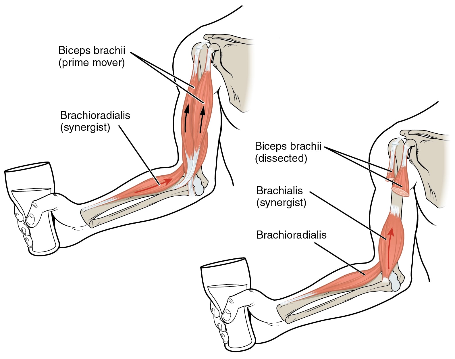
- Superficial muscle plane — biceps brachii (musculocutaneous nerve) is retracted medially; the dissection is developed along its lateral border.
- Musculocutaneous nerve — lies on the deep (posterior) surface of biceps, between biceps and brachialis; protected by retracting biceps medially as a unit. Its terminal sensory branch, the lateral antebrachial cutaneous nerve, emerges lateral to biceps just proximal to the elbow.
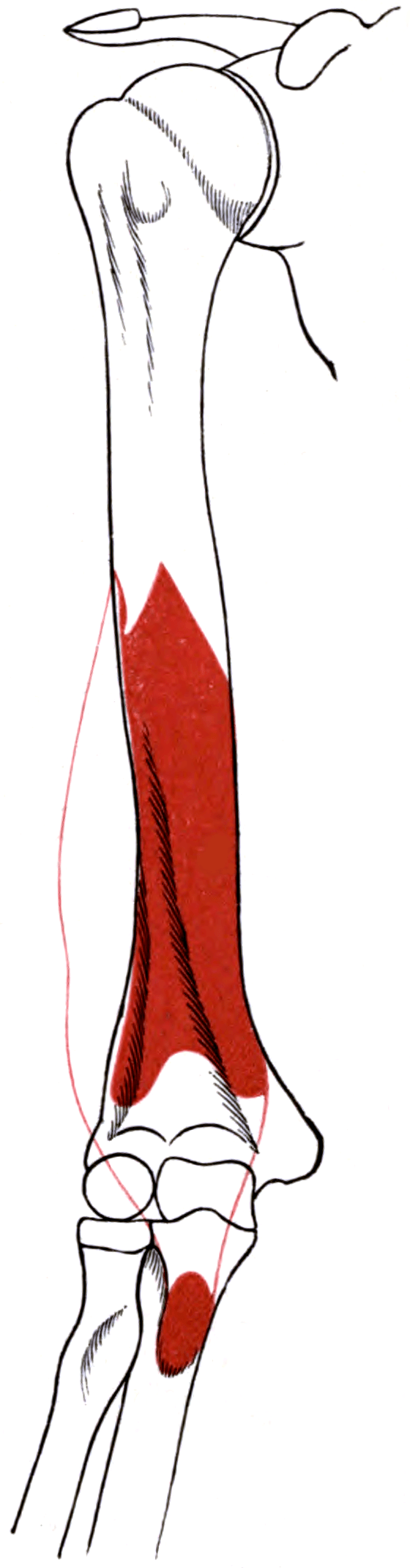
- Deep muscle — brachialis covers the anterior humeral shaft and is split longitudinally; it has dual innervation (musculocutaneous to the medial/larger superficial head, a radial-nerve branch to the inferolateral fibres of the deep head — Leonello 2007).
- Bone — the flat anterior surface of the humeral shaft is reached by sweeping the split brachialis medially and laterally subperiosteally.
- In the mid-shaft, the radial nerve is posterior (in the spiral groove) and is therefore not in the anterior field — a key safety advantage of this approach for proximal/middle-third fractures.
- In the distal third, the nerve has pierced the lateral intermuscular septum (~10 cm proximal to the lateral epicondyle) to enter the anterior compartment, where it lies in the groove between brachialis (medially) and brachioradialis (laterally).
- Splitting brachialis and leaving its lateral fibres on the bone interposes muscle between the instruments/plate and the radial nerve — the nerve is shielded rather than exposed. This is the anatomical basis of the brachialis-splitting approach.
The Approach — Step by Step
- Supine on a radiolucent table with the arm on an arm board or across the chest; a small bump under the scapula assists proximal access. Image intensifier from the head or the contralateral side.
- Surface landmarks: the coracoid process and deltopectoral groove proximally, the lateral border of biceps brachii in the arm, and the lateral epicondyle distally — the full incision lies along the line joining these.
- Tailor incision length to the fracture: a proximal-third fracture needs the deltopectoral-derived proximal segment; a distal-third fracture needs the brachialis-splitting distal segment; MIPO uses short proximal and distal windows only.
Dangers & How to Avoid Them
Structures at risk
For any distal extension or distal bicortical screw, treat the radial nerve as present in the field: it has crossed from posterior to anterior through the lateral intermuscular septum in the distal third. Either positively identify it between brachialis and brachioradialis, or keep the lateral brachialis fibres interposed and confirm screw trajectory away from the posterolateral cortex. Most iatrogenic palsies in anterior humeral surgery occur distally.
Anterolateral vs Posterior Approach to the Humeral Shaft
Choosing the approach
Outcomes & Evidence
Brachialis is dual-innervated — the anatomical basis of the safe split
Humeral shaft approaches & MIPO — the anterior corridor
Based on articles retrieved from PubMed. The cadaveric innervation data are from Leonello et al. (DOI) and the approach/MIPO synthesis from Orapiriyakul et al. (DOI). The layered anatomy, the deltopectoral proximal extension, and the radial-nerve relationships in the distal third reflect standard, well-established surgical-anatomy teaching (Hoppenfeld/AO descriptions) rather than a single cited trial.
Viva Scenarios
Practise clinical reasoning and management decisions out loud
“You are plating a middle-third humeral shaft fracture through an anterolateral approach. The examiner asks: 'What is your internervous plane, and is the brachialis split safe?'”
“A patient develops a radial nerve palsy after anterior plating of a distal-third humeral shaft fracture. The examiner asks how this approach can injure the radial nerve when 'the nerve is posterior'.”
Viva & Exam Focus
LARMBrachialis split safety
Hook:Split the brachialis down the middle: Lateral half is Radial, Medial half is Musculocutaneous — so each half keeps its nerve and the radial nerve stays cushioned.
- The 'internervous plane' question is a trap — name the deltopectoral plane proximally and state plainly that brachialis is SPLIT (dual innervation) distally.
- The radial nerve is the great danger of the DISTAL extension, not the mid-shaft.
- This is the corridor for anterior MIPO of the humerus.