Deformity correction and arthrodesis of the Charcot foot using superconstruct principles | advanced
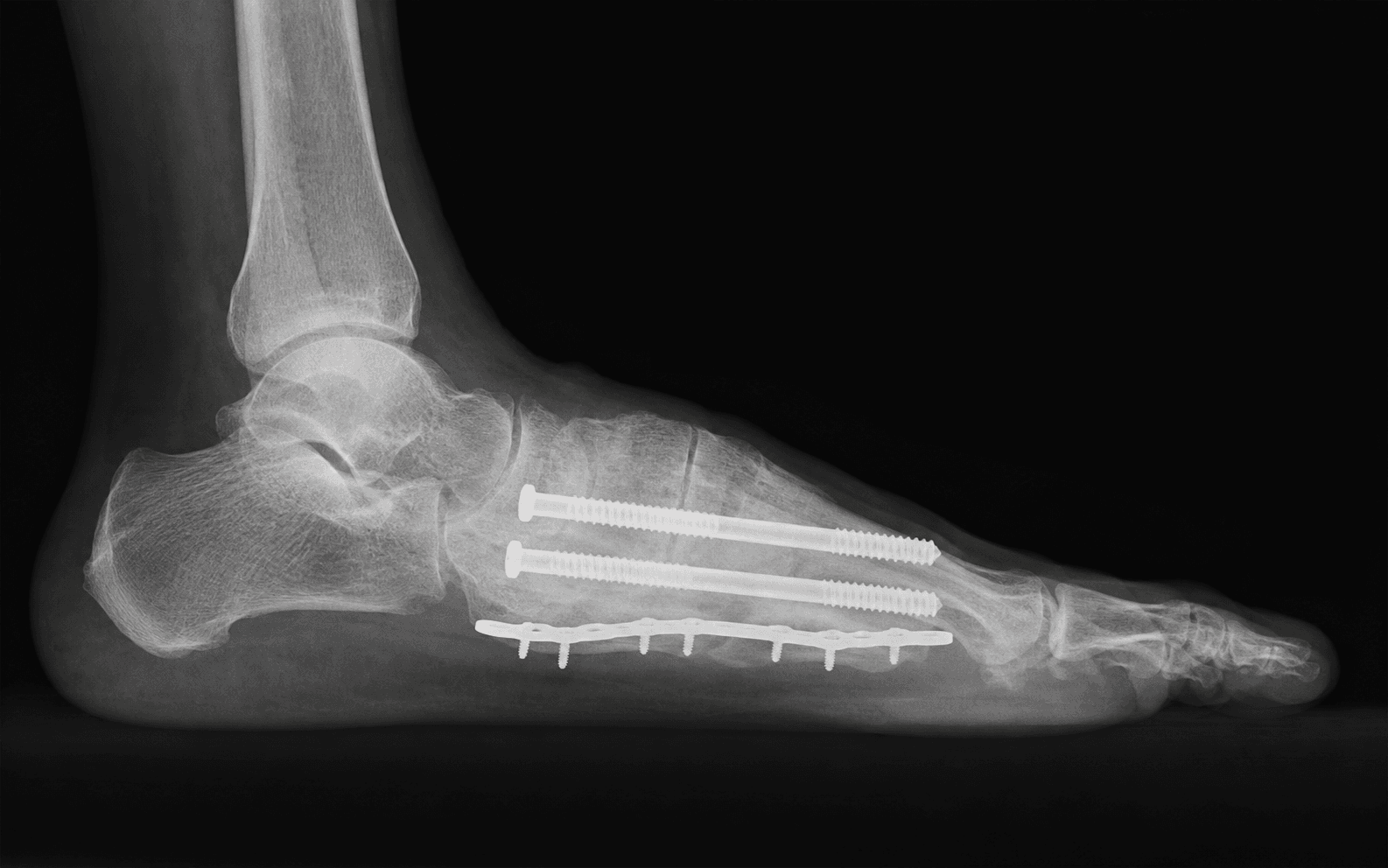
Surgical Imaging
The trap: Operating on a hot, swollen, actively fragmenting Charcot foot (Stage I) in the belief that early fixation will prevent deformity progression.
The reality: Bone is actively resorbing and fragmenting. Fixation placed into this substrate pulls out, the arthrodesis bed has no healing capacity, and the surgery converts a manageable collapse into an infected nonunion with hardware failure. Wait for Eichenholtz Stage II to III (clinical cooling, coalescence or consolidation on imaging) before elective reconstruction.
The problem: The Charcot patient has peripheral neuropathy and cannot feel wound problems. A wound dehiscence that in a sensate foot would cause pain and prompt early review can progress silently to deep infection and osteomyelitis before anyone notices.
Prevention: Meticulous soft tissue handling. Avoid extensive dorsal dissection where the skin is thin and previous ulceration may compromise blood supply. Consider staged procedures (external fixation first, then definitive fusion) in high-risk cases. Weekly wound inspection for the first 4 to 6 weeks post-operatively.
The mechanism: Fixation placed on the plantar surface of the midfoot — for example, plantar plates or protruding screw tips — creates a new bony prominence in the insensate foot that can ulcerate post-operatively, the very problem the surgery was meant to eliminate.
Prevention: Place hardware dorsally or laterally wherever possible. If plantar plate fixation is required, countersink fully and ensure no screw or plate edge protrudes below the plantar cortex. Confirm hardware position fluoroscopically before closure. Plan to remove plantar-sided hardware once fusion is confirmed (often at 6 to 9 months).
The problem: Charcot bone is osteopenic and biologically poor. Standard screw fixation is often inadequate — screws cut through cancellous bone, plates break under cyclic loading, and the construct fails before fusion is achieved.
Prevention: Apply superconstruct principles. Extend the fusion beyond the Charcot zone. Use intramedullary nails or beaming for axial load-sharing. Supplement plate fixation with multiple points of fixation. Accept prolonged non-weight-bearing for 8 to 12 weeks minimum and often longer. Use bone graft (autograft or BMP) in the arthrodesis bed.
The risk: Patients with Charcot foot often have pre-existing or recent plantar ulceration with bacterial colonisation of bone, diabetes with impaired immunity and wound healing, and co-existing vascular disease.
Implications: Deep infection rates after Charcot reconstruction are reported up to 30 percent across series. Hardware removal may be required and the fusion is lost. Two-stage procedures (external fixation to stabilise, then delayed definitive fusion after infection control) may be needed. In some cases, infection leads to amputation.
The trap: A hot, swollen, erythematous diabetic foot is treated as cellulitis with antibiotics when the true diagnosis is acute Charcot neuroarthropathy (Eichenholtz Stage 0 to I). The distinguishing feature is that Charcot produces a painless or only mildly uncomfortable warm swollen foot in an insensate patient, often with a history of prior Charcot episodes. Cellulitis is typically painful.
The fix: Maintain a high index of suspicion. Compare skin temperature with the contralateral foot using a dermal thermometer. MRI shows bone marrow oedema and microfractures in acute Charcot. If clinical Charcot is suspected, immobilise promptly (total-contact cast or removable boot) and obtain MRI — do not just prescribe antibiotics.
C.H.A.R.C.O.TCHARCOT — Eichenholtz Staging and Assessment
S.U.P.E.RSUPER — Superconstruct Principles for Charcot Arthrodesis
O.F.F.L.O.A.DOFFLOAD — Non-Operative Management Principles
Eichenholtz Classification — Staging Determines Timing
The Eichenholtz classification is the foundation for treatment decisions in Charcot neuroarthropathy. Surgery is timed according to the stage.
- Clinical Features
- Warm, swollen, erythematous foot. Pain absent or mild. Early inflammatory phase.
- Radiographic Features
- Normal radiographs or subtle subluxation. Bone scan or MRI shows early marrow oedema.
- Treatment Priority
- Immobilise immediately. Total-contact cast. Do NOT operate unless acute instability threatens skin integrity.
- Clinical Features
- Hot, swollen foot. Temperature greater than 2 degrees above contralateral. Joint effusion.
- Radiographic Features
- Bony fragmentation, subluxation, dislocation. Debris and osseous destruction at involved joints.
- Treatment Priority
- Non-operative. Continue TCC. Surgery is contraindicated for elective reconstruction. Exception: fracture-dislocation threatening the skin — urgent external fixation.
- Clinical Features
- Cooling of foot. Reduced swelling. Temperature difference diminishing.
- Radiographic Features
- Absorption of debris. New bone formation. Coalescence of fragments. Joint surfaces beginning to fuse.
- Treatment Priority
- Consider surgery. Foot is cooling and bone is beginning to consolidate. Elective arthrodesis can be planned for the near term (once temperature normalises).
- Clinical Features
- Normal temperature. Minimal or no swelling. Fixed deformity.
- Radiographic Features
- Consolidated bone. Remodelled deformity. Stable but malaligned architecture.
- Treatment Priority
- Surgery indicated if deformity is unbraceable, ulcer over a prominence, or instability present.
Technical Tip: 'I use a dermal infrared thermometer to compare the Charcot foot against the contralateral side. A temperature difference of greater than 2 degrees Celsius signals active inflammation. I do not plan elective reconstruction until the temperature difference is less than 1 degree for two consecutive measurements at least two weeks apart.'
Sanders Anatomic Classification — Pattern Determines Approach
- Location
- Forefoot (MTP joints)
- Typical Deformity
- Dislocated toes, plantar metatarsal heads
- Common Presentation
- Metatarsal head ulceration, transfer metatarsalgia
- Location
- Tarsometatarsal (Lisfranc)
- Typical Deformity
- Rocker-bottom midfoot, medial column collapse
- Common Presentation
- Plantar midfoot ulcer over the bony prominence
- Location
- Midtarsal (talonavicular, naviculocuneiform)
- Typical Deformity
- Lateral column collapse, abduction of forefoot
- Common Presentation
- Lateral foot ulcer, peritalar subluxation
- Location
- Ankle and subtalar
- Typical Deformity
- Posterior tibial tendon insufficiency, hindfoot valgus, ankle instability
- Common Presentation
- Medial malleolar or lateral hindfoot ulcer
- Location
- Calcaneus
- Typical Deformity
- Posterior calcaneal fragmentation, loss of heel height
- Common Presentation
- Posterior heel ulcer, Achilles tendon dysfunction
Type 2 (TMT/Lisfranc) is the most common pattern and the classic exam case for Charcot reconstruction.
Surgical Indications
Absolute Indications
- Unbraceable deformity — fixed rocker-bottom or hindfoot deformity that cannot be accommodated in a CROW boot, custom AFO, or total-contact cast
- Recurrent or non-healing plantar ulcer over a bony prominence despite adequate offloading (at least 3 months of TCC or CROW boot)
- Deep infection with osteomyelitis in the Charcot foot — requires debridement with or without concurrent stabilisation
- Acute instability or fracture-dislocation threatening the soft tissues (urgent external fixation in Eichenholtz Stage I)
Relative Indications
- Patient declining prolonged non-operative offloading
- Recurrent ulceration despite brace compliance
- Progressive deformity on serial radiographs despite immobilisation
- Inability to wear any footwear due to deformity
Contraindications
Absolute (for elective reconstruction):
- Eichenholtz Stage I (active fragmentation) — catastrophic failure of fixation and worsening of deformity
- Uncontrolled infection with systemic sepsis
- Critical ischaemia (ABI less than 0.45 or absolute toe pressure less than 30 mmHg) without planned revascularisation
Relative:
- Eichenholtz Stage 0 with no instability — treat non-operatively first
- HbA1c greater than 10 percent (optimise glycaemic control before elective surgery)
- Active smoking (increases nonunion and infection risk)
- End-stage renal disease on dialysis (markedly elevated infection and wound complication rates)
- Limited ambulatory potential or non-ambulatory patient (amputation may be more appropriate)
Evidence — Non-Operative vs Surgical Management
Total-Contact Casting
- TCC is the gold-standard non-operative treatment for acute Charcot (Eichenholtz 0 to I) and for offloading plantar ulcers
- A Cochrane review found TCC more effective than removable cast walkers for healing neuropathic plantar ulcers
- TCC for acute Charcot: cast changes every 1 to 2 weeks, continue for 8 to 12 weeks, until the foot cools clinically
- Long-term: transition to CROW boot or custom AFO once the foot is quiescent
Exostectomy
- Simple resection of a plantar bony prominence without arthrodesis
- Indicated for an isolated plantar prominence with a non-healing ulcer in a braceable foot
- Advantage: quick procedure, preserves joint mobility, lower morbidity than arthrodesis
- Recurrence rate: 30 to 50 percent at 2 to 3 years if the underlying deformity is not corrected — the prominence reforms because the deformity persists
Arthrodesis with Deformity Correction
- The definitive procedure for unbraceable Charcot deformity
- Midfoot (TMT) arthrodesis: corrects rocker-bottom, eliminates plantar prominence, provides a stable plantigrade foot
- Hindfoot (tibiotalocalcaneal) arthrodesis: corrects valgus or varus hindfoot deformity
- Ankle arthrodesis: for Charcot ankle destruction
- Fusion rates reported from 60 to 90 percent depending on the site, fixation method, and Eichenholtz stage at surgery
Exostectomy vs Arthrodesis — Decision Guide
Key Evidence
Is the Eichenholtz classification still valid for the diabetic Charcot foot?
Superconstructs in the treatment of Charcot foot deformity: plantar plating, locked plating, and axial screw fixation
Long-term follow-up in diabetic Charcot feet with spontaneous onset
Are the Sanders-Frykberg and Brodsky-Trepman Classifications Reliable in Diabetic Charcot Neuroarthropathy?
Charcot foot reconstruction outcomes: a systematic review
Clinical Decision Scenarios
Practise clinical reasoning and management decisions out loud
“A 56-year-old man with Type 2 diabetes presents with a 3-month history of a non-healing plantar midfoot ulcer. He has a rocker-bottom deformity of the left foot with a 2 cm ulcer directly beneath the midfoot prominence. Radiographs show collapsed TMT joints with debris and early coalescence. Temperature measurement shows a 0.5-degree difference from the right foot. He has been in a CROW boot for 10 weeks with no improvement. How do you manage him?”
“A 62-year-old woman with Type 2 diabetes presents with a 4-week history of a hot, swollen left foot. She noticed swelling after walking through an airport in sandals. She has had diabetes for 18 years with peripheral neuropathy. The left foot is erythematous, swollen, and warm to touch. She says it is not particularly painful. Her GP started oral antibiotics for cellulitis 10 days ago with no improvement. How do you manage this?”
“You are asked to see a 60-year-old man with Type 2 diabetes in the orthopaedic clinic. He had a midfoot Charcot reconstruction with TMT arthrodesis 14 months ago at another hospital. Radiographs show solid fusion but there is a new ulcer on the plantar aspect of the foot, just distal to the arthrodesis site. There is a bony prominence visible on the lateral view that was not present on the immediate post-operative films. How do you explain this and manage him?”