Reverse-Ponseti (Dobbs) serial casting, tendo-Achilles tenotomy and percutaneous talonavicular pinning for the rigid rocker-bottom flatfoot | advanced
Surgical Imaging
The trap: Calling a flexible foot "vertical talus" and sending it to theatre. Oblique talus looks similar at rest (forefoot dorsiflexed, rocker-bottom) but is REDUCIBLE.
The fix: A forced plantar flexion lateral radiograph. In oblique talus the talonavicular joint reduces and the talar axis aligns with the first metatarsal; in true CVT it does not. Only true CVT (rigid) needs the reverse-Ponseti pathway.
The trap: Treating the convex sole as the problem. The rocker-bottom is the RESULT of the vertical talus and the uncorrected hindfoot equinus — the talus pushes into the plantar arch.
The fix: Reduce the talonavicular joint and correct the equinus; the rocker-bottom resolves with reduction, not with any direct 'sole' procedure.
The trap: Operating on the foot in isolation in a child with an undiagnosed neural tube defect or syndrome.
The fix: Examine the spine, hips and the rest of the child. About half of CVT cases are associated with myelomeningocele, arthrogryposis, cerebral palsy, Larsen or Beals syndrome. The underlying diagnosis drives prognosis and bracing strategy.
Location: The talus has a precarious end-arterial blood supply. Extensive peritalar capsular release and dissection around the talar neck jeopardise it.
Risk: AVN of the body of the talus — fragmenting, shortening, pain and a stiff, painful hindfoot years later. The minimally invasive Dobbs method was designed to minimise this dissection.
The trap: Applying standard clubfoot casting mechanics. In clubfoot you abduct and supinate the forefoot around the talar head; in CVT the deformity is the OPPOSITE.
The fix: In CVT you plantarFLEX and adduct the forefoot to bring the navicular dorsally onto the talar head, stretching the tight dorsolateral structures. Then correct the hindfoot equinus last with an Achilles tenotomy.
Why different: Neuromuscular imbalance and abnormal muscle forces (peroneal and extensor over-pull) drive recurrence. Bracing non-compliance compounds this.
Implications: Recurrence and residual deformity are more frequent in syndromic or neuromuscular CVT than in idiopathic feet. Counsel families on mandatory full-time then night-time bracing and on the higher re-operation rate from the outset.
V.E.R.T.I.C.A.LVERTICAL — Recognising Congenital Vertical Talus
R.E.V.E.R.S.EREVERSE — The Dobbs (Reverse-Ponseti) Method
What Is Congenital Vertical Talus?
Congenital vertical talus (CVT), historically called convex pes valgus, is a rigid foot deformity present at birth. It is defined by three fixed elements:
- A vertically orientated talus — its long axis lies almost parallel to the tibial axis, so the talar head projects downward and medially into the plantar arch.
- An irreducible dorsal dislocation of the navicular onto the talar neck — the navicular is locked dorsolaterally and cannot be reduced onto the talar head by manipulation.
- A fixed equinus of the hindfoot — the calcaneus is locked in equinus and will not dorsiflex.
The combination produces the characteristic rigid rocker-bottom flatfoot with a convex plantar surface, a prominent talar head felt in the medial sole, an abducted and dorsiflexed forefoot, and tight dorsolateral structures (the long toe extensors, the peronei, and the talonavicular capsule). It is rare, with an incidence commonly cited at roughly 1 in 10,000 live births, bilateral in about half of cases, and roughly equally distributed between the sexes.
Indications for the Reverse-Ponseti (Dobbs) Pathway
Absolute Indications
- Confirmed true CVT — rigid, with a persistent talonavicular dislocation on a forced plantar flexion lateral radiograph (idiopathic or syndromic)
- Failure of the deformity to correct after an initial trial of manipulation and casting
- An older infant or toddler presenting late, where the deformity is established
Relative Indications
- Mild or partially reducible deformity — a short course of casting may fully correct the talonavicular joint, and only a percutaneous tenotomy and pinning are needed
- Syndromic or neuromuscular CVT — the same pathway applies, but with a lower threshold for limited open reduction and with explicit counselling on a higher recurrence rate
Contraindications
Absolute:
- A flexible oblique talus — this reduces on forced plantar flexion and is NOT a CVT; it is managed non-operatively (observation, stretching, occasional casting)
- A structural positional deformity that is fully correctable (e.g. a severe positional calcaneovalgus foot) — observation and stretching only
- An untreatable life-limiting underlying condition where foot surgery would not be in the child's interest (a rare, multidisciplinary decision)
Relative:
- Severe skin compromise over the dorsum of the foot (defer casting until skin recovers)
- A medically unstable neonate (delay until the child is fit for sedation or anaesthesia)
Differentiating CVT from the Flexible Look-Alikes
This is the single most important decision and the most commonly examined point. The forced plantar flexion lateral radiograph resolves it.
CVT vs Oblique Talus vs Positional Calcaneovalgus Foot
Evidence for the Reverse-Ponseti Method
Why the Shift Away from Extensive Release
Historically, CVT was treated by an extensive single-stage open soft-tissue release — lengthening of the long toe extensors and peronei, release of the talonavicular and calcaneocuboid capsules, a posterior release for equinus, and reduction held with multiple K-wires. While it could correct alignment, it carried real risks: talar avascular necrosis from extensive peritalar dissection, marked stiffness, wound problems over tight dorsal skin, and a not-trivial recurrence rate. The minimally invasive Dobbs method was developed specifically to obtain comparable correction with far less dissection.
The Reverse-Ponseti (Dobbs) Method
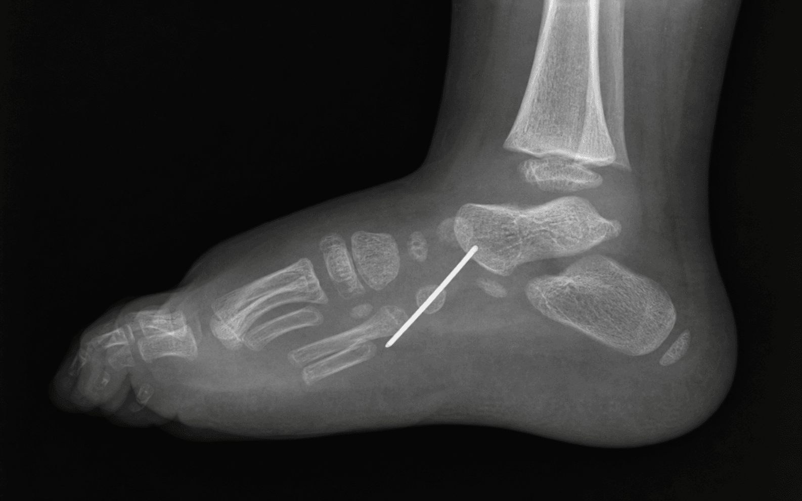
Building on Ponseti principles but applied in the opposite direction, serial weekly casting stretches the dorsolateral structures and seats the navicular onto the talar head. Once the talonavicular joint is reduced, a percutaneous tendo-Achilles tenotomy corrects the residual equinus and a percutaneous K-wire is passed across the talonavicular joint to hold the reduction while the stretched capsule heals. The reported correction rates are high, the soft-tissue dissection is minimal, and the talar AVN and stiffness burden is lower than with extensive releases.
Important Caveats in the Evidence
- The best-reported results are in idiopathic CVT. Outcomes in syndromic and neuromuscular feet are less consistent, with higher recurrence and residual deformity.
- Recurrence is closely tied to orthosis compliance — families who do not maintain full-time then night-time bracing recur more often.
- Long-term follow-up studies are still maturing; mid-term correction is excellent, but lifelong surveillance for recurrence, AVN, and stiffness is appropriate.
Key Evidence
Early results of a new method of treatment for idiopathic congenital vertical talus
Surgical correction of congenital vertical talus under age 2 years
Congenital vertical talus (instructional review)
Treatment of Congenital Vertical Talus: Comparison of Minimally Invasive and Extensive Soft-Tissue Release Procedures at Minimum Five-Year Follow-up
Clinical Decision Scenarios
Practise clinical reasoning and management decisions out loud
“A newborn is referred with a rigid rocker-bottom right foot. The forefoot is abducted and dorsiflexed, the hindfoot is in fixed equinus, and you can feel a firm medial prominence in the sole that does not reduce with manipulation. How do you confirm the diagnosis and establish the initial management?”
“Talk me through the Dobbs reverse-Ponseti method for an idiopathic congenital vertical talus, step by step.”
“A child with a myelomeningocele and a congenital vertical talus has completed the reverse-Ponseti casting and had a percutaneous tenotomy and talonavicular pinning. At 18 months there is clear recurrence of the rocker-bottom deformity. How do you manage this?”
References
-
Dobbs MB, Purcell DB, Nunley R, Morcuende JA (2006). Early results of a new method of treatment for idiopathic congenital vertical talus. J Bone Joint Surg Am. 88(6):1192-200. doi:10.2106/JBJS.E.00402 — The foundational paper describing the reverse-Ponseti method of serial casting followed by percutaneous tendo-Achilles tenotomy and talonavicular pinning, which established the modern minimally invasive standard.
-
Seimon LP (1987). Surgical correction of congenital vertical talus under the age of 2 years. J Pediatr Orthop. 7(4):405-11. doi:10.1097/01241398-198707000-00005 — The influential pre-Dobbs single-stage dorsal open reduction in infants, the historical surgical benchmark against which the minimally invasive method is compared.
-
Drennan JC (1996). Congenital vertical talus. Instr Course Lect. 45:315-22 — Authoritative instructional review defining the pathoanatomy, the rigid rocker-bottom deformity, the radiographic diagnosis and the historical extensive soft-tissue release.
-
Yang JS, Dobbs MB (2015). Treatment of Congenital Vertical Talus: Comparison of Minimally Invasive and Extensive Soft-Tissue Release Procedures at Minimum Five-Year Follow-up. J Bone Joint Surg Am. 97(16):1354-65. doi:10.2106/JBJS.N.01002 — Direct comparison at minimum five years showing the Dobbs method achieves comparable correction with fewer complications and better ankle motion than extensive release.