Distal Radius Corrective Osteotomy (Malunion)

Hand & WristAdvancedCore Procedure

Distal Radius Corrective Osteotomy (Malunion)

Surgical technique guide for corrective osteotomy of distal radius malunion - volar opening-wedge and dorsal closing-wedge extra-articular osteotomy, intra-articular step correction, three-dimensional patient-specific planning, structural bone graft, and concurrent DRUJ management

High-yield overview

Volar opening-wedge or dorsal closing-wedge extra-articular osteotomy + structural bone graft for symptomatic distal radius malunion | advanced

Surgical Imaging

Corrective osteotomy of a distal radius malunion
Corrective osteotomy of a distal radius malunion with an opening wedge and volar locking plate, restoring radial length, inclination and volar tilt.Credit: AI-generated medical illustration Β· OrthoVellum
Critical Danger Structures and Exam Traps
Volar Tilt Under-Correction

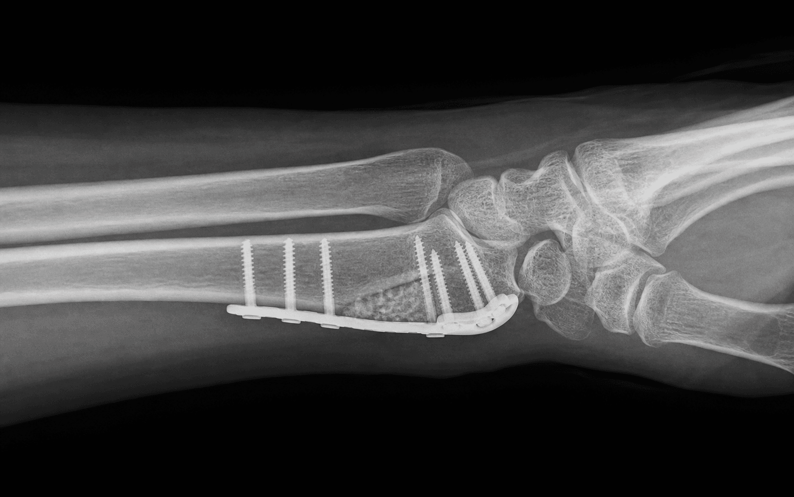
The trap: Accepting residual dorsal tilt of greater than 5-10 degrees after osteotomy because the wedge was undersized or the plate was applied before the desired correction was achieved.

The fix: Confirm the wedge on fluoroscopy matches the pre-op plan. Volar tilt should be restored to roughly 11 degrees (range 8-12). Under-correction causes persistent midcarpal instability, weakness in extension, and ulnocarpal abutment. The wedge height in millimetres approximates the desired correction in degrees for a typical 20-25 mm radial height.

Radial Shortening (Positive Ulnar Variance)

The trap: Failing to recognise that the malunion has produced RADIAL SHORTENING (most often from an impacted dorsal fracture) and that ulnar-sided wrist pain will persist unless the radius is LENGTHENED or the ulna is SHORTENED.

The fix: Calculate pre-op ulnar variance. If greater than 2 mm positive, plan a concurrent ulnar shortening osteotomy or wafer resection. Radial height restoration should bring variance back to neutral β€” intraoperative imaging confirms the correction.

DRUJ Incongruity Left Untreated

The trap: Correcting the radial deformity but leaving a symptomatic DRUJ with restricted pronosupination, a positive ballottement test, or arthrosis. The result is persistent ulnar-sided pain and stiffness.

The fix: Pre-op assess DRUJ clinically (ballottement, piano-key, press test) and radiographically (CT in pronation, supination, and neutral). Plan for concurrent ulnar shortening, wafer resection, or SauvΓ©-Kapandji in the same anaesthetic if DRUJ pathology is significant.

FPL Rupture Over Volar Plate

The trap: Prominent volar plate or distal screws causing attritional rupture of the FPL tendon β€” the most common tendon complication of volar plating in general, and a particular risk when the distal end of the plate is proud or the watershed line is violated.

The fix: Position the distal plate edge PROXIMAL to the watershed line. Check screw lengths under fluoroscopy; ensure no screws penetrate the dorsal cortex into extensor compartments. Use low-profile plates where possible. If a screw is too long, exchange or shorten.

EPL Rupture (Dorsal Approach)

The trap: Attritional EPL rupture after dorsal plating β€” caused by drilling or screw penetration into the third dorsal compartment where the EPL runs, or by prominent screw tips on the dorsal cortex.

The fix: Confirm screw length with fluoroscopy in multiple views; measure so dorsal cortex is not breached. If the dorsal approach is used, release the EPL sheath and protect/retract the tendon. EPL rupture is also reported after non-operatively treated distal radius fractures (not just post-op).

Nonunion of Osteotomy

The trap: A closing- or opening-wedge osteotomy that fails to unite β€” usually due to inadequate fixation, smoking, or absent/non-structural graft.

The fix: Use structural graft in opening-wedge osteotomy (tricortical iliac crest or structural allograft), ensure rigid plate fixation with at least 3 cortical screws on each side of the osteotomy, and counsel smokers on smoking cessation β€” nicotine impairs fracture healing.

Mnemonic

R.A.D.I.U.SRADIUS β€” Pre-operative Planning Parameters

Mnemonic

W.E.D.G.EWEDGE β€” Choosing the Osteotomy Direction

Mnemonic

O.S.T.E.O.T.O.M.YOSTEOTOMY β€” Steps in One Sentence Each

Surgical Indications

Symptomatic Malunion β€” The Three Pillars

A distal radius malunion becomes a surgical indication when ALL THREE are present:

  • Deformity beyond acceptable limits (dorsal tilt greater than 10-15 degrees, radial shortening greater than 3-5 mm, radial inclination loss greater than 5 degrees, intra-articular step greater than 2 mm)
  • Symptoms attributable to the deformity β€” pain, weakness, restricted motion, ulnar-sided wrist pain
  • Failure of non-operative management β€” typically a course of rest, activity modification, hand therapy, and a trial of splinting/medication

Absolute Indications

  • Dorsal angulation greater than 20-25 degrees with significant functional impairment and no radiographic evidence of radiocarpal arthritis
  • Radial shortening greater than 5 mm with positive ulnar variance and ulnocarpal abutment syndrome
  • Intra-articular step greater than 2 mm in a young patient (under 45-50) with a discrete malunited fragment, no radiocarpal arthritis, and preserved cartilage
  • Disabling DRUJ dysfunction (loss of pronosupination, painful clicking, arthrosis) that is at least in part due to the malunion
  • Midcarpal instability (volar intercalated segment instability β€” VISI) secondary to the malunion

Relative Indications

  • Radial shortening 3-5 mm with mild-moderate symptoms
  • Dorsal tilt 10-20 degrees with weakness and functional limitation
  • Concurrent carpal tunnel syndrome attributable to the malunited volar cortex (volar osteotomy decompresses simultaneously)
  • Patient preference for correction after detailed counselling on risks, benefits, and prolonged recovery

Contraindications

Absolute:

  • Established radiocarpal or DRUJ arthritis β€” salvage procedures (partial or total wrist fusion, arthroplasty) are preferred
  • Active infection
  • Medically unfit for anaesthesia with no reversible component

Relative:

  • Heavy smoker unwilling to cease β€” markedly elevated nonunion risk; consider smoking cessation programme before surgery
  • Complex regional pain syndrome (CRPS) β€” proceed cautiously; pre-op CRPS predicts worse outcomes
  • Significant osteopenia that may compromise fixation
  • Low functional demand with tolerable symptoms β€” non-operative management may be appropriate

Evidence Base

Outcomes of Corrective Osteotomy

  • Systematic review (Lozano-Calderon 2006, PENDING PMID) β€” pooled functional outcomes from 15 case series; corrective osteotomy provides approximately 70-90% good-to-excellent results for pain relief and restoration of grip strength
  • Pain reduction β€” most series report 60-80% of patients achieve meaningful pain reduction
  • Grip strength improvement β€” average improvement of 30-50% of contralateral grip after correction
  • Range of motion β€” modest gains in flexion-extension arc (typically 10-20 degrees improvement), more predictable correction of pronosupination if DRUJ is addressed

Patient-Specific Instrumentation

  • 3-D CT templating with custom guides improves accuracy of correction β€” reported standard deviation of angular correction reduced from approximately 5 degrees (free-hand) to 2 degrees (guided) in comparative series
  • Patient-specific plates (PSPs) allow precise pre-contoured fixation, particularly helpful in multiplanar deformity
  • Cost-effectiveness debate β€” higher upfront cost balanced by reduced revision rates and improved functional outcomes in selected series

Concurrent DRUJ Procedures

  • Ulnar shortening osteotomy is the most commonly performed concurrent procedure; transverse or oblique osteotomy with a compression plate
  • Wafer resection (distal ulna arthroplasty) β€” alternative for positive ulnar variance less than 4 mm with TFCC pathology
  • SauvΓ©-Kapandji β€” preferred when there is significant DRUJ arthrosis (creates a distal radioulnar fusion with a proximal pseudarthrosis)

Comparison of Osteotomy Techniques

Extra-articular Corrective Osteotomy β€” Technique Comparison


Key Evidence

Evidence

Outcome after corrective osteotomy for malunited fractures of the distal end of the radius

Level IV
Prommersberger KJ, Van Schoonhoven J, Lanz UB
Clinical implication: Corrective osteotomy provides reliable pain relief and functional improvement in symptomatic distal radius malunion; outcomes are best when performed before radiocarpal arthritis develops.
Source: J Hand Surg Br 2002;27(1):55-60
Evidence

Corrective osteotomy for intra-articular malunion of the distal part of the radius

Level IV
Ring D, Prommersberger KJ, Gonzalez Del Pino J, Capomassi M, Slullitel M, Jupiter JB
Clinical implication: Intra-articular corrective osteotomy is a viable option for young patients with a discrete articular step and no radiocarpal arthritis; outcomes depend on early intervention and preserved cartilage.
Source: J Bone Joint Surg Am 2005;87(7):1503-1509
Evidence

Three-dimensional virtual planning of corrective osteotomies of distal radius malunions: a systematic review and meta-analysis

Level III
de Muinck Keizer RJO, Lechner KM, Mulders MAM, Schep NWL, Eygendaal D, Goslings JC
Clinical implication: Patient-specific instrumentation improves accuracy of correction in complex multiplanar distal radius malunion and may reduce revision rates compared with free-hand technique.
Source: Strategies Trauma Limb Reconstr 2017;12(2):77-89
Evidence

Corrective osteotomy is an effective method of treating distal radius malunions with good long-term functional results

Level III
Mulders MAM, d'Ailly PN, Cleffken BI, Schep NW
Clinical implication: Corrective osteotomy provides durable functional improvement at long-term follow-up; counsel patients about the possibility of late arthritis, particularly with residual intra-articular incongruity.
Source: Injury 2017;48(3):731-737
Evidence

What surgical technique to perform for isolated ulnar shortening osteotomy after distal radius malunion: a systematic review

Level III
Laane CLE, Oude Nijhuis KD, Spil J, Sierevelt IN, Doornberg JN, Jaarsma RL, Verhofstad MHJ, Wijffels MME
Clinical implication: Concurrent ulnar shortening osteotomy is effective for ulnocarpal abutment from positive ulnar variance after distal radius malunion; counsel patients about a potential second procedure for plate removal.
Source: Hand (N Y) 2024;19(6):885-894

Clinical Decision Scenarios

Practise clinical reasoning and management decisions out loud

Viva scenarioAdvanced
Clinical prompt

β€œA 24-year-old right-hand-dominant electrician sustained a distal radius fracture 8 months ago. He was treated non-operatively. He now has dorsal tilt of 25 degrees, radial shortening of 6 mm, and a 3 mm intra-articular step. He has pain, weakness, and can't return to work. How do you manage this?”

Practical approach
This is a young, high-demand patient with a multi-component distal radius malunion β€” significant dorsal tilt, radial shortening, AND an intra-articular step. The combination of all three is at the upper end of the spectrum and requires comprehensive correction. **Pre-operative assessment**: - Bilateral wrist PA, lateral, and oblique radiographs to measure all three parameters and compare with the uninjured side - CT with 3-D reconstruction is mandatory given the multiplanar deformity and the intra-articular component - MRI or arthrogram to assess the TFCC, scapholunate ligament, and chondral surfaces - Wrist arthroscopy at the time of surgery to assess cartilage β€” important because intra-articular osteotomy in a joint with established arthritis gives poor results - DRUJ assessment clinically (ballottement, press test, piano-key) and on CT **Operative plan**: - Volar opening-wedge osteotomy to address the extra-articular deformity (dorsal tilt, radial shortening, inclination) - Intra-articular correction of the 3 mm step β€” given the patient's age and lack of arthritis, this is reasonable - Patient-specific cutting guides and pre-contoured plate based on the 3-D CT β€” will improve accuracy of the complex multiplanar correction - Concurrent DRUJ procedure β€” ulnar shortening osteotomy, given the positive ulnar variance expected after correction - Bone graft: tricortical iliac crest autograft (structural) for the opening-wedge - Arthroscopy to confirm articular reduction and assess for any chondral damage **Post-operative**: - Short-arm splint for 2 weeks, then hand therapy - Out of work for 3-6 months depending on union and function - Plate removal at 6-12 months if symptomatic - Long-term surveillance for radiocarpal arthritis β€” likely given the intra-articular component **Realistic outcomes**: - Pain relief in approximately 70-80% - Grip strength improvement of 30-50% of contralateral - 80% chance of return to heavy manual work, though some patients modify their duties - Risk of post-traumatic arthritis at 10-20 years, particularly from the intra-articular component
Viva scenarioAdvanced
Clinical prompt

β€œA 38-year-old woman had a distal radius ORIF with a volar locking plate 14 months ago. She has been told she has a malunion with 18 degrees of dorsal tilt. She complains of wrist pain, weakness, and cannot return to her work as a chef. What is the most appropriate management?”

Practical approach
This patient has a symptomatic distal radius malunion with significant dorsal tilt and a high functional demand job. She requires a thorough assessment and a corrective osteotomy. **Pre-operative assessment**: - Full history and examination β€” pain pattern, functional limitations, occupation - Radiographs of both wrists (PA, lateral, oblique) β€” measure volar tilt, radial height, radial inclination, ulnar variance - CT with 3-D reconstruction β€” define the deformity in three planes - DRUJ assessment β€” ballottement, press test, supination, pronation - Carpal alignment on lateral β€” DISI pattern expected - Median nerve assessment β€” is there associated carpal tunnel syndrome from the malunited volar cortex? - Hand therapy assessment β€” has non-operative management been optimised? **Decision**: - Dorsal tilt of 18 degrees with significant functional impairment in a high-demand patient is a strong indication for corrective osteotomy - Radial shortening (need to measure) and DRUJ involvement will determine whether a concurrent ulnar shortening osteotomy is needed **Operative plan**: - Volar opening-wedge osteotomy (Henry approach) β€” restores volar tilt, radial height, inclination - Structural bone graft β€” tricortical iliac crest autograft (gold standard for structural support) - Pre-contoured volar locking plate β€” distal edge proximal to watershed line - If pre-op variance is positive or expected to remain positive after correction β€” concurrent ulnar shortening osteotomy **Specific concerns in revision surgery**: - Removal of the original plate β€” careful technique to preserve soft tissue - Identify the median nerve and FPL β€” may be encased in scar tissue - Capsular and periosteal scarring β€” release carefully - Bone quality β€” may be slightly osteopenic in the metaphysis - Risk of nonunion higher in revision β€” strict smoking cessation, structural graft, rigid fixation **Post-operative**: - 2 weeks splint, then hand therapy - Out of work 3-6 months - Plate removal at 9-12 months - Realistic outcomes: significant pain improvement in 70-80%, grip strength improvement, but residual stiffness common in revision cases
Viva scenarioAdvanced
Clinical prompt

β€œA 52-year-old man with a distal radius malunion has 22 degrees of dorsal tilt, radial shortening of 5 mm, and DRUJ symptoms with positive ballottement and a press test. He has tried non-operative management without success. Describe your surgical plan.”

Practical approach
This patient has a classic three-component malunion: dorsal tilt, radial shortening with positive ulnar variance, and DRUJ dysfunction. The corrective osteotomy must address ALL three components at the same operation to achieve a satisfactory outcome. **Pre-operative planning**: - Bilateral wrist radiographs to compare parameters - CT with 3-D reconstruction β€” to plan the osteotomy and assess DRUJ - Patient-specific cutting guide and pre-contoured plate (3-D templated) β€” particularly useful in multiplanar deformity - Calculate the required correction: volar tilt 11 degrees, radial height 11-12 mm, ulnar variance neutral **Operative plan β€” three components**: 1. **Volar opening-wedge osteotomy** β€” to correct the extra-articular deformity: - Volar (Henry) approach - Osteotomy at the metaphysis, perpendicular to the long axis - Open the wedge to restore volar tilt, radial height, and inclination - Structural tricortical iliac crest autograft - Volar locking plate with distal edge proximal to watershed line 2. **Concurrent ulnar shortening osteotomy** β€” to address the positive ulnar variance and DRUJ dysfunction: - Separate incision over the distal ulna - Transverse or oblique osteotomy at the ulnar diaphysis - Shorten to neutral ulnar variance - Compression plate fixation (6-7 hole ulnar shortening plate) 3. **Assessment of TFCC** β€” at the time of surgery, inspect and consider repair if there is a peripheral tear amenable to repair. **Order of procedures**: - I would perform the ulnar shortening osteotomy FIRST, fix it with the plate, then perform the radial osteotomy. This is because the ulnar shortening affects the overall ulnar variance, and fixing the ulnar side first allows me to confirm the radial correction against the final ulnar length. **Post-operative**: - Above-elbow splint for 2 weeks (protects both osteotomies), then short-arm splint - Hand therapy from 2-4 weeks - Both procedures require approximately 6-12 weeks to unite - Plate removal for both sites in 30-40% of cases **Outcomes**: - 70-80% good-to-excellent results with combined radial and ulnar correction - Pain relief is the most predictable outcome - Restoration of pronosupination is critical for functional recovery - Risk of nonunion at either site; risk of persistent DRUJ symptoms in 10-20%
Exam day cheat sheet
Distal Radius Corrective Osteotomy (Malunion) β€” Exam Day Summary
Editorially reviewed β€” transparent references and correction processPublished by OrthoVellum Medical Education TeamEditorial boardMethodologyReview policy
Educational disclosure

Educational content is reviewed for source visibility, editorial coherence, and correction readiness.

No individual clinician credential is claimed unless a named person is shown.

Verify before clinical use; this is not medical advice or a substitute for local guidance.