Reconstruction of fingertip amputations — healing by secondary intention, local flaps, and revision amputation | advanced
Surgical Imaging
The trap: Approaching all fingertip amputations with one technique (e.g. always performing a revision amputation or always attempting a flap). The geometry — dorsal oblique, transverse, or volar oblique — dictates the appropriate reconstruction.
The fix: Use Allen's classification: Type 1 (dorsal oblique, no exposed bone) = secondary intention; Type 2 (transverse, exposed bone) = V-Y advancement or revision amputation; Type 3 (volar oblique, pulp loss deeper) = local flap. Match the technique to the vector of tissue loss, not the habit of the surgeon.
Mechanism: When the distal phalanx tip is amputated distal to the lunula but the nail bed remains partially intact, the unsupported nail plate curls volarly over the fingertip as it grows — this is hook-nail deformity.
Prevention: If the amputation is through the distal phalanx, the nail bed must be shortened or ablated to match the bone length. Leaving a nail bed unsupported by bone will always produce a hook-nail. Options: (a) nail bed ablation with proximal matrix excision, (b) revision amputation at a more proximal level, or (c) reconstruction with a distant flap to provide pulp volume.
Incidence: Reported in 30-80% of patients after fingertip amputation, regardless of reconstruction method — it is the commonest long-term symptom and is under-emphasised in preoperative counselling.
Mechanism: Digital nerve transection, loss of glomus bodies in the pulp, and vasomotor instability. Cold intolerance is worse with revision amputation (sensory end-organs removed) and better with sensate flap reconstruction. It improves over 1-2 years in most patients but rarely resolves completely.
Mechanism: The transected digital nerve end regenerates into unscarred pulp or scar — forming a neuroma that is painful on pressure. This is a significant cause of dissatisfaction after revision amputation.
Prevention: Sharply transect the digital nerve at a level proximal to the amputation scar, allowing the nerve end to retract into unscarred soft tissue. Do not leave a cut nerve end in the pulp scar. If performing a V-Y advancement flap, the nerve is preserved with the flap (the flap is sensate).
V-Y flap necrosis: The volar triangular flap is based on the distal septocutaneous perforators from the digital arteries — if the flap base is narrowed too aggressively or the flap is advanced under excessive tension, tip necrosis occurs.
Cross-finger flap necrosis: More common if the flap is raised too thin (subdermal plane) or if the pedicle is kinked during the two-stage period. The flap should be raised just deep to the subdermal plexus, preserving the underlying paratenon of the donor finger.
Thenar flap: The PIP joint is flexed to reach the thenar donor site — if the flap is inset with the PIP in greater than 60 degrees of flexion, or if the finger is immobilised longer than 2 weeks, the PIP can stiffen in flexion, particularly in older patients.
Cross-finger flap: The PIP and DIP of the donor finger are immobilised for 10-14 days — stiffness is the commonest donor-site morbidity. Prevention: (1) use the middle finger as donor (less stiffness than ring/small), (2) keep immobilisation to less than 14 days, (3) begin immediate range-of-motion exercises after division.
P.U.L.P. R.E.C.O.NPULP RECON — Fingertip Reconstruction Decision Guide
F.L.A.P. T.Y.P.EFLAP TYPE — Matching Flap to Defect
Surgical Indications
Healing by Secondary Intention (Allen Type 1)
- Indication: Clean wounds less than 1 cm squared without exposed bone, dorsal oblique amputations, small pulp-only losses
- Optimal in: Children (excellent healing and sensory recovery), clean sharp wounds, patients who cannot easily attend for flap surgery
- Contraindications: Exposed bone of greater than 2-3 mm, devitalised tissue bed, chronic systemic conditions impairing healing (uncontrolled diabetes, heavy smoking)
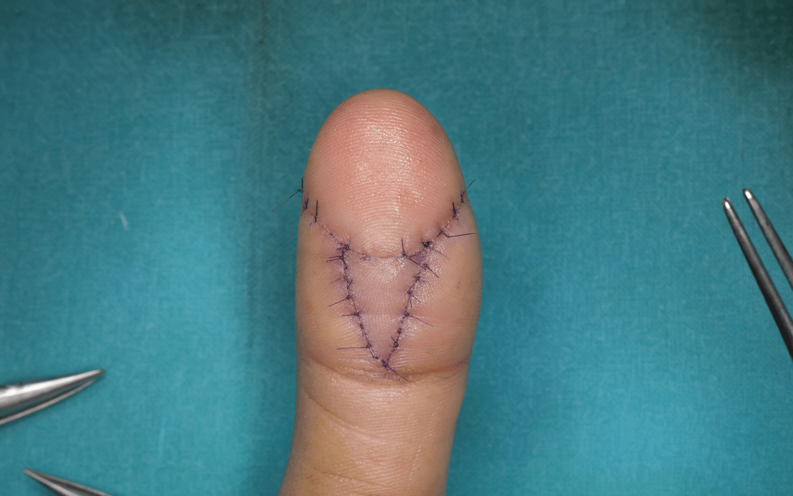
V-Y Advancement Flap (Atasoy Volar Advancement)
- Indication: Transverse or mild volar oblique amputations with exposed bone, pulp loss of less than 1.5 cm of advancement required
- Requirements: Intact digital arteries on both sides of the digit, healthy volar pulp proximal to the defect, skin laxity permitting advancement
- Relative contraindication: Significant dorsal oblique loss (volar tissue preserved but dorsal nail bed/nail lost — this requires a different approach)
Lateral V-Y Advancement Flap (Kutler)
- Indication: Transverse amputations with exposed bone where the surgeon wishes to avoid a single midline volar scar — two triangular flaps advanced from the lateral mid-lateral lines
- Advantage: The midline volar scar of the Atasoy flap is avoided — potentially better sensory recovery
- Disadvantage: Both neurovascular bundles are at risk during flap elevation on each side
Homodigital (Oblique / Unilateral Advancement) Flap
- Indication: Volar oblique pulp defects with exposed bone, particularly on the radial or ulnar side of the digit — a single lateral or volar-lateral flap advanced obliquely
- Key principle: The flap is raised on one neurovascular bundle — preserves the contralateral nerve for sensation of the remaining tip
Cross-Finger Flap
- Indication: Larger volar pulp defects (greater than 1.5 cm squared) with exposed bone, where local advancement is insufficient. Ideal for middle and distal phalangeal volar pulp loss
- Requirements: Healthy adjacent donor finger (usually the middle finger for the index or ring), patient willing to accept staged reconstruction
- Contraindications: Pre-existing PIP stiffness or arthritis in either finger, uncooperative patient, heavy smoker (higher flap failure), older than 50 years (relative — stiffness risk)
Thenar Flap
- Indication: Large pulp defects, particularly in young patients (ideally younger than 30 years) with good PIP flexibility — provides excellent pulp-like tissue
- Contraindications: Older than 40 years (high stiffness risk), pre-existing thenar tenderness or manual work requiring palm pressure, pre-existing PIP flexion contracture
Moberg Volar Advancement Flap (Thumb)
- Indication: Thumb pulp amputations with exposed bone requiring up to 1.5 cm of advancement — uses the entire volar skin of the proximal and middle phalanx of the thumb
- Requirements: Both neurovascular bundles must be intact — the flap is dependent on them
- Contraindication: Injury to one or both digital arteries of the thumb (devascularises the flap)
Revision (Completion) Amputation
- Indication: Severely crushed or degloved tip, significant bone loss where length preservation would give a non-functional digit, patient preference for single-stage procedure with rapid return to work, failed previous flap
- Key surgical objective: Create a sensate, pain-free, well-padded stump with no neuroma — not just a bone cutter
Evidence for Treatment Decisions
ComparisonTable — Treatment Options for Fingertip Amputation
Reconstructive Options — Indications and Outcomes
Key Evidence
Choosing Local Flaps Versus Occlusive Dressings in Fingertip Amputations: A Systematic Review and Meta-Analysis With Proposed Algorithm
Reconstruction of the amputated finger tip with a triangular volar flap. A new surgical procedure
ASPECTS OF SENSATION IN RECONSTRUCTIVE SURGERY OF THE UPPER EXTREMITY
Alternative hand flaps for amputations and digital defects
Treatment and prevention of 'hook nail' deformity with anatomic correlation
Clinical Decision Scenarios
Practise clinical reasoning and management decisions out loud
“A 35-year-old manual worker presents with a transverse amputation of his dominant index finger at the level of the distal phalanx. There is 8 mm of exposed bone with a clean, sharp wound. The pulp is viable proximal to the defect. How do you manage this patient and what factors influence your decision?”
“A 28-year-old woman sustains a volar oblique fingertip amputation of her right ring finger with a 2 cm squared pulp defect and exposed bone. There is not enough local pulp for a V-Y advancement flap. She is otherwise healthy and works as a graphic designer. Describe your reconstructive plan.”
“A 45-year-old man has a thumb pulp amputation with 1 cm of exposed bone. The wound is clean and sharp. The patient is right-hand dominant and works as a carpenter. Describe the surgical approach.”