Calcaneal prominence resection, insertional tendon debridement and reattachment, Zadek osteotomy | advanced
Surgical Imaging
Location: The sural nerve runs in the subcutaneous fat posterolateral to the Achilles tendon, approximately 1-2 cm lateral to the tendon at the level of the calcaneal insertion. It passes from mid-calf (formed by the junction of the medial sural cutaneous nerve and the peroneal communicating branch) to the lateral border of the foot, supplying the lateral heel and the lateral aspect of the fifth toe.
Risk: The nerve is vulnerable during full-thickness skin-flap elevation, especially with a lateral paramedial approach. It can be transected, stretched, or entrapped in scar tissue. Injury produces lateral heel numbness and painful neuroma.
Protection: Use a medial paramedial approach when possible (places the sural nerve safely lateral to the entire operative field). With a lateral approach, identify the nerve in the proximal wound before flap elevation and protect it throughout. Regardless of approach, elevate full-thickness skin flaps sharply on the periosteum to keep the nerve in the subcutaneous layer.
Location: The Achilles inserts on the posterior third of the calcaneal tuberosity, broadly spanning the posterosuperior corner. The central third of the tendon bears the highest tensile load; the medial and lateral margins are thinner and more vulnerable to avulsion with poor anchor placement.
Risk: Inadequate debridement leaves painful degenerate tissue; excessive debridement detaches more than 50% of the insertion, mandating anchor reattachment. Under-anchored repairs risk postoperative avulsion and weakness.
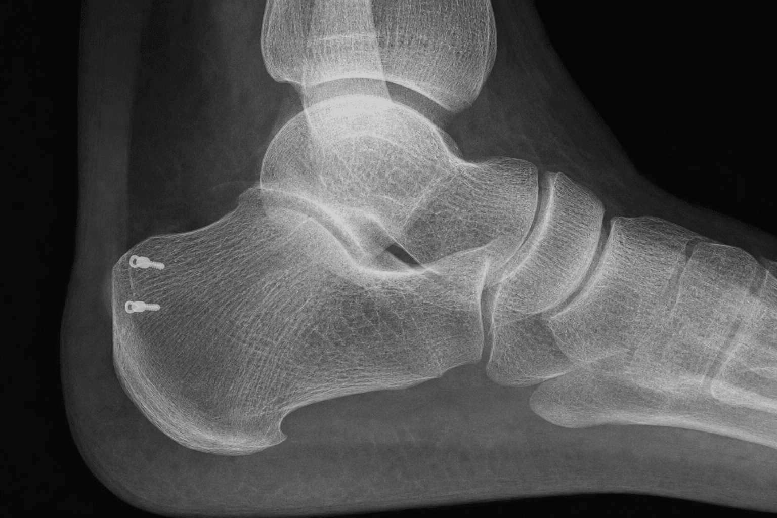
Protection: After debridement, assess the percentage of the insertion that remains attached. If less than 50% remains, use suture anchors. Place anchors in the dense bone of the posterosuperior tuberosity, not in the cancellous body. Ensure anchors are fully seated and purchase is confirmed before tying sutures.
Problem: The skin over the posterior heel is thin, relatively avascular, and subject to significant shear forces from shoe wear. Postoperative swelling, hematoma, and wound tension all increase the risk of wound breakdown and dehiscence — the most common and most troublesome complication of this surgery.
Incidence: Wound complications (breakdown, edge necrosis, infection) reported in 5-15% of cases in published series.
Prevention: Minimise periosteal stripping, achieve meticulous haemostasis, close the wound in layers without tension, consider a relaxing incision or V-to-Y closure if skin deficit is anticipated, and maintain strict elevation for 72 hours postoperatively.
Location: The posterior facet of the subtalar joint lies on the posterosuperior calcaneus, just anterior to the posterosuperior tuberosity. The Zadek osteotomy must be performed entirely posterior to the posterior facet to preserve subtalar joint congruity.
Risk: An osteotomy that violates the posterior facet can produce subtalar arthritis, pain, and loss of hindfoot motion.
Protection: Palpate the posterior facet percutaneously or use image intensification before making the osteotomy cuts. Mark the facet margin. Make the osteotomy at least 1-1.5 cm posterior to the facet. Confirm on fluoroscopy that the osteotomy does not extend into the facet.
Haglund deformity: Bony prominence of the posterosuperior calcaneal tuberosity. Radiographic diagnosis (Fowler-Philip angle, parallel pitch lines). May be asymptomatic until shoe wear or activity irritates the bursa and tendon.
Retrocalcaneal bursitis: Inflammation of the bursa between the Achilles tendon and the posterosuperior calcaneus. Pain is posterolateral, worse with shoe wear, tender lateral to the tendon at the posterosuperior calcaneus.
Insertional Achilles tendinopathy: Degeneration (tendinosis) and often partial tearing of the Achilles at its calcaneal insertion. Pain is directly at the tendon insertion, with possible visible swelling and calcification on radiographs.
Combined pathology: Most surgical patients have all three — the prominence irritates the bursa, chronic bursitis causes tendon degeneration, and the degenerate tendon loses its ability to absorb load. Surgery addresses all three components.
The trap: Resecting some but not all of the posterosuperior calcaneal prominence leaves a residual bump that continues to impinge on the tendon and bursa. This is the most common cause of residual pain and recurrent symptoms.
Verification: After resection, palpate the posterosuperior corner. Flatten the bone with a rongeur and then contour it with a burr. Confirm on fluoroscopy (lateral view) that the posterosuperior corner is flush with the anterior cortex line (parallel pitch lines now negative). Check full passive dorsiflexion — the tendon should clear the calcaneus without impingement.
Intraoperative test: Passively dorsiflex the ankle. No soft-tissue impingement should be palpable between the Achilles and the calcaneus. If impingement persists, resect further until clearance is achieved.
H.A.G.L.U.N.DHAGLUND — Diagnosis and Pre-operative Planning
S.U.R.G.E.O.NSURGEON — Operative Technique Steps
Surgical Indications
Absolute Indications
- Insertional Achilles tendinopathy with Haglund deformity recalcitrant to at least 3-6 months of structured non-operative treatment (heel-lift orthosis, activity modification, eccentric loading programme, at least one image-guided injection)
- Retrocalcaneal bursitis with posterosuperior calcaneal impingement causing refractory posterior heel pain confirmed by positive parallel pitch lines and MRI evidence of bursal wall thickening and tendon involvement
- Partial Achilles tendon avulsion at the insertion (greater than 50% detachment) with associated Haglund prominence, in an active patient
- Symptomatic intratendinous calcification at the Achilles insertion with pain on activity and failure of non-operative management
Relative Indications
- Haglund deformity with mild insertional tendinopathy where non-operative treatment has been partially successful but the patient wishes to return to high-impact sport
- Recurrent symptoms after a previous conservative (partial) bursoscopy or limited bursectomy
- Prominent Haglund deformity in a patient who cannot accommodate shoe modification due to occupational or sporting requirements
- Failure of a Zadek osteotomy to resolve symptoms (revision with tendon repair)
Contraindications
Absolute:
- Active local infection (cellulitis, ulceration, or deep heel wound infection) — surgery deferred until resolved
- Severe peripheral vascular disease with critical limb ischaemia — wound healing is unreliable
- Active inflammatory arthropathy flare at the Achilles insertion — optimise medical management first
Relative:
- Poorly controlled diabetes with significant peripheral neuropathy — elevated wound breakdown risk; optimise glycaemic control first
- Established subtalar arthritis — a Zadek osteotomy may worsen hindfoot pain; consider fusion instead
- Heavy tobacco use (greater than 20 pack-years) — counsel about wound healing risk; cessation before surgery is advisable
- Patient unable to comply with 4-6 weeks non-weight-bearing — this is essential for tendon healing; discuss expectations
Evidence for Non-Operative Treatment
Heel-Lift Orthosis and Shoe Modification
- A heel lift (1-1.5 cm) inside the shoe reduces Achilles tension at the insertion and offloads the retrocalcaneal bursa — this is the first-line conservative measure
- Shoe modification: open-backed shoe or a heel counter with a cut-out at the posterosuperior corner (the Haglund bump area) eliminates direct shoe pressure
- Posterior heel cushioning pads (silicone heel sleeves, felt pads) reduce direct impingement from footwear
- Eccentric loading programme (Alfredson protocol) has moderate evidence for non-insertional Achilles tendinopathy but less evidence specifically for insertional disease — modified protocols that limit dorsiflexion beyond neutral may be needed to avoid exacerbating insertional pain
Injections
- Corticosteroid injection into the retrocalcaneal bursa provides short-term pain relief in 50-70% of cases, but carries a risk of Achilles tendon rupture if injected into the tendon substance — image-guided (ultrasound) injection into the bursa is preferred
- Platelet-rich plasma (PRP) injection: limited evidence for insertional tendinopathy; some case series report symptomatic improvement but the evidence is not robust and it is not considered a replacement for structured rehabilitation
- High-volume injection (saline and steroid) has been described for non-insertional Achilles tendinopathy but has no specific evidence for Haglund deformity
Evidence for Surgery
Prominence Resection with Tendon Debridement
- Resection of the posterosuperior calcaneal prominence combined with debridement of the degenerate tendon and retrocalcaneal bursa provides reliable pain relief in 75-90% of patients at medium-term follow-up (2-5 years)
- Anderson et al. reported satisfactory outcomes in 85% of patients at mean 3.4-year follow-up after calcaneal exostectomy with tendon debridement; patients with greater than 50% tendon detachment requiring anchor reattachment had somewhat lower satisfaction
- The most common reason for residual dissatisfaction is incomplete prominence resection — impingement persists if the posterosuperior corner is not fully flattened
Zadek Osteotomy
- The Zadek dorsal closing-wedge osteotomy is indicated for Haglund deformity with a large posterosuperior prominence (greater than 15 mm), a steep calcaneal inclination angle, or failure of isolated prominence resection
- By removing a dorsal wedge and shifting the tuberosity posteriorly, the osteotomy decompresses the Achilles insertion without requiring an extensive tendon detachment for exposure
- Nielson et al. reported 89% good-to-excellent results at mean 4.5-year follow-up after Zadek osteotomy with concurrent Achilles debridement
- Advantages over isolated exostectomy: avoids extensive tendon detachment in selected cases, addresses the biomechanical cause of the prominence, and allows simultaneous correction of calcaneal malalignment
- Disadvantages: requires internal fixation (screws), longer period of protected weight-bearing (6 weeks non-weight-bearing for osteotomy healing), hardware removal in 10-20% of cases if symptomatic
Isolated Exostectomy vs Zadek Osteotomy with Tendon Debridement
Key Evidence
Surgical treatment of Achilles tendinitis by decompression of the retrocalcaneal bursa and the superior calcaneal tuberosity
Percutaneous Zadek Osteotomy vs Open Haglund Resection for Insertional Achilles Tendinopathy: Early Outcomes and Complication Rates
Comparison of open and endoscopic techniques of isolated calcaneoplasty in the surgical treatment of insertional tendinopathy of the Achilles tendon
Single-Row Repair Versus Double-Row Repair in the Surgical Management of Achilles Insertional Tendinopathy: A Systematic Review
Higher BMI Is Associated With Wound Breakdown Following Resection of Haglund Deformity
Clinical Decision Scenarios
Practise clinical reasoning and management decisions out loud
“A 42-year-old recreational runner presents with a 12-month history of posterolateral right heel pain. She has tried heel lifts, activity modification, a structured eccentric loading programme, and a single ultrasound-guided corticosteroid injection into the retrocalcaneal bursa with temporary relief. Her lateral heel radiograph shows a positive parallel pitch lines test and a Fowler-Philip angle of 74 degrees. MRI shows thickening of the retrocalcaneal bursa, intratendinous calcification at the Achilles insertion, and approximately 60% detachment of the tendon from the calcaneal tuberosity. How do you manage her?”
“A 55-year-old man with a long-standing Haglund deformity underwent an isolated calcaneal exostectomy 18 months ago at another hospital. His initial pain relief lasted 6 months but he has had recurrence of posterolateral heel pain, which is now worse than before surgery. Examination reveals tenderness at the posterosuperior calcaneus, a positive two-pinch test, and pain on full dorsiflexion. Radiographs show a residual posterosuperior prominence. MRI shows recurrent retrocalcaneal bursitis and a new area of intratendinous calcification at the Achilles insertion. How do you proceed?”
“A 35-year-old woman is being counselled for Haglund deformity correction. She asks you about the differences between open and endoscopic surgery. How do you explain the options?”