Mallet Finger — Management and Fixation

Hand & WristIntermediateCore Procedure

Mallet Finger — Management and Fixation

Management of terminal extensor tendon (mallet) injuries at the DIP joint - continuous extension splinting for tendinous and most bony mallets, with operative fixation (extension-block pinning, ORIF, pull-out suture) reserved for large bony fragments with volar subluxation or open injuries

High-yield overview

Terminal extensor tendon disruption at the DIP joint — splinting first, fixation only when the joint has subluxated | intermediate

Surgical Imaging

Mallet finger extension-block pinning
Bony mallet finger treated with extension-block (Ishiguro) K-wire pinning, holding the DIP joint reduced while the dorsal fragment heals.Credit: AI-generated medical illustration · OrthoVellum
Critical Decision Points and Exam Traps
Volar Subluxation — The Real Operative Trigger

The trap: Operating on a bony mallet purely because the dorsal fragment looks large. Fragment size on its own does not predict outcome.

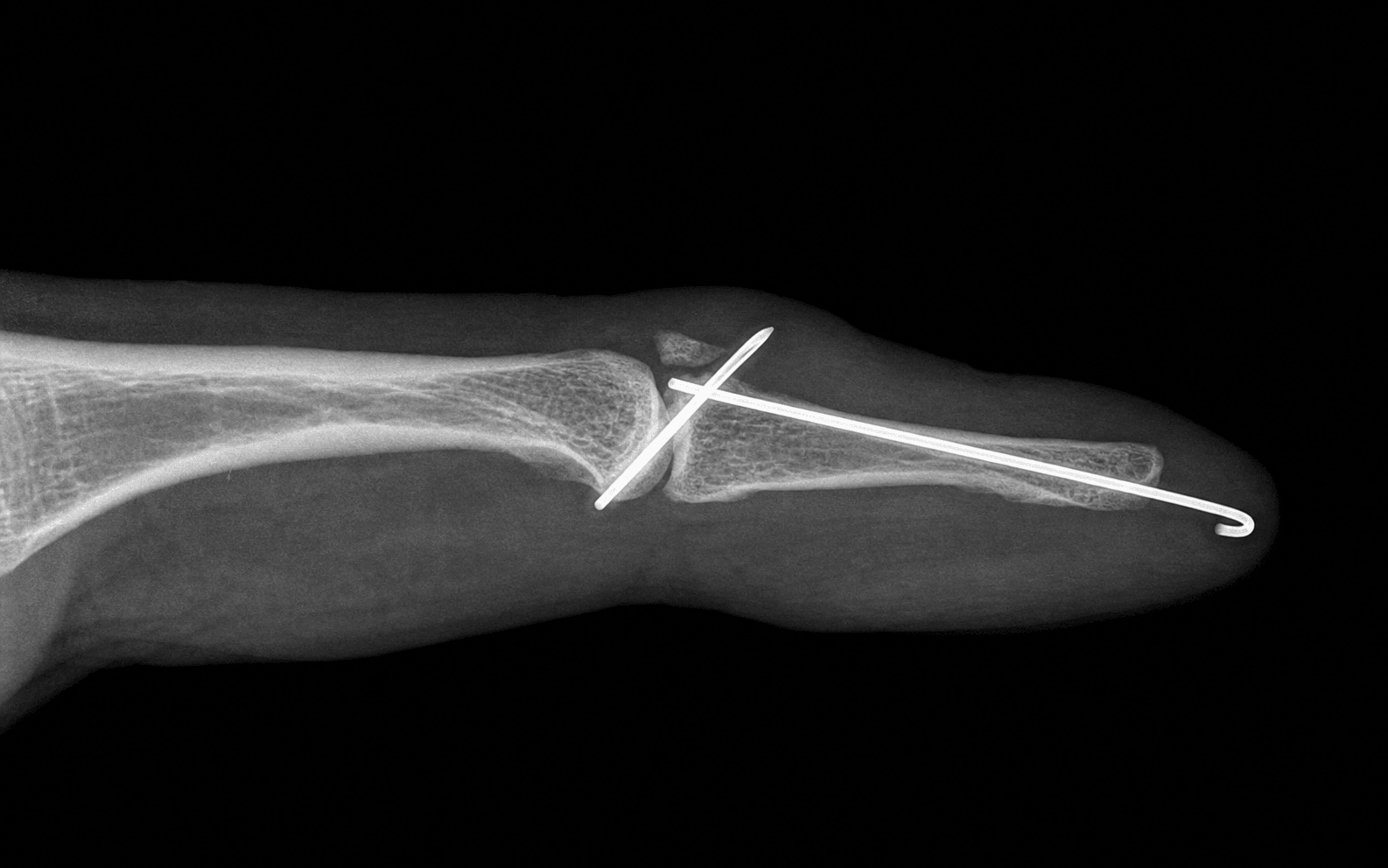
The fix: On a true lateral DIP radiograph, look specifically for volar (palmar) subluxation of the distal phalanx. It is the disordered, subluxated joint — together with a large dorsal fragment — that drives the decision to operate, not the fragment in isolation.

Any Flexion Restarts the Clock

The rule: Splintage must be CONTINUOUS, day and night. If the patient removes the splint and the DIP flexes even once — including during washing — the entire 6 to 8 week count starts over.

The fix: Teach skin care with the finger held flat and the DIP supported on a table so the tip never drops. Audit concordance at every clinic visit; a single break means restarting.

Dorsal Skin Necrosis from Over-extension

The risk: Holding the DIP in marked hyperextension, or a splint that is too tight, can devascularise the thin dorsal skin over the DIP, producing blistering and full-thickness necrosis.

The fix: Splint in SLIGHT hyperextension only; pad bony prominences; review the skin within the first week and at each splint change. Skin blanching on extension means the angle is too great.

Nail-Matrix Injury from Pins and Splints

The risk: A longitudinal K-wire crossing the DIP can injure the germinal or sterile nail matrix, producing a deformed nail. A tight dorsal splint can pressure the eponychial fold.

The fix: When transfixing the DIP, pass the wire along the axis that avoids the nail matrix and keep the entry point clear of the eponychium. Keep splints off the proximal nail fold.

Swan-Neck Deformity — The Classic Sequela

Why it happens: A chronic mallet leaves the terminal tendon lax; extensor force is transmitted proximally to the central slip through the lateral bands, producing PIP hyperextension and a DIP flexion lag.

The fix: Prevent it by achieving and maintaining DIP extension early. An established swan-neck may be managed with a figure-of-eight splint, or in severe symptomatic cases with PIP stabilisation such as a superficialis tenodesis.

Open Mallet — Operate, Do Not Just Splint

The trap: Treating an open (laceration) mallet as a routine closed Type I injury with splintage alone.

The fix: An open injury is a different problem. It needs wound lavage and debridement, direct terminal-tendon repair, and usually a longitudinal K-wire across the DIP to hold extension for about 6 weeks while the tendon heals.

Mnemonic

M.A.L.L.E.TMALLET — Assessment and Management

Mnemonic

S.P.L.I.N.TSPLINT — Principles of Extension Splinting

Definition and Mechanism

A mallet finger is a disruption of the terminal extensor tendon at its insertion on the dorsal base of the distal phalanx, at the DIP joint. The hallmark is loss of active DIP extension with preserved passive extension. The classic mechanism is a sudden forced flexion force applied to an actively extended fingertip — the ball-strike injury that gives it the nickname baseball finger. A direct blow to the dorsum of the DIP or a laceration can also produce it.

The lesion may be:

  • Tendinous (soft-tissue) mallet — rupture of the terminal tendon itself
  • Bony mallet — avulsion of a fragment of the dorsal base of the distal phalanx with the tendon still attached to it

Doyle Classification

I
Description
Closed, soft-tissue (tendinous), with or without a tiny avulsion fragment
Typical Management
Continuous extension splinting
II
Description
Laceration, tendon continuity preserved or lost
Typical Management
Surgical repair
III
Description
Deep abrasion with loss of skin, subcutaneous tissue and tendon substance
Typical Management
Surgical repair, often soft-tissue cover
IVa
Description
Paediatric — transepiphyseal fracture (Salter-Harris pattern)
Typical Management
Closed reduction and splinting if displaced
IVb
Description
Bony fragment of intermediate size, NO volar subluxation
Typical Management
Trial of continuous splinting
IVc
Description
Large bony fragment WITH volar (palmar) subluxation of the distal phalanx
Typical Management
Operative fixation

Indications for Treatment

Non-operative (the mainstay)

  • All closed soft-tissue (tendinous) mallets — Doyle Type I
  • Most bony mallets, including those with a substantial fragment, provided the joint is congruent (no volar subluxation)
  • Delayed presentation — even weeks to months old — still warrants a trial of continuous splinting

Operative indications (selective)

  • A large bony fragment involving more than approximately one third of the articular surface WITH volar (palmar) subluxation of the distal phalanx — the subluxated, unstable joint is the real indication
  • Open injuries — Doyle Types II and III (laceration or tissue loss)
  • A subset of failures of a genuine, concordant trial of splinting
  • Selected paediatric transepiphyseal fractures that are significantly displaced

Relative / cautious indications

  • A fragment involving approximately one third or more of the surface WITHOUT subluxation is increasingly managed non-operatively, because the evidence shows fragment size alone does not dictate outcome
  • Avoid operating simply for cosmesis of a dorsally prominent fragment

Continuous Splinting versus Operative Fixation

Evidence Base

  • Cochrane systematic review: randomised trial evidence for treating mallet finger is limited and insufficient to determine the single best treatment; no trial has shown surgery to be superior to conservative splinting for the typical closed injury.
  • Wehbe and Schneider (1984): in a landmark review of mallet fractures, the size of the fracture fragment did not correlate with the clinical result, supporting conservative management of most bony mallets.
  • Crawford (1984): the molded polyethylene splint (the basis of the Stack-type splint) holds the DIP in extension while allowing skin care, improving patient concordance — the principle that underpins modern non-operative management.
  • Operative series: extension-block (Ishiguro) pinning and ORIF are reported to give good reduction and outcome in bony mallets with volar subluxation, but they are reserved for the minority with an unstable joint.

Clinical Decision Scenarios

Practise clinical reasoning and management decisions out loud

Viva scenarioStandard
Clinical prompt

A 28-year-old cricketer was struck on the tip of his right ring finger by a ball two days ago. He cannot straighten the end of his finger, but he can bend it normally and passive extension of the DIP is full. A true lateral radiograph shows no fracture and a congruent DIP joint. What is your management?

Practical approach
This is a classic closed tendinous mallet finger — Doyle Type I. The mechanism (a forced flexion blow to an extended fingertip), the loss of active DIP extension with preserved passive extension, and the fracture-free, congruent radiograph all point to a soft-tissue terminal tendon rupture. The correct management is non-operative. **Treatment plan**: I would fit a continuous DIP extension splint holding the DIP in slight hyperextension with the PIP left completely free, to be worn day and night for six to eight weeks, followed by a graded wean over a further two to six weeks of night and activity splinting. **The single most important piece of counselling**: the splintage must be continuous. I would explain that if the DIP flexes even once while the splint is off, the entire six to eight week clock restarts. I would teach skin care with the finger held flat on a table so the tip never drops, and review concordance and skin condition within the first week and at each subsequent visit. **Activity**: because he is a cricketer, I would tell him he can return to play with the splint in situ once comfortable, but he must keep the DIP extended at all times. **Why not operate**: there is no bony fragment, no volar subluxation, and the joint is congruent — there is no operative indication. The evidence, including a Cochrane review, shows no benefit of surgery over splinting for the typical closed mallet, and surgery would only add risk in this tiny bone. **Follow-up**: review at two to four weeks to check the skin and splint fit, then at the end of continuous splinting to begin the wean. I would warn him that a small residual extension lag is common and functionally insignificant.
Viva scenarioStandard
Clinical prompt

A 42-year-old labourer injured his middle finger a week ago. He cannot extend the DIP. A true lateral radiograph shows a bony mallet with a dorsal fragment involving approximately half of the articular surface and clear volar (palmar) subluxation of the distal phalanx. How do you manage him, and what operation would you offer?

Practical approach
This is a bony mallet with volar subluxation of the distal phalanx — Doyle Type IVc. The critical finding is not the size of the fragment alone but the volar subluxation, which means the DIP joint is unstable and incongruent. This is one of the few genuine operative indications in mallet finger. **Why operate**: a subluxated joint left unreduced will develop pain, stiffness, deformity and early arthritis. Unlike a congruent bony mallet (which does well in a splint regardless of fragment size), this joint needs to be reduced. **My operation of choice — extension-block (Ishiguro) percutaneous pinning**: with the hand on a radiolucent table under image intensifier, I flex the PIP to about 90 degrees and pass a fine K-wire into the dorsal distal middle phalanx angled distally at about 40 to 45 degrees. This block wire stops the dorsal fragment being driven dorsally as I extend the DIP. I then extend the DIP — the fragment is levered down against the block wire and the joint reduces. I confirm congruity on a true lateral fluoro view, then drive a longitudinal K-wire from the tip of the distal phalanx across the DIP to hold extension and maintain the reduction. I bend and cut the wires and protect the finger in a splint. **Alternative — open reduction and internal fixation**: if the fragment is large and single I might instead use a mini lag screw, or a pull-out suture through the terminal tendon for a fragment too small to capture, always protected by a longitudinal DIP wire for about six weeks. **Post-operative**: the wires remain for about six weeks; I begin active PIP motion immediately. After removal, I start active DIP flexion and extension and buddy-tape for protection. **Counselling**: I would warn him specifically about pin-tract infection, wire migration or breakage, nail-matrix injury and nail deformity, residual extension lag, DIP stiffness, and the possibility of DIP arthritis despite reduction.
Viva scenarioStandard
Clinical prompt

A 60-year-old man presents with a mallet deformity of his index finger that he thinks happened about three months ago. He never sought treatment. He now has a DIP extension lag with early PIP hyperextension — a developing swan-neck posture. The joint is congruent on radiograph. What is your management?

Practical approach
This is a chronic, neglected mallet finger with an early swan-neck deformity. The joint is congruent, which is important — there is no acute operative indication. My first step would still be a genuine, supervised trial of continuous extension splinting, because delayed splinting remains effective for many patients even at three months. **Initial management — continuous splinting**: I would fit a DIP extension splint worn continuously for at least eight weeks, with the PIP left free. I would counsel him carefully on concordance — any flexion restarts the clock — and on skin care. I would review at intervals to check the skin, the splint fit, and the response of the extension lag. **Addressing the swan-neck**: while the DIP is being splinted, the PIP hyperextension is held in check. I would consider a figure-of-eight splint over the PIP to control the hyperextension posture during this phase. If the lag improves and the swan-neck settles, I would wean the DIP splint gradually. **If splinting fails and the swan-neck is symptomatic**: a persistent, functionally limiting swan-neck despite a concordant prolonged splinting trial is the scenario where surgery is considered. Options centre on stabilising the PIP in slight flexion, for example a superficialis (FDS) tenodesis of the PIP, or reconstruction of the oblique retinacular ligament to restore the DIP-to-PIP extension linkage. These are reconstructive procedures with real risks and are reserved for the symptomatic patient. **Counselling and expectation**: I would be honest that a small residual extension lag is common, is often functionally well tolerated, and that the goal of late intervention is a stable, painless, functional finger rather than perfect anatomy. For many patients, a prolonged splinting trial plus a figure-of-eight splint is sufficient and avoids the risks of late surgery.
Exam day cheat sheet
Mallet Finger — Management and Fixation — Exam Day Summary

References

Evidence

Interventions for treating mallet finger injuries

Handoll HH, Vaghela MVCochrane Database Syst Rev
Evidence

Mallet fractures

Wehbe MA, Schneider LHJ Bone Joint Surg Am
Evidence

The molded polythene splint for mallet finger deformities

Crawford GPJ Hand Surg Am
Evidence

Mallet deformity of the finger. Five-year follow-up of conservative treatment

Okafor B, Mbubaegbu C, Munshi I, Williams DJJ Bone Joint Surg Br
Evidence

Acute Mallet Finger Injuries-A Review

Sivakumar BS, Graham DJ, Ledgard JP, Lawson RDJ Hand Surg Am
Editorially reviewed — transparent references and correction processPublished by OrthoVellum Medical Education TeamEditorial boardMethodologyReview policy
Educational disclosure

Educational content is reviewed for source visibility, editorial coherence, and correction readiness.

No individual clinician credential is claimed unless a named person is shown.

Verify before clinical use; this is not medical advice or a substitute for local guidance.