Combined dorsal-volar approach for acute perilunate and lunate dislocations | advanced
Surgical Imaging
Location: The median nerve lies immediately volar to the lunate in its displaced position within the carpal tunnel. Up to 25 percent of perilunate dislocations present with acute median neuropathy.
Risk: Closed reduction can temporarily relieve pressure, but persistent sensory loss or thenar weakness after reduction indicates ongoing compression from haematoma or the lunate itself. This is a surgical emergency — delay greater than 8 hours increases permanent nerve damage risk.
Fix: Document two-point discrimination (normal less than 6 mm) and abductor pollicis brevis strength before any reduction. If deficit persists, proceed directly to combined approach with carpal tunnel release.
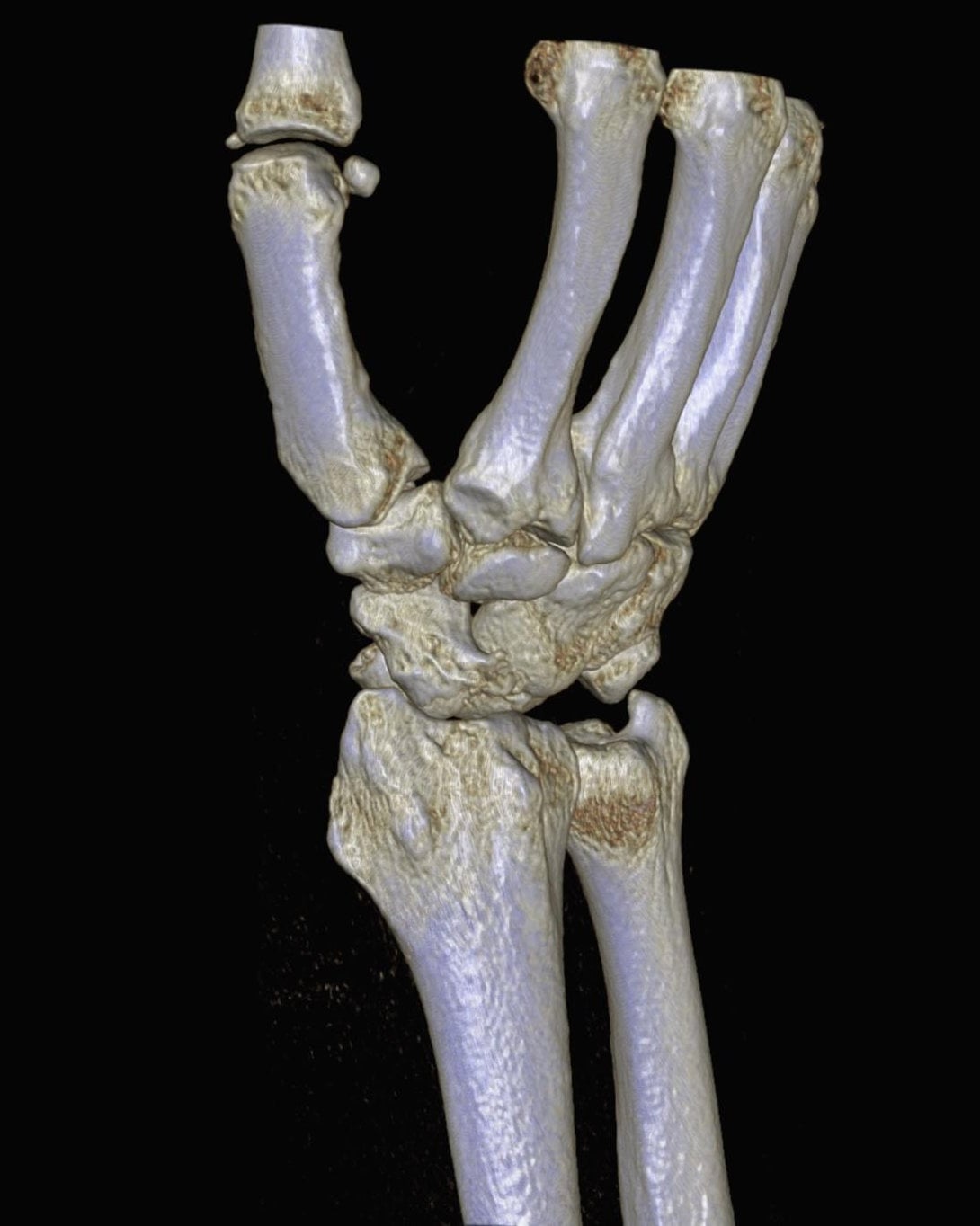
Trap: On the AP radiograph the lunate may appear reduced while the lateral shows complete volar dislocation (spilled-teacup sign). The lunate can rotate 90-180 degrees and lie entirely within the carpal canal.
Risk: Attempting closed reduction of a true Stage IV injury without open visualisation risks further cartilage damage and median nerve contusion. The lunate must be reduced under direct vision through the volar approach.
Fix: Obtain a true lateral radiograph in every high-energy wrist injury. If the capitate lies dorsal to the lunate or the lunate is not concentrically reduced, assume Stage IV and plan open reduction.
Location: The dorsal scapholunate ligament is the critical stabiliser — its repair or reconstruction determines long-term outcome. The lunotriquetral ligament is often torn but less critical if the scaphoid is reduced.
Risk: Failure to anatomically reduce the scapholunate interval (greater than 3 mm gap or greater than 10 degrees scapholunate angle difference) leads to scapholunate advanced collapse (SLAC) within 12-24 months.
Fix: Use K-wires as joysticks to reduce the scaphoid to the lunate under direct dorsal vision. Confirm reduction with fluoroscopy before definitive fixation. Repair the dorsal scapholunate ligament with suture anchors or transosseous sutures.
Location: The space of Poirier is the interval between the radioscaphocapitate and long radiolunate ligaments on the volar surface of the lunate. This is the site of the volar capsular tear in perilunate injuries.
Risk: Leaving the volar rent unrepaired allows recurrent volar intercalated segment instability (VISI) and progressive carpal collapse even if the dorsal ligaments are fixed.
Fix: Through the volar approach, identify the rent, reduce the lunate, and repair the capsule with strong non-absorbable sutures or anchors. This step is as important as the dorsal ligament repair.
Trap: In greater-arc injuries the scaphoid fracture is often oblique and unstable. Simply pinning the proximal and distal poles without compression or bone graft leads to nonunion and humpback deformity.
Risk: Scaphoid nonunion after perilunate dislocation has a 30-50 percent rate if reduction is not anatomic and compression is not achieved. Humpback deformity rapidly progresses to SLAC wrist.
Fix: Reduce the scaphoid anatomically under direct vision (dorsal approach). Use a headless compression screw (preferred) or multiple K-wires with compression. Add cancellous bone graft from the distal radius if comminution or bone loss is present.
Location: After perilunate injury the lunate tends to extend (DISI posture) because the scapholunate ligament is torn. The scaphoid flexes and the lunotriquetral ligament fails to stabilise the triquetrum.
Risk: Persistent DISI (scapholunate angle greater than 70 degrees on lateral radiograph) is the strongest predictor of post-traumatic arthritis and SLAC wrist at 5 years.
Fix: Intraoperative fluoroscopic confirmation of scapholunate angle between 30-60 degrees and scapholunate gap less than 3 mm is mandatory before leaving the operating theatre. Temporary K-wire stabilisation across the reduced intervals protects the repair for 8-12 weeks.
M.A.Y.F.I.E.L.DMAYFIELD — Progressive Perilunar Instability Stages
D.O.R.S.A.L-V.O.L.A.RDORSAL-VOLAR — Combined Approach Principles
C.O.M.P.L.I.C.A.TCOMPLICATION — Long-Term Risks After Perilunate ORIF
Surgical Indications
Absolute Indications
- Acute perilunate or lunate dislocation (Mayfield Stage II-IV) with or without median nerve compression
- Failed closed reduction or recurrent instability after closed reduction
- Open perilunate injury with contamination or associated lacerations
- Acute carpal tunnel syndrome with objective sensory or motor deficit persisting after attempted closed reduction
Relative Indications
- Greater-arc injuries (trans-scaphoid, trans-triquetral, trans-capitate perilunate fracture-dislocations) requiring anatomic scaphoid reduction
- Delayed presentation (greater than 24 hours) with significant swelling where closed reduction is unsafe
- Associated distal radius fracture requiring simultaneous fixation
Contraindications
Absolute:
- Life-threatening polytrauma precluding timely wrist surgery (stabilise patient first)
- Active infection at the surgical site
- Patient refusal or inability to comply with postoperative immobilisation and rehabilitation
Relative:
- Low-demand elderly patient with chronic dislocation greater than 6 weeks and minimal symptoms (consider salvage rather than reconstruction)
- Severe medical comorbidities increasing surgical risk
Evidence for Timing and Approach
Timing of Reduction
- Best outcomes when the carpus is reduced within 6 hours of injury (Herzberg 1993, Level IV)
- Median nerve recovery is time-dependent — permanent sensory loss rises sharply after 8 hours of compression
- Delayed presentation (greater than 24 hours) increases infection risk and technical difficulty of reduction due to soft-tissue swelling and haematoma organisation
Combined Dorsal and Volar Approach
- The combined approach allows complete visualisation of both the dorsal and volar ligamentous injuries and direct median nerve decompression
- Single-approach techniques (dorsal only or volar only) have higher rates of residual instability and missed median nerve compression
- A 2018 systematic review (Mallett 2018) found combined approaches achieved anatomic reduction in 85 percent of cases versus 60 percent with single approaches
Fixation Choices
- Headless compression screws for trans-scaphoid injuries provide superior compression and earlier mobilisation compared with K-wires alone
- Suture-anchor repair of the dorsal scapholunate ligament improves radiographic outcomes at 2 years compared with K-wire stabilisation alone (Pappou 2021, Level III)
Clinical Decision Scenarios
Practise clinical reasoning and management decisions out loud
“A 32-year-old motorcyclist is brought in after a high-speed collision. He has a grossly deformed left wrist with an obvious median nerve sensory deficit (two-point discrimination 12 mm in the index finger, weak abductor pollicis brevis). The lateral radiograph shows a spilled-teacup sign with the lunate lying in the carpal canal. How do you manage this patient?”
“You have performed combined dorsal and volar ORIF for a trans-scaphoid perilunate dislocation in a 28-year-old labourer. At 8 weeks the K-wires are removed and radiographs show anatomic reduction with no gap or step. At 6 months he returns with radial-sided wrist pain and radiographs show early radioscaphoid narrowing. What has happened and how do you manage it?”
“A 45-year-old woman presents 18 months after a perilunate dislocation treated with closed reduction and percutaneous pinning elsewhere. She has chronic wrist pain, reduced grip strength (40 percent contralateral), and radiographs show a 5 mm scapholunate gap with a 85-degree scapholunate angle and early radioscaphoid arthritis. What are your options?”