Staged spanning external fixation then definitive ORIF of the tibial plafond, dictated by the soft-tissue envelope | advanced
- The SOFT-TISSUE ENVELOPE dominates pilon management, not the bone. These are high-energy axial-load injuries that drive the talus into the plafond, with fracture blisters, massive swelling and a high rate of open injury. Early definitive ORIF of a swollen high-energy pilon produced catastrophic wound breakdown and deep infection rates historically reported as high as 40 percent.
- The staged protocol (Sirkin and Patterson) transformed outcomes: STAGE 1 is immediate spanning external fixation across the ankle (with or without fibular ORIF) to restore length, alignment and rotation and rest the soft tissues; STAGE 2 is definitive articular ORIF once swelling settles, signalled by return of skin wrinkles (the wrinkle sign), typically at 7 to 21 days.
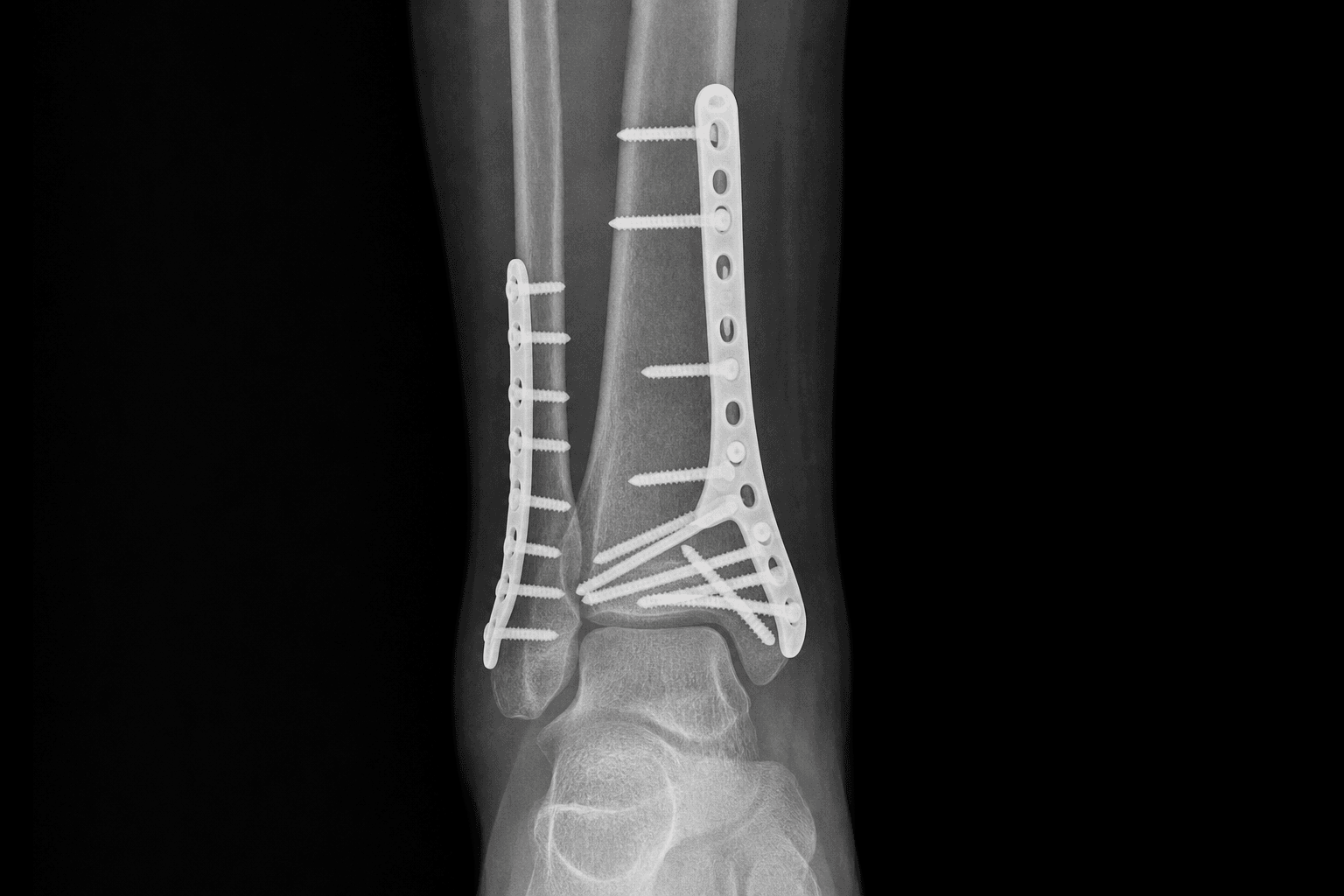
- Reduction follows the Ruedi-Allgower sequence: (1) restore fibular length and rotation, (2) reconstruct the tibial articular surface from posterior to anterior (Volkmann posterolateral, Chaput anterolateral and medial fragments; elevate and bone-graft the impacted central die-punch), (3) buttress the metaphyseal shell, (4) reconnect the reconstructed articular block to the diaphysis.
- Post-traumatic ankle arthritis is extremely common even after an anatomic reduction, because the cartilage is irreversibly injured at the moment of impact. Anatomic articular reduction reduces but does not eliminate this, and the plafond is unforgiving of any residual step or gap. Outcomes overall remain guarded and must be discussed frankly with the patient.
When & Why
Indication. A symptomatic, displaced pilon (tibial plafond) fracture — a high-energy axial-load injury that drives the talus into the distal tibia, producing articular impaction, comminution, metaphyseal extension and a severe soft-tissue injury (AO/OTA type 43, especially the complete-articular C-type). Distinguish it from a lower-energy rotational ankle fracture (Weber / Lauge-Hansen), which has a largely intact plafond and a far better soft-tissue prognosis and must not be managed like a true pilon. The soft tissues, not the bone, dictate the plan. A perfectly reduced plafond beneath dead skin is a disaster. The single most important early decision is therefore not how to fix the bone but how to protect the envelope: span the ankle, elevate the limb, and wait for the wrinkle sign (the return of fine skin creases over the malleoli on dorsiflexion) before any definitive articular surgery. Absent wrinkles mean persistent oedema and a high wound-breakdown risk; present wrinkles mean the envelope has recovered — typically at 7 to 21 days of elevation and spanning fixation.
- Early single-stage ORIF
- Within 24 to 48 hours of injury
- Staged (ex-fix then ORIF)
- Delayed to 7 to 21 days (wrinkle sign)
- Early single-stage ORIF
- Swollen, blistered, high-energy
- Staged (ex-fix then ORIF)
- Recovered, wrinkles returned
- Early single-stage ORIF
- Historically very high (reported up to around 40 percent)
- Staged (ex-fix then ORIF)
- Substantially lower
- Early single-stage ORIF
- High
- Staged (ex-fix then ORIF)
- Lower
- Early single-stage ORIF
- Low-energy patterns with a benign envelope
- Staged (ex-fix then ORIF)
- High-energy C-type pilons with soft-tissue injury
- Early single-stage ORIF
- Not addressed until ORIF
- Staged (ex-fix then ORIF)
- Restored immediately by the spanning fixator
In a high-energy pilon my first decision is not how to fix the bone but how to protect the soft tissues. I span the ankle, elevate the limb and wait for the wrinkle sign — the return of skin creases over the malleoli — before any definitive articular surgery. The skin dictates my timeline, usually 7 to 21 days.
When to deviate from the standard staged plan. For very poor soft tissues, gross contamination, or where formal plating is unsafe, definitive treatment with a ring (circular) fixator plus limited percutaneous articular screws is a recognised alternative that minimises soft-tissue dissection. For a non-reconstructable plafond (severe comminution with unsalvageable articular cartilage), particularly in older or low-demand patients, primary tibiotalar arthrodesis is a legitimate definitive option that avoids a predictable secondary fusion. Consent specifically for wound dehiscence and breakdown over hardware, deep infection and osteomyelitis, post-traumatic ankle arthritis (common even after a perfect reduction), malunion, nonunion, stiffness, and the likely need for a secondary fusion. Be explicit that smoking and diabetes are the dominant modifiable risk factors for wound breakdown, deep infection and nonunion — counsel on smoking cessation and optimise glycaemic control before stage-2 ORIF. Outcomes are guarded and must be discussed frankly. Setup. Supine with a bump under the ipsilateral hip for stage 1; image intensifier from the opposite side; foot at the end of the table. Plan every incision against the planned definitive tibial incisions and skin bridges, keeping at least 7 cm of intact skin between any two surgical incisions. The anterolateral approach is generally preferred for the definitive fixation because it allows healthy muscle to cover the plate.
The Operation
The goal is a safely healed wound over an anatomically reconstructed joint. This is achieved in two stages: stage 1 buys time and length (a spanning external fixator restores length, alignment and rotation and rests the soft tissues); stage 2 buys anatomy (definitive articular ORIF through the approach the CT dictates, once the wrinkle sign is present). The exposure is laid out step by step below.
The staged operative sequence
- Position: supine, bump under the ipsilateral hip, image intensifier from the opposite side, foot at the end of the table.
- Construct: two tibial half-pins in the anteromedial subcutaneous border well proximal to the zone of injury (never within the planned definitive plate footprint); a transcalcaneal transfixion pin through the posterior calcaneal tuberosity (the safe corridor); and frequently a first-metatarsal pin to hold the foot plantigrade.
- Connect with bars in a delta or simple medial frame and apply traction for ligamentotaxis — this pulls the fracture out to length, tensions the soft tissues back to length and indirectly reduces many fragments.
- Performed at stage 1 only if the lateral soft tissues permit, through a direct lateral or posterolateral incision, restoring fibular length and rotation with a one-third tubular or distal fibular plate.
- It re-establishes lateral column length and provides a stable template for the later tibial reduction — but its incision must be planned against the definitive tibial incisions and skin bridges.
- Strict elevation of the limb above the heart; daily wound and pin-site care; watch blister progression.
- Optimise the host: explicit smoking-cessation counselling, glycaemic control, nutrition.
- Obtain CT after the fixator is on (traction CT under ligamentotaxis) — fragments pulled out to length give a far clearer articular map for planning approaches and fragment-specific fixation.
- Do not book definitive surgery until the wrinkle sign returns (return of skin creases over the malleoli), usually at 7 to 21 days.
- Anteromedial: classic, excellent access to the plafond and medial fragments, but the plate lies directly beneath thin subcutaneous skin — higher wound-breakdown risk.
- Anterolateral (generally preferred for high-energy patterns): accesses the Chaput fragment and much of the plafond; muscle (tibialis anterior and the extensor mass) can be brought over the hardware, giving more robust soft-tissue cover.
- Posterolateral: between the peroneals and FHL for the Volkmann (posterolateral) fragment and the fibula through one incision; patient prone or lateral.
- Direct posterior / posteromedial: for posterior fragments; protect the posteromedial neurovascular bundle.
- Keep at least 7 cm of intact skin between any two incisions to avoid devascularising the central skin bridge.
- In the anterolateral approach, identify and protect the superficial peroneal nerve as it pierces the deep fascia in the distal third of the leg and crosses the field — direct laceration or traction causes dorsolateral foot numbness.
- Deepen in the internervous plane and elevate the muscle (tibialis anterior / extensor mass) to expose the plafond, bringing healthy muscle over the eventual plate.
- In the posterolateral approach, work between peroneals and FHL and keep the posterior tibial artery and tibial nerve (posteromedial, behind the medial malleolus) protected during reduction of posterior fragments.
- If not done at stage 1, reduce and plate the fibula to re-establish lateral column length and rotation — the template for the tibial reduction.
- Work from the stable posterior fragment forward. Reduce the posterolateral (Volkmann) fragment first as the keystone reference, then bring the anterolateral (Chaput) and medial fragments to it, recreating the plafond.
- Provisionally hold with K-wires and assess articular congruity directly and on the image intensifier.
- Chase an anatomic articular reduction with no step and no gap — the plafond tolerates incongruity poorly.
- The central impacted (key / die-punch) fragment is elevated from below back to the level of the reconstructed joint line.
- Fill the resulting metaphyseal void with bone graft or substitute to support the elevated articular block.
- Once the articular block is reconstructed and provisionally held, apply a low-profile periarticular (often locking) plate to buttress the metaphyseal shell and reconnect the articular block to the diaphysis.
- Where the articular reduction allows percutaneous reduction of the metaphysis, use minimally invasive plate osteosynthesis (MIPO) to limit soft-tissue insult.
- Confirm articular congruity, restored length, alignment and rotation, fibular reduction and a perfect mortise on the image intensifier in multiple planes.
- Achieve a tension-free closure — if the skin will not close without tension, do not force it; use a negative-pressure wound therapy dressing or arrange delayed closure or flap cover.
Placing tibial half-pins within the planned definitive plate footprint (keep them well proximal to the zone of injury); a stage-1 fibular incision that compromises a later anterolateral or posterolateral skin bridge; and inadequate ligamentotaxis leaving the limb short, so the soft tissues are never tensioned out. Never let articular planning distract from a limb-threatening emergency — compartment syndrome and an open contaminated wound are treated first, with the fixator simply holding the limb while these are addressed.
Leaving a residual articular step or gap (the plafond tolerates incongruity poorly and arthritis follows); failing to elevate the central die-punch fragment (leaves a depressed joint segment); and over-aggressive soft-tissue stripping of fragments, which devascularises them and risks nonunion.
For most high-energy pilons I favour the anterolateral approach because I can lay healthy muscle over the plate rather than leaving hardware under the thin anteromedial skin — while protecting the superficial peroneal nerve as it crosses the field. If I need both anterolateral and posterolateral access, I keep at least 7 cm of skin between the incisions.
I reconstruct the joint surface from posterior to anterior, using the posterolateral Volkmann fragment as my keystone. I bring the Chaput and medial fragments back to it and confirm I have recreated a congruous plafond before I think about the metaphysis. The plafond is unforgiving — I am chasing an anatomic articular reduction with no step and no gap.
My final intra-operative checks are articular congruity, length, alignment, rotation, the fibula and a perfect mortise on the intensifier. Then the most important step in a pilon — a tension-free skin closure. If the skin will not come together easily I will not force it; I will use a negative-pressure dressing or involve plastics for delayed closure or a flap rather than risk breakdown over my plate.
Aftercare & Complications
Rehabilitation | Phase | Timing | Weight-bearing | Therapy & wound care | |-------|--------|----------------|----------------------| | Wound-protective | 0 to ~2 weeks | Strictly non-weight-bearing | Strict elevation; vigilant surveillance for edge necrosis over the plate | | Early motion | Once wound secure | Non-weight-bearing | Gentle active ankle range-of-motion to limit stiffness | | Protected | ~6 to 10 to 12 weeks | Non-weight-bearing until radiographic metaphyseal union | Progressive ankle and hindfoot mobilisation | | Progression | After union (~10 to 12 weeks) | Graded to full weight-bearing under guidance | Proprioception and gait re-education; serial radiographs | Reinforce smoking cessation (impairs wound healing and union) and glycaemic control in diabetics at every stage, with nutritional optimisation and vitamin D or calcium where deficient. Outcomes and prognosis Outcomes after high-energy pilon fractures are guarded. Even with an anatomic reduction and an uncomplicated wound, many patients have residual ankle stiffness, pain and some functional limitation. Post-traumatic arthritis is the dominant long-term issue and relates to the energy and articular damage at the moment of injury more than to surgical technique alone. The staged protocol has dramatically reduced the catastrophic wound complications that historically plagued early ORIF, transforming the safety profile of surgery without making the joint normal. The key prognostic factors are: energy of injury and articular comminution (Ruedi-Allgower III / AO 43-C3 — worse); quality of articular reduction (a residual step or gap predicts faster arthritis); soft-tissue injury and wound complications (the strongest driver of catastrophic outcomes); and host factors (smoking and diabetes worsen wound healing, infection and union).
I counsel patients that a pilon is a serious injury with a guarded prognosis. My aims are a safely healed wound and an anatomically reconstructed joint, but even when I achieve both, post-traumatic arthritis is common because the cartilage is injured at the moment of impact. Staging the surgery is what protects the soft tissues; an anatomic reduction is what gives the joint its best — though not guaranteed — chance.
Complications
- Incidence
- The dreaded complication; historically up to around 40 percent with early ORIF, much lower when staged
- Recognition
- Wound edge necrosis, breakdown, exposed plate, serous or purulent discharge in the early post-operative weeks
- Prevention and management
- Prevention: stage the surgery and operate only after the wrinkle sign; respect 7 cm skin bridges; anterolateral approach to cover the plate with muscle; tension-free closure; smoking cessation and glucose control. Management: early aggressive debridement, negative-pressure wound therapy, antibiotics; early plastic surgery referral for free or local flap cover of exposed bone or hardware
- Incidence
- Markedly higher with early ORIF, smoking, diabetes and open injury
- Recognition
- Persistent pain, swelling, discharge, raised inflammatory markers; lucency or sequestrum on imaging
- Prevention and management
- Prevention: staged protocol, meticulous soft-tissue handling, debridement and antibiotics for open injury, host optimisation. Management: debridement, deep cultures, targeted antibiotics; remove hardware if loose or once union achieved; staged reconstruction or fusion for established osteomyelitis
- Incidence
- Very common even after anatomic reduction (high-energy cartilage injury)
- Recognition
- Progressive ankle pain and stiffness, loss of joint space and osteophytes on radiographs over months to years
- Prevention and management
- Prevention: anatomic articular reduction reduces but cannot eliminate it — the cartilage injury occurs at impact. Management: activity modification, analgesia, bracing, intra-articular injection; tibiotalar arthrodesis (or selected total ankle replacement) for end-stage symptomatic arthritis
- Incidence
- Recognised, higher with comminution and indirect reduction
- Recognition
- Varus or valgus or procurvatum or recurvatum deformity, articular step, shortening; symptomatic gait disturbance
- Prevention and management
- Prevention: restore fibular length first, anatomic articular reduction, confirm alignment on the intensifier in multiple planes. Management: corrective osteotomy for symptomatic extra-articular malunion; intra-articular malunion may necessitate fusion
- Incidence
- More common at the metaphyseal-diaphyseal junction, with comminution, smoking, infection
- Recognition
- Persistent pain and motion at the fracture, lack of bridging callus, hardware loosening or failure
- Prevention and management
- Prevention: preserve fragment blood supply (limit stripping), bone graft metaphyseal voids, stable buttress fixation, no smoking. Management: bone grafting and revision fixation; treat any underlying infection; consider fusion if the articular surface is destroyed
- Incidence
- Common — the ankle and subtalar joints
- Recognition
- Reduced dorsiflexion and plantarflexion and hindfoot motion, gait alteration at follow-up
- Prevention and management
- Prevention: stable fixation enabling early ankle range of motion once the wound is secure; physiotherapy. Management: structured physiotherapy; rarely arthrolysis; manage as part of overall guarded functional expectations
- Incidence
- A recognised end point in severe pilons
- Recognition
- Disabling post-traumatic arthritis or failed reconstruction unresponsive to non-operative measures
- Prevention and management
- Prevention: best initial reduction and infection avoidance. Management: tibiotalar arthrodesis is the salvage workhorse; consider primary arthrodesis up front for a non-reconstructable plafond in a low-demand patient
Viva & Exam Focus
PILONPILON — staged management principles
RUEDIRUEDI — articular reduction sequence
The trap: treating a pilon as a fracture to be fixed urgently. Operating definitively on a swollen, blistered, high-energy plafond leads to wound dehiscence and deep infection, historically reported up to 40 percent with early ORIF. The fix: stage the management — span the ankle, elevate the limb, and wait for the wrinkle sign (return of skin creases) before definitive ORIF, typically at 7 to 21 days. The skin dictates the timeline, not the radiograph.
Multiple approaches may be needed (anteromedial, anterolateral, posterolateral) plus a separate fibular incision. Closely spaced incisions devascularise the intervening skin bridge and cause central wound necrosis. Maintain at least 7 cm of intact skin between incisions, and choose the approach (often anterolateral) that places healthy muscle over the hardware.
The superficial peroneal nerve pierces the deep fascia in the distal third of the leg and crosses the field of the anterolateral approach. Direct laceration or traction neuritis causes dorsolateral foot numbness. Identify and protect it as it emerges from the fascia before deepening the dissection.
The posterior tibial artery and tibial nerve run in the posteromedial corner behind the medial malleolus; the posterolateral approach works between peroneals and FHL with the bundle medial. Posterior dissection for the Volkmann or posteromedial fragments can injure the bundle — know its position for every posterior approach and protect it during reduction of posterior fragments.
Pilon: high-energy axial load drives the talus into the plafond — articular impaction, comminution, metaphyseal involvement, severe soft-tissue injury (AO/OTA type 43). Rotational ankle fracture: lower-energy torsional injury (Weber / Lauge-Hansen) — malleolar fractures with a largely intact plafond and a far better soft-tissue prognosis. Do not manage a true pilon like a rotational ankle fracture.
High-energy pilons can have an associated leg compartment syndrome and are frequently open. Tense compartments and out-of-proportion pain mandate urgent assessment. Treat open wounds with early debridement, antibiotics and tetanus cover; have a low threshold for fasciotomy. The external fixator stabilises the limb while these are addressed — do not let articular planning distract from a limb-threatening emergency.
Clinical Decision Scenarios
Practise clinical reasoning and management decisions out loud
“A 35-year-old man falls from a height and sustains a closed high-energy pilon fracture (AO 43-C3) of the right distal tibia with marked swelling and haemorrhagic fracture blisters over the medial malleolus. How do you manage him from presentation?”
“You are planning the definitive ORIF for a C-type pilon. Talk me through how you choose your surgical approach and how you protect the soft tissues and key structures.”
“Two years after a staged ORIF for a C-type pilon, a 50-year-old patient has disabling ankle pain and stiffness. Radiographs show severe tibiotalar joint space loss with a healed, well-aligned reconstruction. How do you explain this and what are the options?”
The core concept
- High-energy axial load drives the talus into the plafond — articular impaction, comminution, severe soft-tissue injury
- The soft-tissue envelope dominates management, not the bone
- Early definitive ORIF through swollen skin caused wound breakdown and deep infection historically up to around 40 percent
- Staged protocol (Sirkin / Patterson): stage 1 spanning ex-fix, stage 2 definitive ORIF after the wrinkle sign
- Distinguish a true pilon (AO 43) from a low-energy rotational ankle fracture
Classification
- Ruedi-Allgower: I undisplaced; II displaced congruous; III comminuted and impacted (worst)
- AO/OTA 43: A extra-articular; B partial articular; C complete articular (true high-energy pilon, C3 fully comminuted)
- Tscherne for closed soft-tissue injury; Gustilo-Anderson for open injury
- Bony grade predicts reconstructability; soft-tissue grade predicts complications and timing
Assessing the soft tissues
- Fracture blisters: clear equals partial thickness; haemorrhagic equals deeper dermal injury, worse — do not incise through them
- The wrinkle sign (return of skin creases over the malleoli) is the operative green light for stage 2
- Wrinkle sign typically returns at 7 to 21 days of elevation and spanning fixation
- Always exclude compartment syndrome and treat open injuries (debridement, antibiotics, tetanus)
Stage 1 — spanning ex-fix (day 0)
- Goals: restore length, alignment, rotation; rest and decompress the soft tissues
- Tibial half-pins proximal to injury, transcalcaneal pin, often a first-metatarsal pin; traction for ligamentotaxis
- Fibular ORIF optional at stage 1 — restores lateral column length, but plan against definitive incisions
- Elevate the limb; obtain CT after the fixator (traction CT) to map fragments
Stage 2 — definitive ORIF (Ruedi-Allgower)
- 1. Restore fibular length and rotation
- 2. Reconstruct the articular surface posterior to anterior (Volkmann, then Chaput and medial fragments)
- 3. Elevate and bone-graft the impacted central die-punch fragment
- 4. Buttress the metaphysis and reconnect the articular block to the diaphysis (low-profile periarticular plate / MIPO)
- Tension-free closure is the single most important final step
Approaches and danger zones
- Anteromedial: great access but plate under thin skin (higher breakdown risk)
- Anterolateral: muscle cover over hardware (preferred for high-energy) — protect the superficial peroneal nerve
- Posterolateral: Volkmann fragment and fibula via one incision — between peroneals and FHL
- Maintain at least 7 cm skin bridges between incisions; protect the posteromedial neurovascular bundle
Complications
- Wound dehiscence and necrosis: the dreaded complication — stage surgery, anterolateral cover, tension-free closure, early flap if it fails
- Deep infection: worse with early ORIF, smoking, diabetes, open injury
- Post-traumatic arthritis: very common even after anatomic reduction (cartilage injured at impact)
- Malunion, nonunion (metaphyseal-diaphyseal junction), stiffness, and need for secondary fusion
Alternatives and counselling
- Definitive ring (circular) fixator plus limited internal fixation for very poor soft tissues
- Primary tibiotalar arthrodesis for a non-reconstructable plafond in a low-demand patient
- Smoking cessation and glycaemic control are key modifiable risk factors — counsel explicitly
- Counsel that outcomes are guarded and post-traumatic arthritis is common despite a perfect reduction
Background & Evidence
Mechanism and pathoanatomy. A pilon fracture is a high-energy axial-compression injury in which the talus is driven proximally into the tibial plafond, impacting and comminuting the articular surface and the metaphysis while inflicting severe damage on the soft-tissue envelope. CT-based fragment mapping (Topliss) defines the principal fragments that guide approach selection: the anterolateral (Chaput) fragment, the posterolateral (Volkmann) fragment, the medial malleolar fragment and the central impacted die-punch (key) fragment. The cartilage is injured at the moment of impact, which is why post-traumatic arthritis remains common even after an anatomic reconstruction. The soft tissues predict the outcome, not the bony grade. The bony classification predicts the reconstructive challenge, but the soft-tissue injury predicts the complications that actually determine outcome. Assess: - Fracture blisters: clear-fluid blisters are partial-thickness; blood-filled (haemorrhagic) blisters indicate deeper dermal injury and a worse prognosis — do not incise through them.
- Massive swelling: pitting oedema obliterating the normal skin creases over the malleoli.
- Skin integrity: any open wound upgrades urgency (debridement, antibiotics, tetanus).
- The wrinkle sign: return of fine skin creases over the malleoli on dorsiflexion indicates the swelling has resolved enough to permit definitive surgery — the single most useful clinical sign for timing. Classification. Two complementary systems describe the bone; two further systems grade the soft tissues.
- Categories
- I undisplaced, II displaced congruous, III comminuted impacted
- Basis
- Articular displacement and comminution
- What it predicts
- Reconstructability and broad prognosis (III worst)
- Categories
- A extra-articular, B partial articular, C complete articular
- Basis
- Relationship of articular surface to shaft
- What it predicts
- Surgical complexity; C-type is the true high-energy pilon
- Categories
- C0 to C3 by soft-tissue injury
- Basis
- Closed soft-tissue damage
- What it predicts
- Wound and infection risk; timing of ORIF
- Categories
- I to IIIC by wound and contamination
- Basis
- Open wound severity and contamination
- What it predicts
- Need for debridement, flap cover, infection risk
Key evidence. Ruedi and Allgower (1979) defined the classic four-step reconstruction principle (fibula, articular surface, bone graft, buttress to diaphysis) and the classification that bears their name. Sirkin and colleagues (1999) and Patterson and Cole (1999) established the staged protocol — spanning external fixation followed by delayed ORIF — showing a dramatic reduction in wound complications versus early aggressive ORIF. Bonar and Marsh (1994) reviewed the pivotal shift away from early ORIF toward soft-tissue-respecting strategies, and Topliss and colleagues (2005) provided the CT-based fragment anatomy that underpins modern approach selection.
References
The operative treatment of intra-articular fractures of the lower end of the tibia
Original description of the four-step reconstruction principle and the Ruedi-Allgower classification of plafond fractures.
A staged protocol for soft tissue management in the treatment of complex pilon fractures
Landmark staged protocol (spanning external fixation then delayed ORIF) showing dramatically reduced wound complications versus early ORIF.
Two-staged delayed open reduction and internal fixation of severe pilon fractures
Two-stage delayed ORIF series supporting soft-tissue rest before definitive articular reconstruction.
Tibial plafond fractures: changing principles of treatment
Review of the shift away from early aggressive ORIF towards soft-tissue-respecting and staged strategies.
Anatomy of pilon fractures of the distal tibia
CT-based fragment mapping (anterolateral Chaput, posterolateral Volkmann, medial and central die-punch fragments) informing approach selection.