Proximal Interphalangeal Joint Fracture-Dislocation

Hand & WristAdvancedCore Procedure

Proximal Interphalangeal Joint Fracture-Dislocation

Surgical technique guide for PIP joint fracture-dislocations: extension block pinning, ORIF, dynamic external fixation, and volar plate arthroplasty.

High-yield overview

Management of volar base middle phalanx fractures with dorsal subluxation | advanced

Surgical Imaging

PIP fracture-dislocation pinning
PIP fracture-dislocation stabilised with a K-wire (extension-block or transarticular pinning) to hold the reduced joint while the volar base fragment heals.Credit: AI-generated medical illustration · OrthoVellum
Critical Danger Zones and Traps
The V-Sign on True Lateral

The Trap: Accepting a lateral radiograph where the joint space is not perfectly parallel. A V-shaped joint space opening dorsally indicates persistent dorsal subluxation of the middle phalanx.

The Fix: This is unacceptable and leads to rapid joint destruction. The reduction must be concentric. If closed reduction and splinting cannot maintain parallel joint surfaces, surgical stabilisation is required.

Prolonged Immobilisation

The Trap: Immobilising the PIP joint for longer than 3 to 4 weeks to wait for fracture union.

The Fix: The PIP joint tolerates immobilisation poorly. Stiffness is the most common complication. All treatments (splinting or surgical) aim to allow early, controlled active motion within a safe arc, usually starting within days of the injury or surgery.

Over-Reliance on ORIF

The Trap: Attempting to internally fix a highly comminuted volar base fracture with multiple tiny screws.

The Fix: Comminuted fragments often lose blood supply and fail with ORIF. Dynamic external fixation (e.g., Suzuki frame) uses ligamentotaxis to restore joint space and allows motion, making it superior for comminuted pilon type fractures.

Missed Central Slip Injury

The Trap: Focusing solely on the volar fracture and missing a concomitant dorsal soft tissue injury (central slip rupture).

The Fix: Dorsal PIP fracture-dislocations (volar lip fractures) are most common, but volar dislocations (dorsal lip fractures) occur and disrupt the central slip. Always assess the mechanism and the specific fracture pattern.

Inadequate Pin Placement

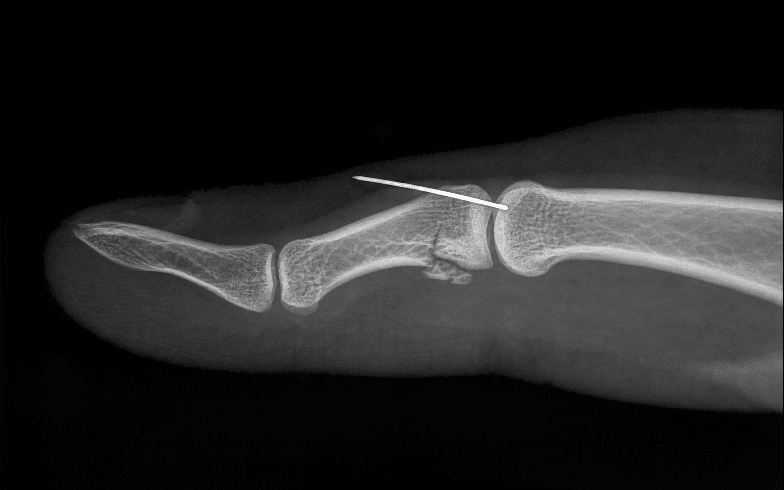
The Trap: Placing an extension block pin too far distally or proximally, failing to block the middle phalanx at the correct angle.

The Fix: The pin must be driven into the head of the proximal phalanx, intra-articularly but avoiding the central articular load-bearing area, to physically block extension at the angle where the joint is concentrically reduced.

Approaching from the Wrong Side

The Trap: Using a dorsal approach for a volar base fracture.

The Fix: Volar base fractures require a volar (Bruner zigzag) or midaxial approach to directly visualise the volar plate, the fracture fragments, and the articular surface without disrupting the extensor mechanism.

Mnemonic

S.T.A.B.L.EAlgorithm for PIP Fracture-Dislocations

Mnemonic

P.I.N.SSuzuki Frame Components

Surgical Indications

Absolute Indications

  • Joint irreducible by closed means (soft tissue interposition, typically volar plate or lateral band).
  • Persistent dorsal subluxation (V-sign) despite flexing to 30 to 40 degrees.
  • Unstable fracture pattern requiring more than 40 degrees of flexion to maintain reduction.
  • Open fracture-dislocations.
  • Chronic or missed fracture-dislocations (greater than 3 weeks).

Relative Indications

  • Large, single volar fragment representing greater than 40 percent of the articular surface (amenable to ORIF).
  • Patient inability to comply with a strict splinting and supervised therapy protocol.
  • Significant articular step-off (greater than 2 mm) in a large, fixable fragment.

Contraindications

  • Stable joint concentricity maintained in less than 30 degrees of flexion (manage with dorsal block splinting).
  • Extremely sedentary patient with low functional demands (relative).
  • Severe peripheral vascular disease or active infection in the digit.

Evidence for Treatment Modalities

Dorsal Block Splinting

  • Indication: Stable fracture-dislocations (typically less than 30 percent of the articular surface).
  • Protocol: Joint reduced and splinted in 20 to 30 degrees of flexion. Active flexion is encouraged immediately. The splint extension block is gradually reduced by 10 degrees per week.
  • Outcomes: Excellent functional results if concentric reduction is maintained and early motion is initiated. The literature strongly supports non-operative management for stable patterns, showing comparable or superior outcomes to surgery due to the avoidance of surgical trauma and subsequent stiffness.

Extension Block Pinning

  • Indication: Unstable joints where the fragment is too small or comminuted for ORIF, but the joint can be reduced in flexion.
  • Technique: A K-wire is driven into the proximal phalanx head to mechanically block extension past the point of instability. Allows active flexion.
  • Evidence: Provides reliable stability while permitting the crucial early flexion necessary for cartilage nutrition and preventing stiffness. Originally described by McElfresh, this technique remains a workhorse for unstable, unfixable dorsal fracture-dislocations.

Dynamic External Fixation (Suzuki / Compass Hinge)

  • Indication: Highly comminuted, pilon type fractures of the middle phalanx base.
  • Mechanism: Utilises ligamentotaxis to pull the comminuted fragments into alignment and distract the joint space, preventing collapse while allowing active motion.
  • Outcomes: Associated with a high rate of pin-tract infections, but functional outcomes are generally good to excellent for otherwise un-reconstructable injuries. Studies show average arcs of motion of 70 to 80 degrees, which is highly functional for a pilon fracture.

Volar Plate Arthroplasty (Eaton)

  • Indication: Comminuted volar base fractures or chronic fracture-dislocations where the volar restraint is lost.
  • Mechanism: The fracture fragments are excised, and the volar plate is advanced into the defect and sutured into the middle phalanx to restore stability and provide a smooth gliding surface.
  • Evidence: Reliable for restoring stability and preventing subluxation. Patients often lose 10 to 15 degrees of terminal extension, but this is a small price for a stable, pain-free, mobile joint.

Hemi-Hamate Autograft

  • Indication: Large volar base defects (greater than 50 percent), acute or chronic, not amenable to primary repair.
  • Mechanism: A graft taken from the distal articular surface of the hamate (which perfectly matches the concavity of the middle phalanx base) is fixed into the defect.
  • Evidence: Technically demanding but can restore near-normal anatomy and motion in complex cases. Long-term studies show good graft incorporation and maintenance of joint space, though donor site morbidity (hamate pain) can occur.

Clinical Decision Scenarios

Practise clinical reasoning and management decisions out loud

Viva scenarioAdvanced
Clinical prompt

A 25-year-old basketball player presents with a dorsal PIP joint dislocation of his middle finger, sustained 2 weeks ago. X-rays show a volar base fracture of the middle phalanx involving approximately 45 percent of the articular surface. The joint remains dorsally subluxated despite splinting. How will you manage this?

Practical approach
This is a subacute, unstable PIP joint fracture-dislocation with a large articular fragment. The persistent dorsal subluxation is unacceptable and will lead to rapid joint destruction. Because the fragment is large (45 percent) and the presentation is delayed, non-operative management or simple extension block pinning is unlikely to succeed. **Pre-operative counselling**: I would explain the need for surgery to restore joint congruity. I would warn him that the PIP joint will remain permanently thicker than normal, and he will likely lose some terminal extension or full flexion, regardless of the treatment. **Surgical Plan**: I would proceed with an open reduction and internal fixation (ORIF). - I will use a volar Bruner incision over the PIP joint, retracting the neurovascular bundles into the skin flaps. - I will divide the A3 pulley and retract the flexor tendons laterally to expose the volar plate and the fracture. - Given the 2-week delay, there will be early callus and scar tissue. I will carefully debride the fracture site to define the articular margins without sacrificing bone stock. - I will reduce the volar fragment and temporarily fix it with a K-wire. - Under fluoroscopy, I will confirm a concentric joint reduction on a true lateral view. - I will secure the fragment with two 1.0 mm or 1.2 mm lag screws, ensuring they are countersunk beneath the cartilage surface. **Fallback**: If the fragment fragments during fixation or is not amenable to ORIF due to comminution, I would convert to a volar plate arthroplasty (Eaton) or a hemi-hamate autograft. **Post-operative**: I will place him in a dorsal blocking splint and initiate early active motion to prevent stiffness, removing the splint completely by 4 weeks.
Viva scenarioStandard
Clinical prompt

Explain the biomechanical principle of extension block pinning for PIP fracture-dislocations. Where exactly does the pin go?

Practical approach
The biomechanical principle of extension block pinning relies on the fact that PIP fracture-dislocations (involving the volar base) are unstable in extension but reduce concentrically in flexion. By preventing the joint from extending past the critical angle of instability, the middle phalanx is maintained in a reduced position against the proximal phalanx condyles. This allows the volar fracture fragments and the volar plate to heal in an anatomic position while permitting active flexion, which is essential for cartilage nutrition and preventing joint stiffness. **Pin Placement**: - The pin does NOT cross the PIP joint. It is a single K-wire. - It is inserted dorsally into the head of the proximal phalanx. - It is angled distally, protruding from the bone to act as a physical bumper or block against the dorsal aspect of the middle phalanx. - The exact angle and position of the pin depend on the degree of flexion required to maintain a concentric reduction. If the joint is stable at 40 degrees of flexion, the pin is placed to block extension beyond 40 degrees. Intraoperatively, fluoroscopy is used to confirm that as the patient or surgeon extends the finger, the middle phalanx hits the pin and stops, and the joint remains perfectly concentric at that maximum permitted extension.
Viva scenarioAdvanced
Clinical prompt

A 40-year-old manual worker presents with a 6-week-old PIP fracture-dislocation of the index finger. The joint is stiff, swollen, and dorsally subluxated on X-rays. The volar base fragment involves 60 percent of the articular surface. What is your surgical plan?

Practical approach
This is a chronic PIP fracture-dislocation with a massive articular defect. The joint is chronically subluxated and the cartilage is likely damaged. Due to the delay and the size of the fragment (60 percent), ORIF is impossible. Volar plate arthroplasty (Eaton) is also relatively contraindicated for defects greater than 50 percent, as the volar plate cannot bridge such a large gap without significant instability or severe flexion contracture. **Surgical Plan**: The best option for a young, active manual worker with a 60 percent defect is a hemi-hamate autograft reconstruction. - I would use a volar Bruner approach to expose the PIP joint, excising the scarred volar plate and the malunited/comminuted volar base fragments, creating a clean rectangular defect. - I would then expose the distal articular surface of the hamate via a dorsal longitudinal incision over the 4th and 5th CMC joints. - The ridge between the 4th and 5th metacarpal facets on the hamate perfectly mimics the median ridge of the middle phalanx base. - I would harvest an osteochondral graft from the hamate matching the exact dimensions of the PIP defect using an oscillating saw. - The graft is transferred to the PIP joint and secured with two 1.0 mm or 1.2 mm lag screws, ensuring the articular surface is flush. **Post-operative**: Early protected motion is initiated in a dorsal blocking splint to mould the joint and prevent stiffness. I would counsel the patient to expect permanent enlargement of the joint and some loss of terminal extension, but this procedure provides the best chance for a stable, functional arc of motion.
Exam day cheat sheet
PIP Fracture-Dislocation — Exam Day Summary

References

Evidence

Volar plate arthroplasty of the proximal interphalangeal joint: a review of ten years' experience

Level IV
Eaton RG, Malerich MMJ Hand Surg Am. 1980;5(3):260-8
Clinical implication: Volar plate arthroplasty remains the gold standard salvage procedure for chronic or severely comminuted volar base PIP fracture-dislocations.
Evidence

Unstable fracture dislocations of the proximal interphalangeal joint of the fingers: a preliminary report of a new treatment technique

Level IV
Agee JMJ Hand Surg Am. 1978;3(4):386-9
Clinical implication: Dynamic external fixation is the treatment of choice for comminuted pilon-type fractures that are unamenable to internal fixation.
Evidence

Management of fracture-dislocation of the proximal interphalangeal joints by extension-block splinting

Level III
McElfresh EC, Dobyns JH, O'Brien ETJ Bone Joint Surg Am. 1972;54(8):1705-11
Clinical implication: Extension block pinning is a simple, effective, and minimally invasive technique for unstable fracture-dislocations with small volar fragments.
Evidence

The pins and rubbers traction system for treatment of comminuted intraarticular fractures and fracture-dislocations in the hand

Level IV
Suzuki Y, Matsunaga T, Sato S, Yokoi TJ Hand Surg Br. 1994;19(1):98-107
Clinical implication: The Suzuki frame is an elegant and accessible method for applying ligamentotaxis to complex PIP pilon fractures, enabling early motion.
Editorially reviewed — transparent references and correction processPublished by OrthoVellum Medical Education TeamEditorial boardMethodologyReview policy
Educational disclosure

Educational content is reviewed for source visibility, editorial coherence, and correction readiness.

No individual clinician credential is claimed unless a named person is shown.

Verify before clinical use; this is not medical advice or a substitute for local guidance.