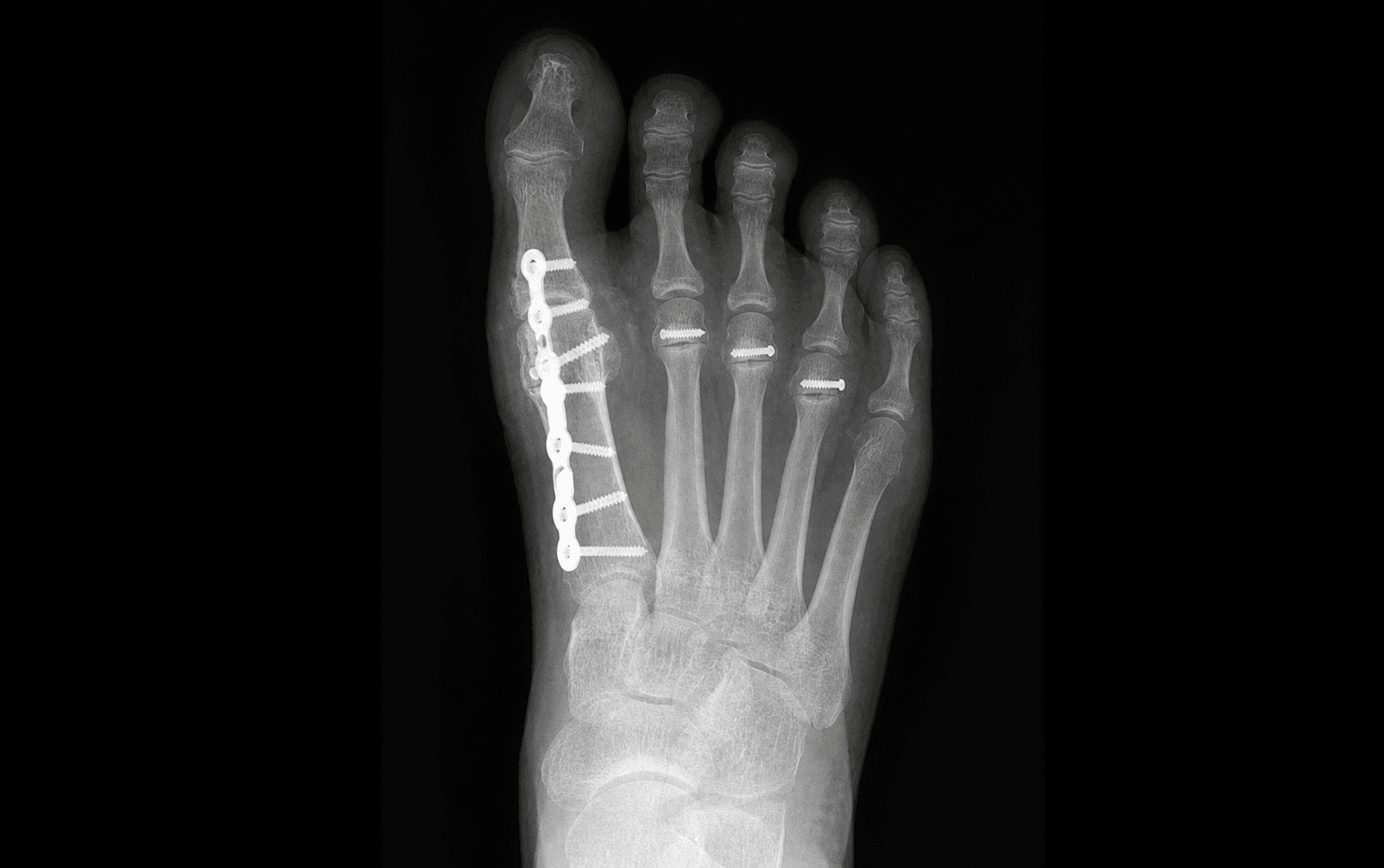
First MTP arthrodesis combined with lesser metatarsal surgery (Hoffmann or Weil) and claw toe correction | advanced
Surgical Imaging
Location: The dorsalis pedis artery runs in the interval between extensor hallucis longus (EHL) and extensor digitorum longus (EDL) in the proximal third of the foot, then passes lateral to EHL towards the first webspace.
Risk: The dorsal longitudinal incision for first MTP arthrodesis and the dorsal transverse incision for lesser metatarsal surgery both cross the dorsalis pedis. Identify and protect it at the start of every dorsal approach. Laceration produces significant intraoperative bleeding and risks compromising the already tenuous vascularity of rheumatoid toes.
Location: The deep peroneal nerve lies lateral to the dorsalis pedis artery in the same intertendinous interval (between EHL and EDL). It supplies the first webspace sensation.
Risk: Retraction or careless dissection during dorsal approaches can injure the deep peroneal nerve, producing numbness in the first webspace or neuroma formation. Identify the nerve with the artery at the start of each approach and protect with vessel loops.
Location: The medial (tibial) and lateral (fibular) sesamoids sit within the flexor hallucis brevis tendons beneath the first metatarsal head, articulating with the sesamoid grooves.
Risk: During resection of the first MTP joint articular surfaces, the sesamoids must be preserved. Disruption or excision of the sesamoid apparatus removes the plantar fulcrum for hallux push-off and causes significant functional impairment. The flexor hallucis brevis must remain intact.
The trap: Over-resection of the metatarsal head in the Hoffmann procedure or excessive shortening with the Weil osteotomy shifts load to the adjacent ray, producing a new focal plantar keratosis — transfer metatarsalgia. This is the most common reason for reoperation after rheumatoid forefoot reconstruction.
The fix: Preserve the metatarsal parabola. The second metatarsal should remain longest. Resect a consistent 4-6 mm of metatarsal head in the Hoffmann procedure. In the Weil osteotomy, translate the capital fragment dorsally by the same amount at each ray. Check the plantar profile intraoperatively with manual palpation — no single metatarsal head should be prominent.
Keller arthroplasty: Excision of the proximal half of the proximal phalanx and the metatarsal head. Simple, quick, reliable pain relief, but produces a weak unstable hallux, loss of push-off power, and transfer metatarsalgia from loss of the first ray weight-bearing function. Reserved for low-demand or elderly non-ambulatory patients.
First MTP arthrodesis: Gold standard for ambulatory rheumatoid patients. Provides a stable plantigrade first ray, corrects hallux valgus at its source, restores weight-bearing, and offloads the lesser metatarsals. Union rates 85-95%. In the exam, arthrodesis is the correct answer for functional patients.
Why it matters: Rheumatoid arthritis causes pannus formation and ligamentous laxity at the craniocervical junction — atlantoaxial subluxation, subaxial subluxation, and basilar invagination are present in up to 30-40% of long-standing RA patients. Intubation can compress the cervical cord, causing quadriplegia or death.
The rule: Before any procedure under general anaesthesia in rheumatoid arthritis, obtain flexion-extension cervical spine radiographs. If instability is present, either use regional (spinal or popliteal) anaesthesia or proceed with awake fibreoptic intubation with cervical spine precautions. Never assume this has been done — in the exam, always mention it before proceeding to theatre.
F.O.R.E.F.O.O.TFOREFOOT — Rheumatoid Forefoot Deformity Recognition
F.U.S.I.O.NFUSION — First MTP Arthrodesis Key Points
W.E.I.LWEIL — Lesser Metatarsal Weil Osteotomy
Surgical Indications
Absolute Indications
- Failed conservative management after at least 6 months of appropriate footwear modification, custom orthoses, orthopaedic shoes, and optimised medical management (DMARDs and biologics)
- Intractable plantar metatarsalgia with painful callosities beneath dislocated or subluxated lesser MTP joints causing significant functional limitation
- Severe hallux valgus deformity with first MTP joint destruction (radiographic joint space loss, erosions, subluxation) producing pain unresponsive to shoe modification
- Fixed lesser toe deformities (claw toes or hammer toes with MTP dislocation) interfering with footwear or causing ulceration
Relative Indications
- Progressive hallux valgus with early medial column collapse despite medical management
- Lesser MTP joint synovitis with dorsal subluxation but not yet fully dislocated — surgical correction before fixed dislocation yields better outcomes
- Patient preference for surgical reconstruction to enable return to enclosed footwear and improve mobility
- Painful callosities with recurrence despite regular podiatric care
Contraindications
Absolute:
- Active infection of the foot or systemic sepsis
- Inadequate soft tissue envelope (skin ulceration, active rheumatoid vasculitis)
- Critical ischaemia: absent pedal pulses not restorable by vascular intervention
- Medical unfitness for anaesthesia or prolonged rehabilitation
Relative:
- Poor bone stock with severe osteopenia or fractures through osteoporotic metatarsals (consider staged surgery)
- Active rheumatoid flare — optimise medical management before elective surgery
- Non-compliance with non-weight-bearing restrictions or post-operative rehabilitation
- Non-ambulatory or bedbound patient (conservative management preferred)
Pathophysiology of the Rheumatoid Forefoot
The Cascade of Deformity
- Synovitis of the MTP joints is the initiating event — pannus formation erodes the articular cartilage, distends the joint capsule, and destroys the collateral ligaments and plantar plate
- Volar plate attenuation allows the proximal phalanx to subluxate dorsally; the extensor tendons (previously dorsal) migrate into a subluxated dorsal-lateral position, reinforcing the deformity
- MTP dorsal subluxation progresses to dislocation — the proximal phalanx rides dorsally over the metatarsal head, the plantar fat pad is dragged distally with it, and the metatarsal head becomes exposed to direct plantar pressure
- Metatarsal head prominence beneath the thinned plantar fat pad produces intractable plantar keratoses, metatarsalgia, and pain
- Claw toe deformity: the intrinsic muscles lose their stabilising function at the MTP joint (interossei and lumbricals act as MTP flexors and IP extensors), so as the MTP extends, the IP joints flex, producing the classic claw toe posture (MTP extension, PIP flexion, DIP flexion)
- Hallux valgus of the first ray is driven by MTP synovitis and medial capsular failure, producing lateral deviation of the hallux and medial prominence of the metatarsal head
- First ray instability (loss of first-ray weight-bearing) transfers load to the lesser metatarsals, worsening the metatarsalgia cycle
Evidence for Non-Operative Management
Conservative Treatment Options
- Footwear modification: wide toe-box, rocker-bottom sole, soft upper, minimal heel height — effective early in the disease course but insufficient once fixed deformity develops
- Custom moulded orthoses: metatarsal dome or bar to offload prominent metatarsal heads; accommodative insoles with total contact design
- Medical management optimisation: coordinate with rheumatology to ensure the patient is on the most appropriate DMARD/biologic regimen; ongoing synovitis in the forefoot while on maximum medical therapy supports earlier surgical referral
- Podiatric care: regular debridement of callus, padding, and monitoring for ulceration in patients with vascular compromise or neuropathy
- Intra-articular corticosteroid injection: temporary symptom relief for acute MTP synovitis flares, but repeated injection provides diminishing returns and does not correct structural deformity
Evidence for Surgery
First MTP Arthrodesis vs Keller Arthroplasty
First MTP arthrodesis is now the gold standard for rheumatoid hallux valgus in ambulatory patients:
- Provides a stable, plantigrade medial column that bears weight normally
- Corrects the hallux valgus deformity at the joint level, eliminating the bunion prominence
- Offloads the lesser metatarsals by restoring first-ray function
- Long-term studies report sustained pain relief and functional improvement in 85-95% of patients
- Nonunion rate 5-10%; risk increased by smoking, poor bone stock, and excessive resection
Keller excision arthroplasty is reserved for elderly, low-demand, or non-ambulatory patients:
- Resection of the metatarsal head and the proximal half of the proximal phalanx
- Reliable pain relief but produces a shortened, weak hallux with loss of push-off power
- Transfer metatarsalgia is common (15-30%) because the first ray no longer bears its share of load
- Risk of hallux malleus (floppy hallux), cock-up deformity, and progressive lesser toe overload
First MTP Arthrodesis vs Keller Arthroplasty — Evidence Summary
Hoffmann Procedure vs Weil Osteotomy for Lesser Rays
Hoffmann procedure (metatarsal head resection):
- Excision of the lesser metatarsal heads through a dorsal approach
- Relieves plantar pressure by removing the bony prominence
- Simple, reliable, quick — particularly suited to multiple dislocated MTP joints in advanced disease
- Risk of transfer metatarsalgia from uneven resection; loss of the MTP joint with no articulating surface
- Long-standing standard for the severely deformed rheumatoid forefoot
Weil shortening osteotomy:
- Dorsal oblique osteotomy of the metatarsal neck with dorsal translation of the capital fragment
- Preserves the MTP joint articular surface — preferred for lesser MTP joints that are subluxated but not fully dislocated
- More technically demanding; requires precise osteotomy angle, controlled translation, and fixation
- Risk of avascular necrosis of the capital fragment (2-10%), stiffness, floating toe, and over-shortening
- Growing evidence supporting Weil osteotomy over Hoffmann in less severe disease, but Hoffmann remains appropriate for advanced dislocation
Hoffmann vs Weil — Lesser Ray Surgery Comparison
Key Evidence
Rheumatoid forefoot reconstruction: a long-term follow-up study
Arthrodesis of the first metatarsophalangeal joint for hallux valgus in rheumatoid arthritis
Weil's metatarsal osteotomy in the treatment of metatarsalgia
Rheumatoid forefoot deformity: a comparison study of 2 functional methods of reconstruction
Clinical Decision Scenarios
Practise clinical reasoning and management decisions out loud
“A 52-year-old woman with seropositive rheumatoid arthritis on adalimumab and methotrexate presents with progressive forefoot pain, hallux valgus of 35 degrees, and painful plantar callosities beneath the second and third metatarsal heads. Radiographs show first MTP joint destruction with erosions, dorsal subluxation of the second and third MTP joints, and periarticular osteopenia. She has failed 12 months of orthopaedic shoes and custom orthoses. She asks what surgery you would recommend.”
“You have just completed a first MTP arthrodesis with Hoffmann lesser metatarsal head resections for a 65-year-old woman with rheumatoid arthritis. After tourniquet deflation, you notice the second and third toes are pale with delayed capillary refill. What do you do?”
“A 58-year-old man with rheumatoid arthritis had a first MTP arthrodesis and lesser metatarsal Weil osteotomies 9 months ago. He presents with ongoing pain beneath the first MTP joint, worse with push-off. Radiographs show no bridging trabeculae at the fusion site with a visible radiolucent gap and a broken screw. How do you manage this?”
References
-
Coughlin MJ (2000). Rheumatoid forefoot reconstruction. A long-term follow-up study. J Bone Joint Surg Am, 82(3):422-431. — Long-term outcomes of combined first MTP arthrodesis with lesser metatarsal head resection in rheumatoid patients.
-
Mann RA, Thompson FM (1984). Arthrodesis of the first metatarsophalangeal joint for rheumatoid arthritis. Foot Ankle, 4(4):173-178. — Seminal paper establishing first MTP arthrodesis as the gold standard for rheumatoid hallux valgus.
-
Cracchiolo A, Swanson A, Swanson GD (1985). The arthritic great toe metatarsophalangeal joint: a review of Keller arthroplasty, silicone implant arthroplasty, and arthrodesis. Foot Ankle, 5(6):249-259. — Comparative review demonstrating arthrodesis superiority over excisional arthroplasty.
-
Barouk LS (1996). Weil's metatarsal osteotomy in the treatment of metatarsalgia. Orthopade, 25(4):338-344. — Description and clinical application of the Weil osteotomy for metatarsalgia and lesser ray correction.
-
Mulcahy D, Daniels TR, Lau JT, Boyle E, Bogoch ER (2003). Metatarsal head resection versus dorsal closing wedge osteotomy in rheumatoid forefoot reconstruction. Foot Ankle Int, 24(12):912-916. — Comparative study of Hoffmann resection versus Weil osteotomy for lesser ray correction.
-
Shi K, Tomita T, Hayashi A et al. (2000). Surgical treatment of rheumatoid forefoot deformities. Mod Rheumatol, 10(2):120-125. — Clinical series of rheumatoid forefoot reconstruction outcomes and patient satisfaction.