Anterior radical debridement, anterior column reconstruction and posterior instrumented fusion for Pott disease | advanced
Surgical Imaging
Early onset paraplegia (within 2 years) is usually due to active inflammatory compression (abscess, caseous tissue, oedema) and has good recovery potential with decompression and ATT. Late onset paraplegia (greater than 2 years) is due to mechanical compression by the internal kyphosis (bony ridge), dural fibrosis or vascular insufficiency — recovery is less reliable and surgery is more hazardous.
Timing: A patient with progressive neurological deficit on adequate ATT requires surgical decompression within 24-48 hours. The longer the deficit is complete (Frankel A/B), the poorer the motor recovery prognosis. A patient who presents with paraplegia that is improving on ATT may be treated non-operatively if deformity and instability are absent.
A paravertebral or psoas cold abscess is a collection of caseous material and pus that tracks along fascial planes. It is NOT an emergency unless it is causing significant compression.
Indications for drainage: (1) Large abscess causing significant thecal sac compression, (2) Abscess extending into the spinal canal, (3) Failed resolution after 4-6 weeks of ATT (persistent large collection), (4) Need for tissue diagnosis.
Technique: Drain via the same surgical approach used for debridement. Psoas abscesses can often be drained percutaneously under CT or ultrasound guidance. Do NOT drain a cold abscess without starting ATT first — there is a risk of sinus formation and secondary infection.
Children (especially under 10 years) have a growing spine and can develop progressive kyphosis that worsens as they grow — this is called the 'spine at risk' phenomenon. The vertebral body anterior growth plate is destroyed; the posterior elements continue to grow, producing a progressive kyphosis.
Adults have a stable deformity once healed — the angle of kyphosis does not typically progress after bony fusion. However, adjacent segment breakdown or graft failure can cause late progression.
Implication: In children, a more aggressive surgical approach (anterior-posterior reconstruction) is indicated even for moderate kyphosis because of the risk of progression during growth. Rajasekaran's 'spine at risk' signs guide decision-making.
Graft or cage subsidence into the adjacent vertebral bodies causes loss of correction, kyphosis progression, and possible neurological compromise.
Risk factors: (1) Incomplete debridement leaving necrotic bone at the graft-host interface, (2) Endplate preservation is inadequate — aggressive curettage that removes all healthy structural endplate, (3) Posterior tension band disruption (non-instrumented posterior column), (4) Multi-level disease with long graft spanning more than two levels.
Prevention: Use a posterior instrumented construct to offload the anterior column. Preserve the healthy endplate (subchondral bone) of the adjacent vertebrae at the recipient site. Use a wide footplate cage or graft to distribute load. Extend reconstruction at least one level above and below the diseased segment where possible.
The most common cause of treatment failure in spinal tuberculosis is non-compliance with anti-tubercular therapy — not the surgery itself. Patients may feel well after 2-4 months and stop medication.
Consequences: Drug resistance, reactivation, progression of disease, implant infection (catastrophic — requires implant removal and prolonged second-line ATT).
Protocol: Standard short-course chemotherapy for spinal TB: 2 months of rifampicin, isoniazid, pyrazinamide and ethambutol (intensive phase), followed by 10 months of rifampicin and isoniazid (continuation phase) — total 12 months. In surgically treated patients, some centres extend to 12-18 months. Directly observed therapy (DOT) is recommended where compliance is a concern.
Thoracic (transthoracic): The segmental vessels (intercostal arteries and veins) cross the mid-lateral vertebral body and must be ligated before reaching the spine. The azygos vein on the right, the aorta and hemiazygos on the left — all at risk with aggressive retraction. The thoracic duct on the left at T4-T6.
Thoracolumbar (T10-L2): The diaphragm is divided 2-3 cm from its costal insertion. The crus of the diaphragm can be divided if needed but the aortic hiatus must be respected. The great vessels (aorta and IVC) lie directly on the anterior spine.
Lumbar (retroperitoneal): The ureter lies on the psoas muscle and must be identified and protected. The iliac vessels cross anterior to the L4-L5 disc. The sympathetic chain runs along the anterolateral vertebral bodies — injury causes a warm leg.
Prevention: Meticulous stepwise dissection with identification and protection of each structure. Pre-operative contrast-enhanced CT to map the vascular anatomy relative to the diseased level.
P.O.T.T.SPOTT'S — Management Principles of Spinal Tuberculosis
S.P.I.N.ESPINE — Approach Selection for Pott Disease
Surgical Indications
Absolute Indications
- Progressive or severe neurological deficit (Frankel C or worse) despite adequate ATT — surgical decompression is urgent
- Neurological deterioration while on ATT — the most urgent surgical indication
- Failure of medical management: progression of deformity or increase in pain after 4-6 weeks of adequate ATT
- Severe kyphosis (Cobb angle greater than 40 degrees) at presentation or progressive kyphosis on treatment
- Instability: translational movement on dynamic radiographs or segmental collapse with loss of normal sagittal alignment
- Large abscess not resolving on ATT with significant thecal sac compression or causing dysphagia/airway compromise (cervical)
Relative Indications
- Moderate kyphosis (20-40 degrees) in children — risk of progression with growth; combined anterior-posterior surgery is recommended
- Multi-level disease (more than 2 contiguous vertebral bodies) — associated with instability and deformity progression
- Persistent severe pain attributed to mechanical instability despite adequate ATT
- Tissue diagnosis required — when imaging is atypical or malignancy cannot be excluded
- Late onset paraplegia (greater than 2 years after quiescent disease) — mechanical compression from internal kyphosis
Contraindications
Absolute:
- Active pulmonary TB with positive sputum — treat with ATT first until sputum conversion (usually 4-8 weeks) before elective spinal surgery. Urgent decompression for neurological deficit overrides this
- Multi-drug resistant (MDR) TB without effective drug regimen — surgery is extremely high risk for implant infection and non-union
- Poor nutritional status (albumin less than 25 g/L, severe cachexia) — correct nutrition before surgery
- Active miliary TB — prioritise medical treatment
Relative:
- Advanced age with multiple comorbidities
- Severe pulmonary compromise — thoracotomy may not be tolerated; consider costotransversectomy or posterior approach
- Irreversible complete paraplegia of more than 6 months duration (Frankel A) — surgical decompression rarely improves motor function; surgery is for deformity and pain only
Evidence for Non-Operative Treatment
Anti-Tubercular Chemotherapy (ATT)
ATT is the foundation of medical management for spinal tuberculosis. The standard regimen follows the same principles as pulmonary TB:
Standard short-course regimen:
- Intensive phase (2 months): four drugs — rifampicin (R), isoniazid (H), pyrazinamide (Z), ethambutol (E)
- Continuation phase (10 months): two drugs — rifampicin (R), isoniazid (H)
- Total duration: 12 months minimum; 12-18 months in the presence of extensive disease, neurological involvement, or after surgery
Why duration is longer than pulmonary TB (6 months): The vertebral body is a relatively poorly perfused site and has a higher organism burden. Drug penetration into caseous material and bone is variable. Relapse rates are higher with shorter courses (less than 9 months) in spinal TB.
Monitoring: Monthly clinical assessment (pain, neurology, weight, ESR/CRP). Radiographs at 3, 6, 12 months. MRI if neurological deterioration occurs or if response is unsatisfactory.
Outcome of Non-Operative Management
The Medical Research Council Working Party on Tuberculosis of the Spine conducted a landmark series of multi-centre randomised trials comparing ambulatory chemotherapy with surgical treatment:
- Chemotherapy Alone
- 85-90%
- Debridement Alone
- 88-92%
- Radical Resection + Graft
- 92-96%
- Chemotherapy Alone
- Mean 15 degrees
- Debridement Alone
- Mean 10 degrees
- Radical Resection + Graft
- Mean less than 5 degrees
- Chemotherapy Alone
- 70-85%
- Debridement Alone
- 75-85%
- Radical Resection + Graft
- 85-95%
- Chemotherapy Alone
- 5-10%
- Debridement Alone
- 4-8%
- Radical Resection + Graft
- less than 3%
Data from MRC Working Party trials on tuberculosis of the spine (multiple reports, 1973-1999).
Key conclusion: Chemotherapy alone is sufficient for patients without neurological deficit, without significant deformity (kyphosis less than 20 degrees), and with disease limited to one or two vertebral bodies. Surgical intervention improves kyphosis correction and provides more reliable neurological recovery in patients with deficit.
Evidence for Surgery
The Hong Kong Operation (Hodgson & Stock, 1956)
The landmark contribution of Hodgson and Stock at the University of Hong Kong was the recognition that:
- The disease is primarily anterior (vertebral body and disc)
- An anterior approach gives direct access to the pathology
- Radical debridement followed by autogenous bone grafting achieves both disease clearance and mechanical reconstruction
- The graft incorporates rapidly in a well-vascularised environment after debridement
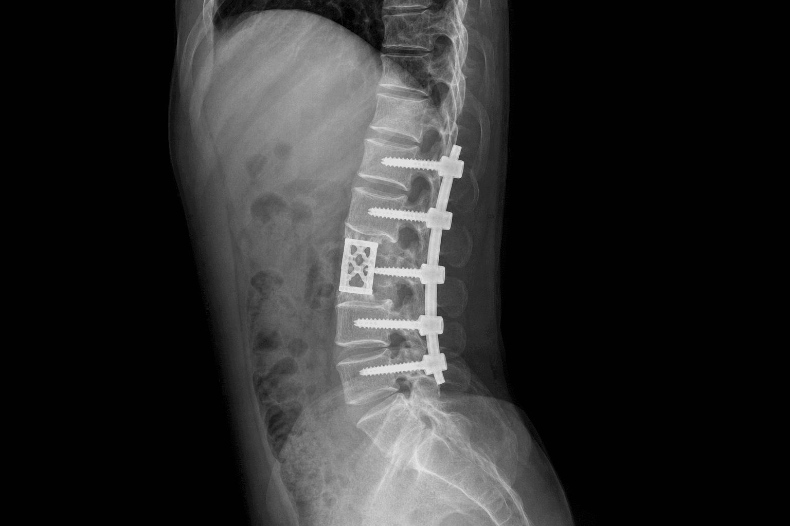
Their original description: Transthoracic approach, resection of the affected vertebral body and adjacent discs back to healthy bleeding bone, and placement of a tricortical iliac crest autograft under compression. No posterior instrumentation was used.
Long-term outcomes (Upadhyay, 1996): At a mean follow-up of 15 years, 94% of patients had solid bony fusion. Kyphosis correction achieved at surgery was partially lost during graft incorporation (mean loss of 6 degrees). Late neurological deterioration was rare (less than 2%).
Modern Surgical Approach: Anterior Debridement with Posterior Instrumentation
The addition of posterior pedicle screw instrumentation to the Hong Kong operation has significantly improved kyphosis correction and maintenance:
- Immediate rigid stabilisation allows early mobilisation without external bracing
- The posterior construct offloads the anterior graft, reducing subsidence and graft fracture
- Kyphosis correction is improved (mean 15-20 degrees correction vs 5-10 degrees with anterior alone)
- Fusion rates are higher (greater than 95% in modern series)
Surgical Approach Comparison for Spinal Tuberculosis
Implication for practice: Combined anterior-posterior surgery is the modern standard for patients with:
- Kyphosis greater than 30 degrees
- Involvement of more than 2 vertebral bodies
- Neurological deficit
- Significant instability
Anterior alone is reserved for single-level disease with good bone stock, no significant kyphosis, and no neurological compromise. Posterior alone has a limited role — mainly for disease confined to the posterior elements or when an anterior approach is contraindicated.
Key Evidence
Anterior spinal fusion for tuberculosis of the spine — the Hong Kong operation
Prediction of the angle of gibbus deformity in tuberculosis of the spine
A 15-year assessment of controlled trials of the management of tuberculosis of the spine in Korea and Hong Kong — Thirteenth Report of the Medical Research Council Working Party on Tuberculosis of the Spine
Duration of anti-tuberculosis chemotherapy in conjunction with radical surgery in the treatment of spinal tuberculosis
Clinical Decision Scenarios
Practise clinical reasoning and management decisions out loud
“A 45-year-old man from a high-TB-burden country presents with a 3-month history of worsening back pain, low-grade fevers and progressive lower limb weakness over 2 weeks. He is now Frankel C (motor useless but sensory preserved). MRI shows T7-T8 vertebral body destruction with a large paravertebral abscess and thecal sac compression. He has been on a 4-drug ATT regimen for 10 days from the referring hospital. How do you manage him?”
“An 8-year-old child presents with a 6-month history of back pain and a progressive 45-degree kyphosis at T10-T11 on standing radiograph. MRI shows T10-T11 vertebral body destruction with a small paravertebral abscess but no significant thecal sac compression. Neurologically the child is normal (Frankel E). She has been on ATT for 8 weeks without improvement in the kyphosis. Her parents are worried about the deformity. How do you manage this?”
“A 55-year-old woman underwent anterior debridement and iliac crest strut grafting for T11-L1 Pott disease 18 months ago. She completed 12 months of ATT. She now presents with recurrent back pain and a 15-degree increase in her kyphosis over the past 6 months. She has no neurological deficit. Standing radiographs show the graft has subsided into the L1 vertebral body by 8 mm and there is halo formation around the graft. CT confirms non-union at the distal graft-host interface. What do you do?”