Subtrochanteric Femoral Fracture Fixation

TraumaAdvancedCore Procedure

Subtrochanteric Femoral Fracture Fixation

Surgical technique guide for fixation of subtrochanteric femoral fractures using cephalomedullary nailing, including Russell-Taylor classification, deformity correction, reduction techniques, implant selection, management of atypical bisphosphonate-related fractures, and prophylactic fixation of impending lesions

High-yield overview

Cephalomedullary (intramedullary) nailing of subtrochanteric femoral fractures | advanced

Surgical Imaging

Subtrochanteric femur fracture fixed with a cephalomedullary nail
Subtrochanteric femur fracture fixed with a long cephalomedullary nail; the cephalic screw into the head and distal interlocking control the strong deforming forces on the proximal fragment.Credit: AI-generated medical illustration Β· OrthoVellum
Critical Danger Structures and Exam Traps
Apex-Anterolateral Angulation β€” The Defining Deformity

The trap: Attempting to nail without first correcting the characteristic apex-anterolateral deformity. If the proximal fragment remains flexed, abducted and externally rotated, the guidewire and nail will follow the deformed proximal fragment and create varus malreduction with the distal fragment.

The fix: Reduce the deformity before reaming. On a fracture table, apply traction, slightly flex the distal fragment, and internally rotate the limb to align the distal fragment with the flexed proximal fragment. Use percutaneous Schanz pins or reduction clamps if closed reduction fails. Confirm AP and lateral alignment with fluoroscopy before committing to reaming.

Varus Malreduction β€” The Commonest Error

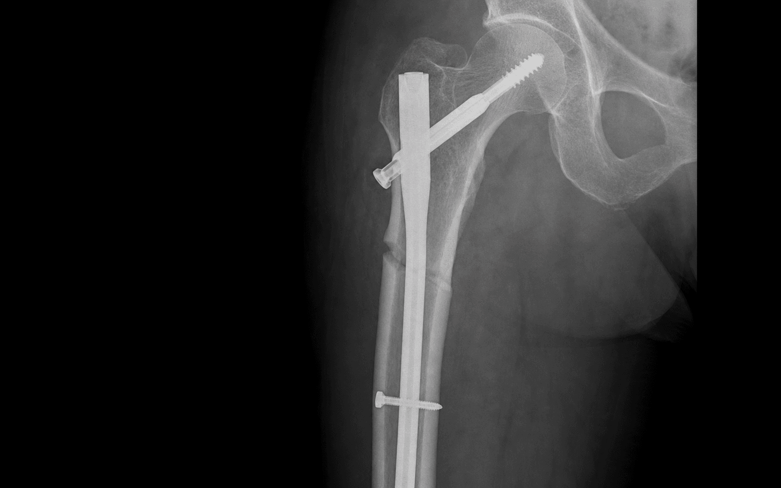
Recognition: On the postoperative AP radiograph, compare the neck-shaft angle with the contralateral side. Varus is present when the medial cortex of the proximal fragment overrides the distal fragment medially, the calcar sits proud, and the neck-shaft angle is reduced below approximately 125-130 degrees.

Consequences: Varus shifts the mechanical axis medially, increases bending stress on the implant (particularly at the fracture site and distal locking bolts), and predisposes to implant failure and nonunion.

Prevention: Correct entry point at the greater trochanteric tip (not lateral); verify reduction in both planes before reaming; use a lateral decubitus position for difficult reductions to let the proximal fragment sag into extension.

Iatrogenic Proximal Femur Fracture During Reaming

Mechanism: Reaming over a guidewire that is not truly intra-medullary, or reaming a small proximal fragment with significant comminution, can split the proximal femur posterolaterally or propagate the fracture into the piriformis fossa.

Prevention: Confirm the guidewire is central in the canal on both AP and lateral fluoroscopy before reaming. In comminuted proximal fragments, use hand reamers or flexible reamers first. Reduce the fracture adequately before reaming. In osteoporotic bone, consider a trochanteric entry nail (wider proximal profile) rather than a standard antegrade nail.

Management: If a split occurs during reaming, stop. Assess on fluoroscopy. A stable crack may be managed by proceeding with a larger-diameter nail that bypasses the split. An unstable split may require conversion to plate fixation with cerclage protection.

Atypical Bisphosphonate Fracture β€” Do Not Miss It

The trap: Treating prodromal pain as muscle strain or tendinopathy, or fixing a completed atypical fracture with a plate (which has a high failure rate in this pathological bone) instead of a cephalomedullary nail.

Recognition: Prodromal deep thigh or groin pain in a patient on long-term bisphosphonate therapy (typically greater than 3-5 years). Radiographs show lateral cortical thickening, beaking or periosteal reaction at the subtrochanteric or mid-diaphyseal femur. Completed fractures are transverse, with medial cortical spiking and minimal comminution.

Management: When the cortical breach reaches 50 percent or greater with pain, prophylactic cephalomedullary nailing. For completed fractures, intramedullary nailing is the fixation of choice. Plate fixation has substantially higher failure rates in this setting. Drug holiday and teriparatide consideration are adjuncts, not alternatives.

Russell-Taylor Type II β€” Piriformis Fossa Destroyed

The trap: Attempting to place a standard antegrade nail through a comminuted piriformis fossa in a Russell-Taylor Type II fracture. The entry point is destroyed, and forcing the guidewire creates malreduction and further comminution.

The fix: Recognise Type II fractures preoperatively (comminution extending into the piriformis fossa on AP radiograph). Use a trochanteric entry cephalomedullary nail (entry at the tip of the greater trochanter) or a 95-degree blade plate/DCS. Type IIB fractures with lesser trochanter involvement have medial cortex disruption and need particular attention to calcar restoration.

Exam tip: Russell-Taylor Type IA fractures can be nailed through the piriformis fossa with indirect reduction. Type IB, IIA and IIB fractures require either a trochanteric entry nail or plate-based fixation, and the more medial cortex is lost, the more important medial support strategies become.

Loss of Medial Calcar Support

Why it matters: The medial femoral cortex (calcar) is the primary load-bearing surface in the proximal femur under normal walking. In subtrochanteric fractures with lesser trochanter detachment or comminution, the medial buttress is lost and the entire load passes through the lateral implant under bending β€” a mechanical environment that favours varus collapse, nonunion and hardware failure.

Prevention and management: Restore medial contact wherever possible through reduction. Use cerclage wires to hold comminuted medial fragments. In Type IIB fractures with extensive medial bone loss, consider indirect bone grafting or a medial buttress plate in addition to the IM nail. A well-placed nail with good distal purchase can compensate for some medial loss, but not for complete medial cortical disruption.

Mnemonic

S.U.B.T.R.OSUBTRO β€” Subtrochanteric Fracture Core Principles

Mnemonic

R.E.D.U.C.EREDUCE β€” Reduction Techniques for Subtrochanteric Fractures

Mnemonic

N.A.I.LNAIL β€” Cephalomedullary Nail Key Technical Points

Surgical Indications

Absolute Indications

  • Displaced subtrochanteric femoral fracture (within 5 cm distal to the lesser trochanter) in an adult β€” virtually all displaced fractures in this region require surgical fixation due to the powerful deforming forces and poor outcomes with non-operative treatment
  • Open subtrochanteric fracture β€” surgical fixation after wound debridement and antibiotic therapy
  • Polytrauma with femoral shaft or subtrochanteric fracture β€” early fixation for damage control and mobilisation
  • Pathological subtrochanteric fracture (metastatic disease) β€” stabilisation for pain relief and mobilisation
  • Impending pathological fracture (cortical breach greater than 50 percent with pain) β€” prophylactic fixation before completion

Relative Indications

  • Atypical bisphosphonate-related subtrochanteric fracture with prodromal pain and radiographic cortical changes β€” prophylactic cephalomedullary nailing to prevent completion
  • Non-united subtrochanteric fracture after previous failed fixation β€” revision with exchange nailing or plate augmentation
  • Periprosthetic fracture around a well-fixed femoral stem with subtrochanteric extension β€” requires specific implant strategies

Contraindications

Absolute:

  • Patient unfit for anaesthesia due to severe medical comorbidity or terminal illness where the burden of surgery exceeds the benefit
  • Active deep infection at the surgical site (not an open fracture with contamination β€” that is a different scenario requiring staged treatment)

Relative:

  • Very poor bone stock with no restorable medial cortex β€” consider plate-based construct with medial augmentation or megaprosthesis in elderly patients
  • Ipsilateral femoral neck fracture plus subtrochanteric extension β€” may require separate cervical fixation or a reconstruction-type nail with separate neck screw capability

Russell-Taylor Classification

The Russell-Taylor classification (1986) guides implant selection based on whether the piriformis fossa and the lesser trochanter/medial cortex are involved:

Russell-Taylor Classification of Subtrochanteric Fractures


Clinical significance: Type I fractures allow a standard piriformis entry antegrade nail, which provides the best biomechanical axis. Type II fractures destroy the piriformis fossa and require either a trochanteric entry nail (entered at the tip of the greater trochanter, slightly medial) or a plate-based construct. The sub-classification by lesser trochanter involvement (A vs B) reflects the integrity of the medial cortex β€” Type IB and IIB fractures have medial disruption and higher nonunion rates.

Evidence for Implant Selection

Cephalomedullary (Intramedullary) Nail vs Plate Fixation

Cephalomedullary nail (implant of choice):

  • Load-sharing device that exploits the femoral canal biomechanics
  • Minimally invasive insertion with percutaneous technique
  • Allows immediate distal interlocking for rotational and axial stability
  • Long reconstruction nails (up to 480 mm) reach the distal femoral isthmus
  • Proximal cephalomedullary locking screws at 130 degrees enter the femoral head/neck
  • Biomechanically superior in cyclic loading compared to plate constructs
  • Lower blood loss, shorter operative time, and lower infection rate than plate fixation in most series

95-degree blade plate (alternative):

  • Provides fixed-angle fixation with a blade seated in the femoral neck
  • Requires more extensive lateral soft-tissue dissection
  • Better suited when the piriformis fossa is destroyed (Russell-Taylor Type II)
  • Can be combined with cerclage wires for medial fragment control
  • Direct visualisation of the fracture site for anatomical reduction
  • Higher blood loss and infection risk compared to IM nailing
  • Slower rehabilitation due to plate reliance on cortical contact

Dynamic condylar screw (DCS, 95 degrees):

  • Similar indications to the blade plate but with a barrel/screw mechanism
  • Allows compression across the fracture site after seating
  • Bulkier and requires more lateral exposure than a blade plate
  • Useful in distal subtrochanteric fractures extending towards the supracondylar region

Cephalomedullary Nail vs 95-Degree Blade Plate β€” Evidence Summary


Key Evidence

Evidence

Treatment of subtrochanteric fractures. A comparison of the Gamma nail and the dynamic hip screw: short-term outcome in 58 patients

Level III
SaarenpÀÀ I, Heikkinen T, Jalovaara P β€’ International Orthopaedics
Clinical implication: Cephalomedullary nailing is preferred for subtrochanteric fractures, particularly when medial cortical support is compromised β€” the load-sharing biomechanics of an intramedullary device outperform a plate construct in this high-stress zone.
Evidence

Reduction Techniques for Trochanteric and Subtrochanteric Fractures of the Femur: a Practical Guide

Level V
Falkensammer ML, Benninger E, Meier C β€’ Acta Chirurgiae Orthopaedicae et Traumatologiae Cechoslovaca
Clinical implication: Reduction of subtrochanteric fractures demands deliberate opposition of the three deforming forces (flexion, abduction, external rotation of the proximal fragment) before any implant insertion β€” failure to correct the deformity before reaming is the commonest cause of varus malreduction.
Evidence

Subtrochanteric fractures

Level V
Fielding JW β€’ Clinical Orthopaedics and Related Research
Clinical implication: This foundational series established the principle that all displaced subtrochanteric fractures in adults require surgical fixation and that restoring the medial cortex is critical to preventing varus collapse and nonunion.
Evidence

Atypical subtrochanteric and diaphyseal femoral fractures: report of a task force of the American Society for Bone and Mineral Research

Level III
Shane E, Burr D, Ebeling PR, et al. β€’ Journal of Bone and Mineral Research
Clinical implication: Recognition of atypical bisphosphonate fractures is essential β€” prodromal lateral thigh pain in a patient on long-term bisphosphonates warrants full-length femoral radiographs. Prophylactic cephalomedullary nailing prevents completion and is preferred over plate fixation.
Evidence

Defining Cephalomedullary Nail Breakage Rates: A Systematic Review and Meta-Analysis

Level III
Lambers AP, D'Alessandro P, Yates P β€’ Journal of Orthopaedic Trauma
Clinical implication: Cephalomedullary nail breakage at the subtrochanteric level is driven by nonunion under high bending forces β€” restoring medial calcar support and ensuring adequate distal locking are the key preventive strategies.

Clinical Decision Scenarios

Practise clinical reasoning and management decisions out loud

Viva scenarioStandard
Clinical prompt

β€œA 42-year-old man is brought to the emergency department after a high-speed motorcycle crash. Radiographs show a transverse subtrochanteric fracture of the right femur, 3 cm distal to the lesser trochanter. The piriformis fossa appears intact. The proximal fragment is flexed and abducted. How do you classify, reduce and fix this fracture?”

Practical approach
This is a Russell-Taylor Type IA subtrochanteric fracture: the piriformis fossa is intact and the lesser trochanter is not comminuted (it is a simple transverse pattern 3 cm distal to the lesser trochanter). The characteristic deformity β€” proximal fragment flexed, abducted and externally rotated β€” confirms the diagnosis. **Classification and planning**: Type IA means I can use a standard antegrade intramedullary nail entering through the piriformis fossa, which provides the best biomechanical alignment along the femoral shaft axis. I would also confirm that a long nail is available that reaches well past the fracture to the distal isthmus for two distal locking bolts. **Reduction strategy**: I position the patient supine on a fracture table with skeletal traction through a distal femoral Steinmann pin. The key manoeuvre is to flex the distal fragment on the fracture table to match the flexed proximal fragment, and internally rotate the limb so the distal fragment aligns with the externally rotated proximal fragment. I check reduction on AP and lateral fluoroscopy. If closed reduction is inadequate, I insert a Schanz pin into the greater trochanteric region as a joystick to manipulate the proximal fragment into extension while counter-pressure is applied at the fracture site. **Fixation technique**: I enter the piriformis fossa with a guidewire, confirm central position on both AP and lateral views, pass the wire down the canal, ream 1-2 mm greater than the planned nail, and insert a long cephalomedullary nail. I place a proximal cephalomedullary lag screw into the femoral head (confirmed central on both views, tip-apex distance less than 20 mm) and two distal interlocking bolts. I verify the final neck-shaft angle against the contralateral side to exclude varus. **Postoperative**: Toe-touch weight-bearing for 6 weeks, progressive to full weight-bearing by 12 weeks, given the young age, simple pattern and expected good bone quality.
Viva scenarioStandard
Clinical prompt

β€œA 71-year-old woman with a 7-year history of alendronate therapy presents with a 3-month history of deep right thigh pain. She has no history of trauma. Radiographs show lateral cortical thickening in the right subtrochanteric region with a cortical breach of approximately 60 percent, but the femur is not yet completely fractured. What do you recommend?”

Practical approach
This presentation is an impending atypical femoral fracture in a patient on long-term bisphosphonate therapy. The prodromal thigh pain (3 months duration), lateral cortical thickening, and cortical breach of approximately 60 percent without complete fracture fulfil the ASBMR criteria for an impending atypical fracture. **Drug management**: I would immediately stop the alendronate. I would start calcium and vitamin D supplementation. I would consider teriparatide to promote fracture healing, though the evidence is not definitive and I would discuss this with the patient and her endocrinologist. A formal bone health review and DEXA scan are indicated. **Surgical management**: Given the cortical breach is greater than 50 percent with persistent pain, I would recommend prophylactic cephalomedullary nailing. The nail protects the entire subtrochanteric-diaphyseal femur from completing the fracture and from further propagation. Prophylactic nailing in intact (impending) bone has lower complication rates than fixation of a completed fracture, and avoids the morbidity of a displaced fracture requiring emergency surgery. **Technique**: A long cephalomedullary nail is inserted through a standard trochanteric entry. The key technical point is to ensure the nail spans the entire length of the lateral cortical thickening region to protect all stress-risers. Distal interlocking is placed distal to the thinned cortical segment. **Postoperative**: Weight-bearing as tolerated from day 1 β€” the bone is not fractured, so early mobilisation is safe. Expect slower healing than a typical fracture if the cortical breach deepens; monitor with serial radiographs at 6 and 12 weeks. **Additional consideration**: I would image the contralateral femur. Bilateral involvement is common in atypical femur fractures β€” if the left femur also shows cortical thickening or breach, prophylactic nailing should be discussed for that side too, either staged or in the same sitting depending on the patient's fitness. **Patient counselling**: I would explain the relationship between long-term bisphosphonate use and atypical fractures, the rationale for stopping the drug, the role of teriparatide, and the reason for prophylactic nailing. I would emphasise that without prophylactic fixation the risk of completing the fracture is high and the consequences are significant.
Viva scenarioAdvanced
Clinical prompt

β€œYou have just completed cephalomedullary nailing for a subtrochanteric femoral fracture. On the postoperative AP radiograph, the neck-shaft angle measures 118 degrees on the injured side compared to 130 degrees on the contralateral side. The fracture pattern was a comminuted Russell-Taylor Type IIB with lesser trochanter detachment. How do you recognise and manage this varus malreduction?”

Practical approach
The difference in neck-shaft angle (118 degrees injured vs 130 degrees contralateral) indicates approximately 12 degrees of varus malreduction. In a Type IIB fracture with lesser trochanter detachment and medial cortical comminution, varus is a recognised complication because the loss of medial buttress predisposes the proximal fragment to drift into varus under the pull of the abductors and body weight. **Recognition**: The key radiographic signs of varus in this setting are: reduced neck-shaft angle (confirmed at 12 degrees less than contralateral), medial cortex override where the proximal fragment sits medially proud relative to the distal fragment, and the calcar is not in contact with the distal medial cortex. **Assessment**: I would assess whether the fracture pattern and implant construct allow this degree of varus to be accepted. In a Type IIB fracture with medial comminution, some varus is difficult to avoid completely. A well-positioned nail with good distal purchase can tolerate mild varus (less than 10 degrees) and may still unite. However, 12 degrees is at the upper limit of what I would accept. **Immediate revision**: If recognised in the immediate postoperative period (before the patient leaves theatre), I would consider returning to theatre for revision. The options are: (1) re-reduce the fracture by manipulating the proximal fragment out of varus using a Schanz pin joystick, then re-lock the proximal cephalomedullary screws; (2) supplement with a medial buttress plate to prevent further varus collapse; (3) exchange to a larger-diameter nail if the construct is unstable. **Conservative management**: If I accept the 12-degree varus (and this is a borderline decision), I would protect the limb: non-weight-bearing for 6-8 weeks, close radiographic surveillance for progressive varus or nonunion, and counsel the patient that revision surgery may be required if the fracture does not unite or the varus worsens. **Late revision**: If the fracture goes on to nonunion or the varus progresses beyond 15-20 degrees, corrective osteotomy with exchange nailing or plate augmentation and bone grafting would be required. A well-planned valgus-producing osteotomy at the fracture site, fixed with a longer reconstruction nail or a 95-degree blade plate, can restore the mechanical axis. **Exam principle**: The management decision depends on (1) the magnitude of varus, (2) the fracture stability (comminution, bone quality), (3) whether the problem is recognised immediately or late, and (4) the patient's functional demands. A young active patient with 12 degrees of varus in an unstable Type IIB pattern warrants a lower threshold for early revision than an elderly low-demand patient.
Exam day cheat sheet
Subtrochanteric Femoral Fracture Fixation β€” Exam Day Summary

References

Evidence

Treatment of subtrochanteric fractures. A comparison of the Gamma nail and the dynamic hip screw: short-term outcome in 58 patients

Level III
SaarenpÀÀ I, Heikkinen T, Jalovaara P β€’ International Orthopaedics
Clinical implication: Cephalomedullary nailing is preferred for subtrochanteric fractures, especially those with medial cortical compromise.
Evidence

Subtrochanteric fractures

Level V
Fielding JW β€’ Clinical Orthopaedics and Related Research
Clinical implication: Foundational evidence for operative management of all displaced subtrochanteric fractures in adults.
Evidence

Atypical subtrochanteric and diaphyseal femoral fractures: report of a task force of the American Society for Bone and Mineral Research

Level III
Shane E, Burr D, Ebeling PR, et al. β€’ Journal of Bone and Mineral Research
Clinical implication: Recognition and management of atypical bisphosphonate fractures, including prophylactic fixation criteria.
Evidence

Reduction Techniques for Trochanteric and Subtrochanteric Fractures of the Femur: a Practical Guide

Level V
Falkensammer ML, Benninger E, Meier C β€’ Acta Chirurgiae Orthopaedicae et Traumatologiae Cechoslovaca
Clinical implication: Early cephalomedullary nail designs had a risk of iatrogenic fracture, emphasising correct trochanteric entry.
Evidence

Defining Cephalomedullary Nail Breakage Rates: A Systematic Review and Meta-Analysis

Level III
Lambers AP, D'Alessandro P, Yates P β€’ Journal of Orthopaedic Trauma
Clinical implication: Cephalomedullary nail breakage is driven by nonunion under high bending forces β€” medial calcar support and adequate distal locking are key preventives.
Editorially reviewed β€” transparent references and correction processPublished by OrthoVellum Medical Education TeamEditorial boardMethodologyReview policy
Educational disclosure

Educational content is reviewed for source visibility, editorial coherence, and correction readiness.

No individual clinician credential is claimed unless a named person is shown.

Verify before clinical use; this is not medical advice or a substitute for local guidance.