Medial opening-wedge or lateral closing-wedge osteotomy of the distal tibia for asymmetric ankle OA | advanced
Surgical Imaging
The trap: Measuring and correcting the wrong angle. The tibial anterior surface (TAS) angle is the distal tibial joint surface orientation on the AP ankle radiograph — this is what the osteotomy corrects. The tibial plateau angle (medial proximal tibial angle, MPTA) is the proximal joint surface and is relevant to proximal tibial osteotomies (e.g. HTO), not SMO.
The fix: On the pre-operative planning AP radiograph, measure the TAS angle specifically. Normal is 93 degrees plus or minus 4. Plan your wedge size to correct this angle back to approximately 93 degrees. Do not base your calculation on the MPTA or the hip-knee-ankle mechanical axis alone.
Location: At the level of the tibial osteotomy, 4-6 cm proximal to the ankle joint, through the fibula.
Risk: Failure to osteotomise or adequately shorten the fibula is the single most common technical cause of under-correction in medial opening-wedge SMO. The intact fibula acts as a mechanical block preventing the lateral cortex from opening. In lateral closing-wedge technique, a tight fibula prevents the tibial fragments from closing and translating.
Fix: In medial opening-wedge: perform an oblique osteotomy of the fibula and remove a 5-10 mm segment if needed to allow lateral opening. In lateral closing-wedge: perform a fibular osteotomy at the same level with Z-osteotomy or oblique technique. Confirm intraoperatively that the fibula is not blocking correction before plating.
Location: The tibialis anterior tendon, deep peroneal (fibular) nerve, and anterior tibial neurovascular bundle run anteromedially just distal to the osteotomy site. The saphenous vein and nerve run along the medial subcutaneous border of the tibia.
Risk: Direct injury from the oscillating saw or osteotome if the anterior tibial cortex is breached excessively. The deep peroneal nerve is at particular risk during lateral closing-wedge osteotomy performed through a lateral approach, where it lies in the anterior compartment.
Fix: Use Hohmann retractors subperiosteally. Stay subperiosteal on the anterior tibial surface. Protect the deep peroneal nerve with a retractor in the anterior compartment. The saphenous nerve and vein require careful subcutaneous dissection on the medial side.
Over-correction: Producing an unintended valgus tilt when correcting varus OA (or vice versa). The newly overloaded compartment (lateral, in the case of varus over-correction) rapidly degenerates. This is one of the commonest reasons for revision to arthrodesis within 2-3 years.
Under-correction: Leaving the TAS angle in residual varus after medial opening-wedge. The medial compartment continues to bear disproportionate load, the patient's pain persists, and arthritis progresses. Both scenarios represent failure of the procedure to achieve its purpose.
Fix: Pre-operative templating with measured wedge angle (typically 5-15 degrees). Intraoperative fluoroscopic check of the corrected TAS angle before final plate fixation. Accept neutral alignment — do not aim for slight valgus over-correction at the ankle (unlike the knee, where a few degrees of valgus is accepted in HTO).
Why critical: The subtalar joint (STJ) provides compensatory inversion/eversion motion. After SMO, residual malalignment or incomplete correction at the tibiotalar joint is partially compensated by the STJ. A stiff STJ cannot perform this function, and the patient will experience persistent pain regardless of how well the osteotomy is performed.
Assessment: Clinically assess STJ range of motion (inversion/eversion in sitting, heel neutral position). Check for tenderness over the sinus tarsi. On weight-bearing radiographs: assess the STJ for degenerative changes (subchondral sclerosis, cysts, joint space narrowing). Broden views and CT if equivocal.
Implications: Significant subtalar arthritis or stiffness is a relative contra-indication to SMO. Consider ankle arthrodesis or total ankle replacement instead.
Incidence: Nonunion after SMO is reported in approximately 2-8% of cases. Risk is higher in smokers, diabetics, patients with poor bone quality, and in opening-wedge technique where the defect is filled with bone graft and the biomechanical environment is less stable than a closing-wedge (where bone apposition is inherent).
Recognition: Persistent pain at the osteotomy site beyond 4-6 months, failure of trabeculae to cross the osteotomy on serial radiographs, hardware breakage or loosening, visible fracture line that persists without progressive bridging.
Prevention: Stable plate fixation, adequate bone graft for opening-wedge defects, protected weight-bearing for 6-8 weeks, smoking cessation, optimise nutrition and vitamin D. Management: revision fixation with bone grafting if established nonunion by 6-9 months.
A.L.I.G.N.E.DALIGNED — Supramalleolar Osteotomy Planning
F.I.X.A.T.EFIXATE — SMO Fixation Principles
Surgical Indications
Absolute Indications
- Asymmetric (eccentric) ankle osteoarthritis with a correctable deformity: varus OA with medial compartment overload and preserved lateral compartment cartilage, or valgus OA with lateral compartment overload and preserved medial cartilage
- Stage 2-3 ankle OA (Takakura stage II or III): joint space narrowing in one compartment with partial loss of cartilage but salvageable opposing surface
- Varus ankle with TAS angle less than 90 degrees (or valgus with TAS greater than 96 degrees) and symptomatic despite non-operative treatment
- Young, active patient (typically under 55-60 years) who wishes to delay or avoid ankle fusion or replacement
- Mobile subtalar joint with at least 30 degrees of sagittal ankle motion
Relative Indications
- Post-traumatic malunion of the distal tibia or plafond contributing to asymmetric ankle loading
- Ankle OA secondary to distal tibial physeal arrest (e.g. juvenile arthritis, growth disturbance)
- Periarticular deformity with ankle joint subluxation that is correctable
- Patient unsuitable for arthrodesis (bilateral disease, contralateral hindfoot fusion) or TAR (young age, high activity demands)
Contra-Indications
Absolute:
- End-stage (stage 4) concentric ankle OA with complete loss of joint space bilaterally — no compartment to offload to
- Rigid subtalar arthritis (the STJ cannot compensate for residual malalignment)
- Active infection (osteomyelitis, septic arthritis) at or near the ankle
- Severe osteoporosis with insufficient bone stock for plate fixation
- Charcot neuroarthropathy or significant peripheral neuropathy with loss of protective sensation
Relative:
- Rheumatoid arthritis with inflammatory disease activity (optimise medically before surgery)
- Active smoking (significantly elevated nonunion risk — insist on cessation)
- BMI greater than 35 (increased wound complication and nonunion rates)
- Previous ankle trauma with extensive scarring compromising surgical approach
- Diabetes mellitus with peripheral vascular disease
Evidence for Non-Operative Treatment
Non-Operative Management
- Activity modification: Limit impact activities, use a rocker-bottom shoe or ankle-foot orthosis to reduce hindfoot loading
- Physiotherapy: Strengthening of ankle dorsiflexors, plantarflexors, and inversion/eversion muscles; maintain ROM
- Analgesia: Paracetamol and topical or oral NSAIDs; intra-articular corticosteroid injections provide temporary relief (weeks to months) but do not alter disease progression
- Orthotics: Lateral wedge insole for medial compartment overload (varus ankle) to shift load laterally; custom hindfoot orthoses for valgus deformity
- Evidence: Non-operative treatment effectively manages symptoms in early-stage asymmetric OA but does not halt progression. In eccentric OA, the biomechanical abnormality (angulated joint surface, eccentric loading) persists and drives progressive degeneration of the overloaded compartment. SMO addresses the mechanical cause rather than just the symptoms.
Evidence for Supramalleolar Osteotomy
Rationale and Biomechanical Basis
The supramalleolar osteotomy redistributes joint contact pressures by realigning the distal tibial articular surface relative to the talus. In varus ankle OA, the medial compartment bears disproportionate load; after medial opening-wedge correction, contact pressure shifts laterally towards the preserved cartilage. Biomechanical studies show that a 10-degree correction of the TAS angle can shift the centre of contact pressure by 10-20% of the tibiotalar surface area.
Opening-Wedge vs Closing-Wedge
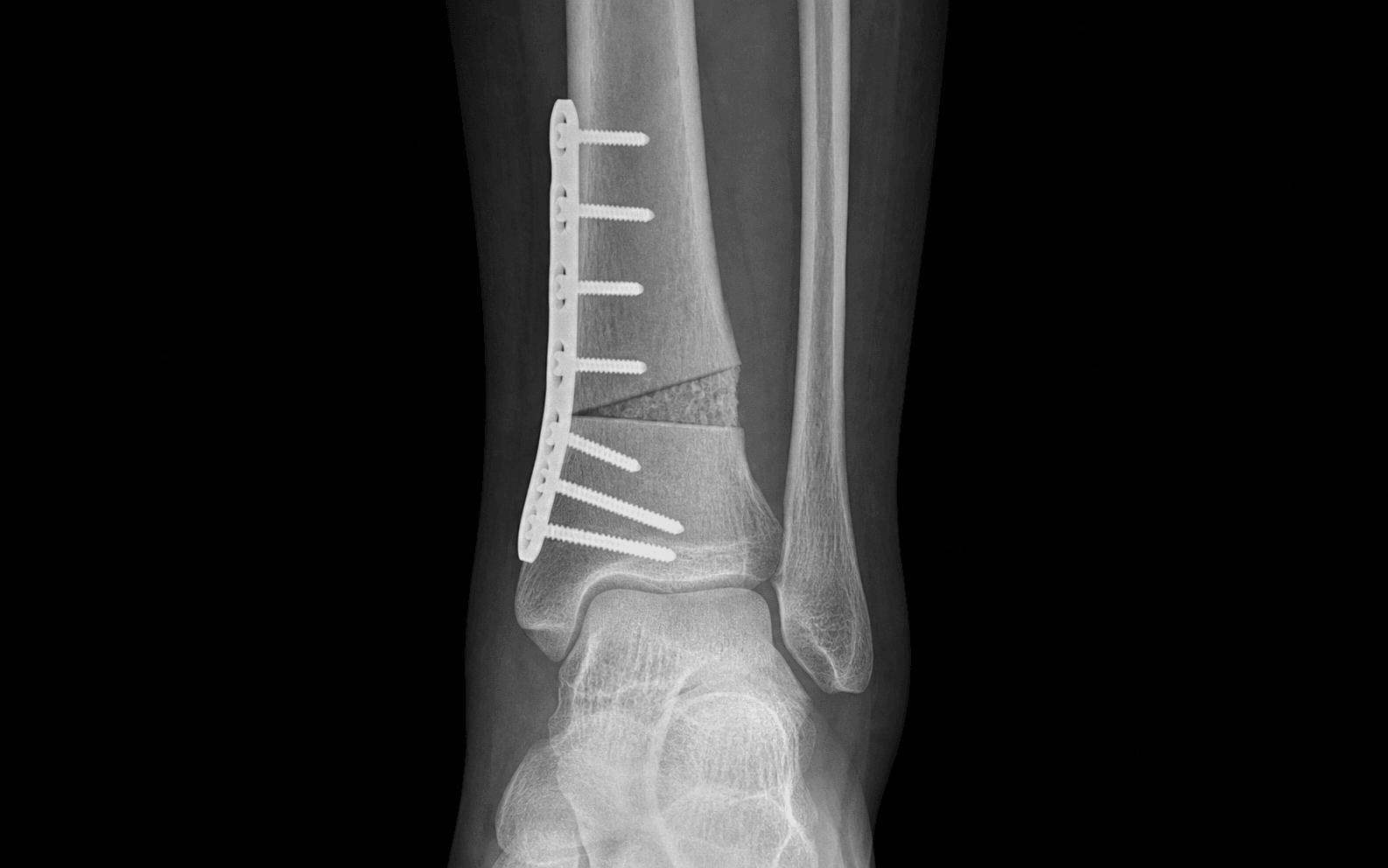
Medial opening-wedge osteotomy:
- Performed through a medial approach to the distal tibia
- Tibialis anterior tendon is retracted anteriorly or partially released
- A wedge of predetermined size is opened medially, the lateral cortex acts as a hinge (or is perforated in a controlled fashion)
- The defect is filled with bone graft (tricortical iliac crest autograft or allograft)
- Fixed with an anatomically contoured anteromedial locking plate
- Preserves bone stock, allows precise correction with a single osteotomy, but requires bone graft and has a theoretically slower union than closing-wedge
Lateral closing-wedge osteotomy:
- Performed through an anterolateral approach
- A calculated wedge of bone is removed from the lateral cortex
- The osteotomy is closed and the tibia translates medially
- Inherent bony contact at the osteotomy site (no graft required)
- Requires fibular osteotomy and more extensive lateral dissection
- Historically associated with risk of transient peroneal nerve palsy from lateral approach
Medial Opening-Wedge vs Lateral Closing-Wedge — Comparison
Key Evidence
Mid- to Long-term Results of Supramalleolar Osteotomy
Realignment surgery as alternative treatment of varus and valgus ankle osteoarthritis
Supramalleolar osteotomy for the treatment of ankle osteoarthritis leads to favourable outcomes and low complication rates at mid-term follow-up: a systematic review
Supramalleolar osteotomy for the treatment of distal tibial angular deformities and arthritis of the ankle joint
Low tibial osteotomy for varus-type osteoarthritis of the ankle
Clinical Decision Scenarios
Practise clinical reasoning and management decisions out loud
“A 48-year-old man presents with a 3-year history of progressive right ankle pain worsened by walking. Examination reveals a varus hindfoot alignment. Standing AP radiographs show medial compartment joint space narrowing with a TAS angle of 83 degrees and a talar tilt of 8 degrees. The lateral compartment is preserved. He has 35 degrees of sagittal ankle motion and a mobile subtalar joint. How do you manage him?”
“You are performing a medial opening-wedge supramalleolar osteotomy. During the procedure, after placing the bone graft and applying the plate, you check the fluoroscopy and find that the TAS angle is only 88 degrees — your target was 93 degrees. The fibula has been osteotomised. What has likely happened and what do you do?”
“A 55-year-old woman with valgus ankle osteoarthritis presents with lateral compartment pain. Her TAS angle is 98 degrees (valgus). Standing radiographs show lateral joint space narrowing and medial compartment preservation. She has a BMI of 38, is a smoker (20 pack-years), and has well-controlled type 2 diabetes. How do you counsel her regarding SMO?”
References
-
Krähenbühl N, Zwicky L, Bolliger L, Schädelin S, Hintermann B, Knupp M (2017). Mid- to Long-term Results of Supramalleolar Osteotomy. Foot Ankle Int;38(2):124-132. doi:10.1177/1071100716673416. PMID 27765869. — Retrospective review of 46 SMOs with mean 9.5-year follow-up; 71% good/excellent outcomes, 79% survivorship at 10 years.
-
Pagenstert GI, Hintermann B, Barg A, Leumann A, Valderrabano V (2007). Realignment surgery as alternative treatment of varus and valgus ankle osteoarthritis. Clin Orthop Relat Res;462():156-168. doi:10.1097/BLO.0b013e318124a462. PMID 17563701. — Prospective series of 35 patients; significant AOFAS improvement at 5.6 years; stage IV OA predictably poor outcome.
-
Butler JJ, Azam MT, Weiss MB, Kennedy JG, Walls RJ (2023). Supramalleolar osteotomy for the treatment of ankle osteoarthritis leads to favourable outcomes and low complication rates at mid-term follow-up: a systematic review. Knee Surg Sports Traumatol Arthrosc;31(2):701-715. doi:10.1007/s00167-022-07144-7. PMID 36151410. — Systematic review of 18 studies (547 patients); mean AOFAS improvement 33.2 points, complication rate 12.1%.
-
Stamatis ED, Cooper PS, Myerson MS (2003). Supramalleolar osteotomy for the treatment of distal tibial angular deformities and arthritis of the ankle joint. Foot Ankle Int;24(10):754-764. doi:10.1177/107110070302401004. PMID 14587989. — 42 patients with distal tibial angular deformity; significant alignment and pain improvement at 4.5 years.
-
Tanaka Y, Takakura Y, Hayashi K, Taniguchi A, Kumai T (2006). Low tibial osteotomy for varus-type osteoarthritis of the ankle. J Bone Joint Surg Br;88(7):909-913. doi:10.1302/0301-620X.88B7.17325. PMID 16798994. — 26 ankles with varus OA; 73% excellent/good results at mean 8.3 years; outcomes correlated with Takakura stage.