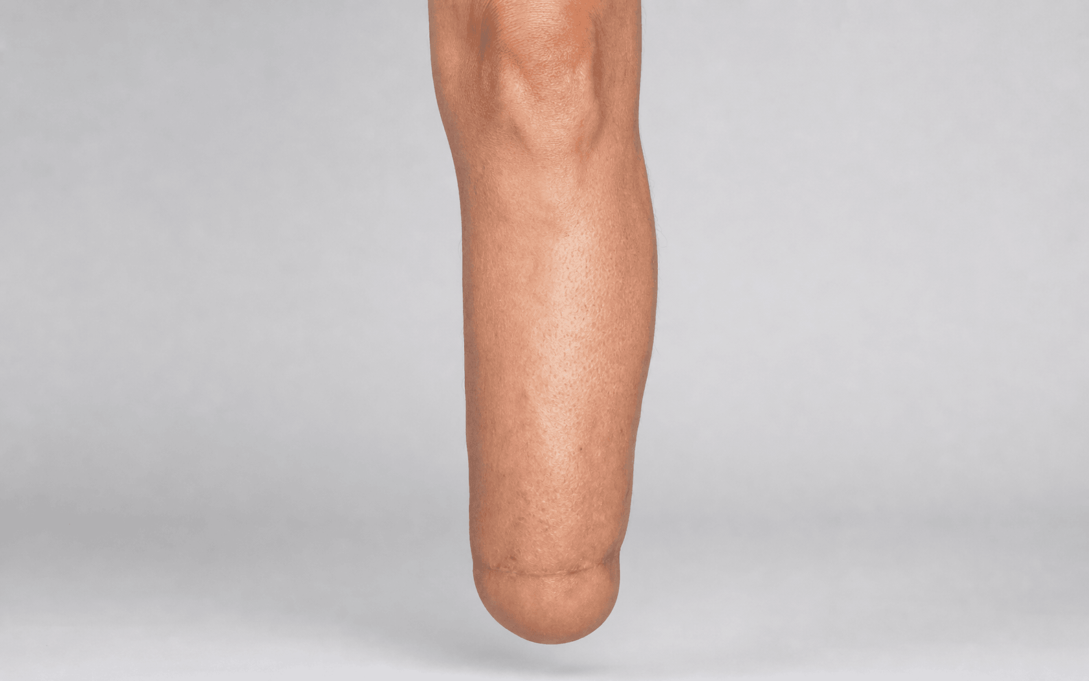
End-bearing amputation at the ankle level preserving the heel pad | advanced
Surgical Imaging
The danger: The posterior tibial artery gives off the medial calcaneal branches which supply the heel pad.
The fix: A patent posterior tibial artery is an absolute prerequisite. Avoid dissection in the posteromedial corner behind the medial malleolus. If Doppler shows no posterior tibial flow, a Syme amputation is contraindicated.
The danger: Damage to the fat pad lobules or the blood supply leads to pad necrosis, destroying the weight-bearing surface.
The fix: Dissect the calcaneus entirely subperiosteally. Hug the bone tightly with a sharp blade or periosteal elevator from superior to inferior. Never incise directly into the fat pad.
The danger: Without bony attachments, the Achilles tendon and weight-bearing forces will pull the heel pad posteriorly, leaving the distal tibia covered only by thin skin.
The fix: Anchor the deep fascia of the heel pad to drill holes in the anterior aspect of the distal tibia and fibula. Use heavy non-absorbable sutures to secure it squarely beneath the bones.
The danger: The ankle has five major sensory nerves. Failure to address them leads to painful neuromas trapped within the weight-bearing prosthesis.
The fix: Identify the posterior tibial, sural, saphenous, superficial peroneal, and deep peroneal nerves. Apply gentle distal traction, cleanly transect them, and allow them to retract well away from the stump end.
The danger: Resecting too much tibia converts the procedure to an essentially very short transtibial amputation, losing the metaphyseal flare needed for end-bearing and prosthesis suspension.
The fix: Resect only the malleoli and a maximum of 1 centimetre of the distal tibial articular surface. The cut must be perfectly parallel to the ground in both coronal and sagittal planes.
The danger: Performing a definitive one-stage closure in the presence of severe forefoot sepsis leads to stump infection and failure.
The fix: Use the two-stage technique. Stage one: disarticulate the ankle and loosely approximate the pad over the intact malleoli. Stage two: resect the malleoli and perform definitive closure after the infection is completely cleared.
S.Y.M.ESYME — Key Technical Principles
P.A.DPAD — Heel Pad Management
Surgical Indications
Absolute Indications
- Unsalvageable forefoot or midfoot infection / gangrene (e.g., severe diabetic foot infection that cannot be managed with a partial foot amputation)
- Severe distal foot trauma (crush injuries or blast injuries where the forefoot cannot be reconstructed but the heel pad is pristine)
- Tumours of the forefoot or midfoot (where adequate oncological margins can be achieved by ankle disarticulation)
- Congenital foot deformities (e.g., fibular hemimelia, severe untreated clubfoot) where a Syme amputation provides a superior base for a prosthesis compared to the native deformed foot
Prerequisites for Success
- Vascular: Palpable posterior tibial pulse, Doppler biphasic flow, or TcPO2 greater than 30 mmHg. The posterior tibial artery is the sole supply to the heel pad via the medial calcaneal branches.
- Soft Tissue: The heel pad skin must be completely intact, without deep ulceration, necrosis, or active infection. It must be mobile and healthy enough to bear the entire body weight.
- Functional: The patient must have the cognitive and physical capacity to participate in prosthetic rehabilitation.
Contraindications
Absolute:
- Absent posterior tibial artery flow (unless reconstructable via surgical or endovascular bypass)
- Heel pad necrosis or deep ulceration (the pad cannot serve as a weight-bearing surface)
- Inadequate tissue oxygenation (TcPO2 less than 20 mmHg universally predicts failure)
- Ascending gas gangrene (requires a higher level, open amputation for life salvage)
Relative:
- Severe neuropathy leading to unrecognised heel pad trauma (requires strict patient compliance and specialized prosthetic care)
- Bedbound non-ambulatory status (though a Syme is end-bearing and can assist with pivot transfers, a transtibial amputation often heals more reliably and faster in severe dysvascular bedbound patients)
Biomechanics of the Syme Amputation
- Energy Expenditure: Ambulation with a Syme amputation increases energy expenditure by only 10 to 15 percent compared to a normal gait. In contrast, a transtibial amputation increases energy expenditure by 20 to 40 percent. This is particularly crucial for elderly or dysvascular patients who have limited cardiopulmonary reserve.
- Proprioception: Preservation of the natural heel pad provides excellent proprioceptive feedback to the patient. The thick fibrous septa and fat lobules compress and distribute load uniformly across the distal tibial metaphysis.
- Lever Arm: The Syme stump provides a very long lever arm for the prosthesis, reducing the force required by the quadriceps and hip extensors to control the limb during the stance phase of gait.
Evidence for One-Stage vs Two-Stage Syme
One-Stage Syme
- The classical approach described by James Syme in 1843.
- Indicated in trauma, congenital deformities, and controlled dysvascular cases without active infection.
- Definitive bony resection and soft tissue closure are performed in a single operation.
- Excellent outcomes in non-infected patients but carries an unacceptably high failure rate if performed in the presence of severe forefoot sepsis due to contamination of the broad cancellous bone surface of the distal tibia.
Two-Stage Syme (Pinzur and Smith)
- Developed specifically for the diabetic patient with an infected forefoot.
- Stage One: Ankle disarticulation, removal of infected forefoot, but the malleoli are left INTACT. The oversized heel pad is loosely approximated over the malleoli. This avoids exposing cancellous bone to infection and provides a biological dressing over the joint.
- Stage Two: Performed 2 to 6 weeks later when the infection is clinically resolved and inflammatory markers are normal. The wound is reopened, the malleoli are resected, and definitive closure is performed.
- Evidence shows the two-stage technique significantly reduces stump infection and revision to transtibial amputation in the diabetic population, making it the standard of care for infected cases.
Syme Amputation vs Transtibial Amputation (BKA)
Clinical Decision Scenarios
Practise clinical reasoning and management decisions out loud
“A 65-year-old diabetic male with a chronic, infected midfoot ulcer and gangrene of the toes is referred for amputation. He was previously mobile. Pulses are absent. Explain your workup to determine if he is a candidate for a Syme amputation.”
“During a classical Syme amputation, you are preparing to resect the calcaneus from the heel pad. What is the most critical technical principle at this stage, and what anatomical structure are you trying to protect?”
“Six months following a successful one-stage Syme amputation for trauma, your patient presents to clinic complaining of severe, sharp, shooting pain at the anterolateral aspect of the stump every time they don their prosthesis. The pain radiates proximally and is exquisitely tender to light tapping. What is the most likely diagnosis, and how would you manage it?”