Syndesmotic screw or suture-button fixation of the disrupted distal tibiofibular joint in Weber C fractures and high ankle sprains | advanced
Surgical Imaging
The trap: Relying on indirect reduction (clamping the fibula to the tibia) and checking only AP and mortise fluoroscopy views to confirm reduction. CT studies show indirect reduction produces malreduction in up to 52% of cases.
The fix: Directly visualise the fibular reduction within the incisura — either through the existing fibular fracture site, through a small anterolateral arthrotomy to expose the AITFL and anterior incisura, or use intra-operative CT. Obtain a contralateral ankle CT pre-operatively as a reference template for the native incisura anatomy.
The trap: Placing syndesmotic fixation before confirming anatomic fibular length and rotation. A shortened or malrotated fibula forces the talus into an abnormal position within the mortise and produces persistent syndesmotic malreduction even with perfect screw placement.
The fix: Restore fibular anatomy with plate osteosynthesis FIRST. Confirm fibular length (radiographic: the fibular shadow should align within 1-2 mm of the tibial shadow on the AP view, and the talocrural angle should match the contralateral side), rotation (check the relationship of the fibula to the lateral tibial groove), and the incisura relationship before any syndesmotic fixation.
The trap: Allowing the patient to weight-bear on a syndesmotic screw without clear instructions. Syndesmotic screws are positional screws, not lag screws — they are designed to hold reduction while the ligaments heal, not to bear load. Weight-bearing on a rigid screw fixation leads to screw breakage in 10-30% of cases.
The fix: Strict non-weight-bearing for 6-8 weeks with a below-knee plaster or boot for syndesmotic screw fixation. If earlier weight-bearing is desired, use a suture-button (TightRope) construct, which is dynamic and permits physiological micromotion. Document the fixation type and weight-bearing protocol clearly.
The trap: Fixing the lateral side only without recognising a significant posterior malleolar fracture (greater than 25% of articular surface) with attached PITFL. The PITFL is the primary restraint to posterior tibiofibular translation — an unrepaired PITFL injury leaves the syndesmosis unstable regardless of anterior screw placement.
The fix: Assess the posterior malleolus on CT in all Weber C fractures and high ankle sprains. Fix posterior malleolar fractures involving the PITFL attachment and greater than 25% of the articular surface through a posterolateral approach. In some cases, posterior malleolar fixation alone restores syndesmotic stability without a separate syndesmotic screw.
The trap: Tightening syndesmotic screw(s) with the ankle in maximum dorsiflexion (tightens the syndesmosis around the wider anterior talar dome) without accounting for individual ankle anatomy. This can over-compress the syndesmosis, restrict talar motion, and cause impingement or early arthritis.
The fix: Clamp and fix the syndesmosis in neutral or slight dorsiflexion (10-15 degrees) rather than maximum dorsiflexion. Verify the medial clear space on the mortise view after fixation — it should equal the contralateral side (normally 2-4 mm). Excessive tightening of a suture-button construct can also over-compress; use finger-tight approximation and confirm fluoroscopically.
The trap: Performing the hook test or external rotation stress test BEFORE achieving anatomic fibular fixation. An unfixed, shortened, or malrotated fibula gives a false-positive instability test — the syndesmosis appears unstable when the real problem is the fibula.
The fix: Always reduce and fix the fibula with plate osteosynthesis FIRST. Then, with the fibular plate in situ, reassess syndesmotic stability using the hook test, external rotation stress test, and Cotton test under fluoroscopy. Only proceed to syndesmotic fixation if instability is confirmed AFTER anatomic fibular restoration.
R.E.D.U.C.EREDUCE — Syndesmotic Reduction Principles
F.I.X.E.DFIXED — Fixation Selection and Aftercare
M.A.L.R.E.DMALRED — Syndesmotic Malreduction Recognition
Surgical Indications
Absolute Indications
- Weber C or pronation-external rotation (Lauge-Hansen) ankle fracture with confirmed intra-operative syndesmotic instability after anatomic fibular plate fixation — demonstrated by the hook test, external rotation stress test, or Cotton test under fluoroscopy
- High ankle sprain (isolated syndesmotic disruption) with Grade III instability — complete disruption of the AITFL, PITFL, and interosseous ligament confirmed on MRI or stress radiography, and clinical or radiographic talar shift
- Syndesmotic diastasis greater than 6 mm on the AP radiograph (compared with the contralateral side) with clinical instability
Relative Indications
- Maisonneuve fracture (proximal fibular fracture with syndesmotic disruption and medial injury) — fixation of the syndesmosis after anatomic fibular reduction or fixation
- Chronic syndesmotic instability with persistent pain and confirmed diastasis on weight-bearing CT or stress radiography — delayed reconstruction using suture-button or screw with ligamentous repair
- Ankle fracture with associated posterior malleolar fracture involving greater than 25% of the articular surface and PITFL disruption where posterior fixation alone does not restore syndesmotic stability on intra-operative testing
Contraindications
Absolute:
- Syndesmotic instability that resolves after anatomic fibular plate fixation — do not over-fix a stable syndesmosis
- Active deep infection or open fracture contamination at the planned fixation site
- Significant tibial plafond or pilon fracture that precludes accurate syndesmotic assessment
Relative:
- Poor skin condition or significant soft tissue compromise at the lateral ankle (delay fixation until soft tissue recovery)
- Severe osteoporosis where screw purchase is unreliable (consider suture-button or larger diameter screws with bicortical purchase)
- Patient non-compliance with weight-bearing restrictions (suture-button may be preferred over screw fixation)
Intra-operative Syndesmotic Instability Tests
Hook Test
- Technique: After anatomic fibular plate fixation, insert a bone hook or clamp around the distal fibula and apply lateral traction while viewing the syndesmosis on fluoroscopy
- Positive finding: Lateral translation of the fibula away from the tibia (widening of the tibiofibular clear space greater than 5 mm compared to the fixed position) with lateral traction
- Limitations: Operator-dependent force; does not detect pure rotational instability; may give false-negative if AITFL is partially intact
- Evidence: Widely used and accepted as a standard intra-operative test; sensitivity and specificity vary by study but it is a reliable practical bedside tool
External Rotation Stress Test
- Technique: Apply external rotation force to the foot (or the talus directly) while the ankle is held in neutral or slight dorsiflexion, under fluoroscopic imaging of the mortise view
- Positive finding: Widening of the medial clear space greater than 5 mm (or greater than 2 mm compared to the contralateral side) on the mortise view with external rotation
- Limitations: Requires adequate imaging quality; medial clear space interpretation depends on the quality of the medial-sided repair (if deltoid is ruptured); a valgus stress component may produce a false-positive in pure deltoid insufficiency
- Evidence: Considered the most specific test for syndesmotic instability when performed after fibular fixation; best assessed on the mortise view
Cotton Test
- Technique: Apply lateral or medial talar shift force (manual or with a laminar spreader) under fluoroscopy while viewing the mortise view
- Positive finding: Asymmetric talar shift within the mortise; widening of the medial clear space suggesting residual instability despite lateral fixation
- Limitations: Does not isolate syndesmotic instability from pure deltoid incompetence; lateral talar shift may occur from deltoid rupture alone
- Evidence: Useful adjunct but least specific of the three tests — interpret in conjunction with the hook test and external rotation stress test
Trampoline Test (Ballottlement)
- Technique: With the ankle in neutral, palpate the anteroinferior tibiofibular ligament (AITFL) region. Press on the fibula laterally — a springy or bouncy feel (like a trampoline) indicates disruption of the AITFL and syndesmotic instability
- Positive finding: Loss of normal firm resistance; a spongy, springy feel when the fibula is depressed laterally
- Limitations: Subjective and examiner-dependent; does not quantify the degree of instability
- Evidence: Described in clinical practice as a useful adjunct; most valuable when combined with fluoroscopic stress testing
Evidence for Fixation Methods
Syndesmotic Screws
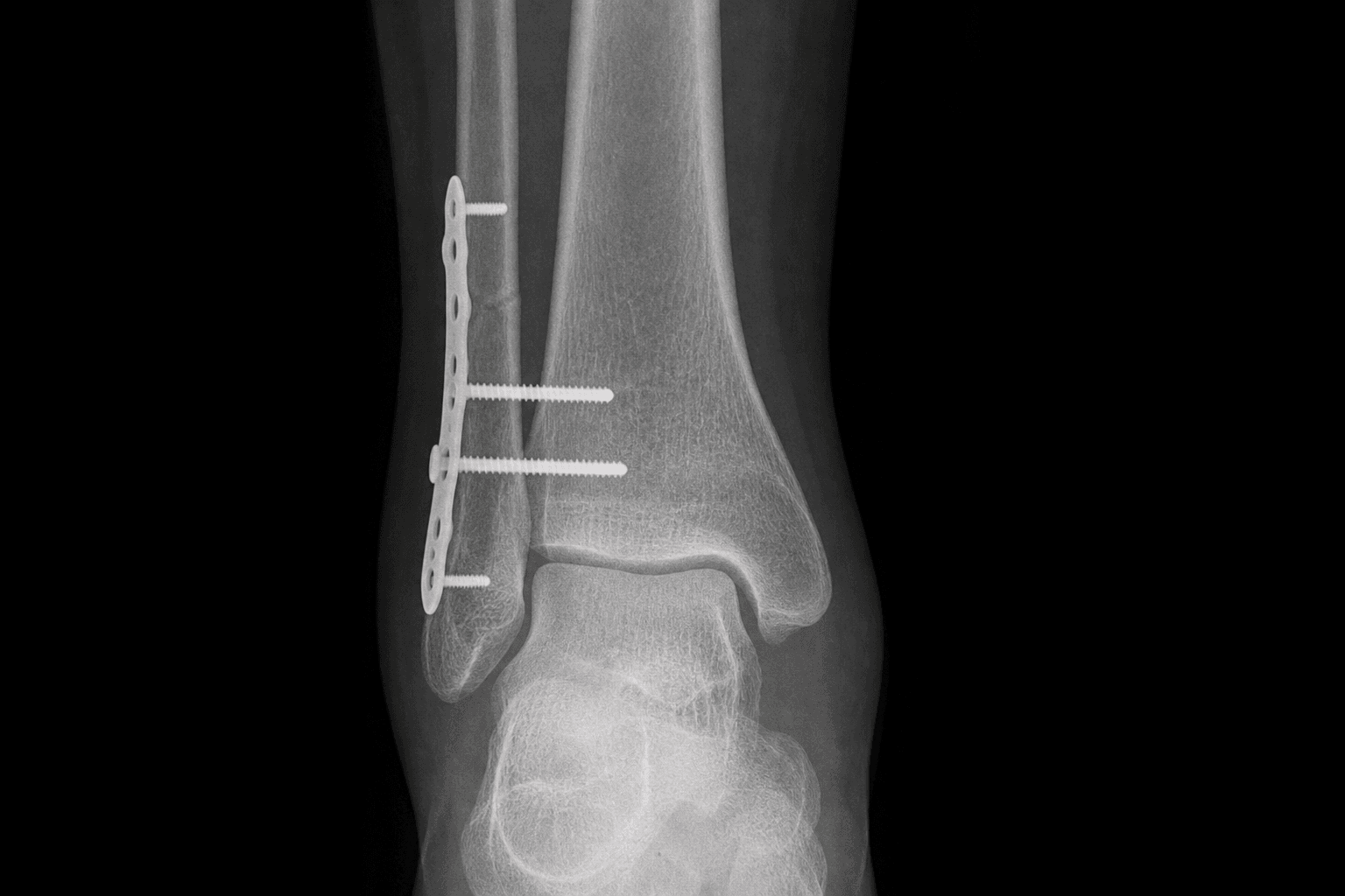
Positional screw fixation (3.5 or 4.5 mm cortical screw) is the historical gold standard for syndesmotic stabilisation. The screw is placed through the fibula into the tibia at a level 2-4 cm above the tibial plafond (above the synovial reflection), angled 25-30 degrees anteriorly in the coronal plane.
- Quadricortical fixation (4 cortices): Screw engages both tibial cortices and both fibular cortices — provides rigid fixation but eliminates all physiological tibiofibular motion. Screws frequently break with weight-bearing (10-30% rate).
- Tricortical fixation (3 cortices): Screw engages both fibular cortices and the medial tibial cortex only — leaves the lateral tibial cortex free, permitting some tibiofibular motion. Theoretically lower breakage rate but higher risk of loosening. Less commonly used.
- Number of screws: Biomechanical studies show two screws (placed at 2 cm and 3-4 cm above the plafond) provide greater torsional stability than one screw. One screw is acceptable in smaller frames or less severe disruptions; two screws are preferred in larger patients and high-energy injuries.
- Screw removal: Typically removed at 8-12 weeks post-operatively to restore tibiofibular motion and reduce the risk of breakage. The evidence for routine removal is debated — some studies show no difference in outcome with retained screws, but breakage and subsequent removal difficulty are practical concerns. Removal carries a small risk of re-diastasis.
Suture-Button Fixation (TightRope)
A suture-button device (e.g. TightRope, ZipTight) passes a fibre-wire loop through 4-cortex drill holes (fibula to tibia) and is secured with metallic or bioabsorbable buttons on the lateral fibula and medial tibia.
- Advantages: Does not require routine removal; allows earlier weight-bearing (protocols vary but many permit partial weight-bearing from 2-4 weeks); provides dynamic flexible fixation that permits physiological tibiofibular micromotion while maintaining reduction
- Biomechanics: Suture-button constructs match or exceed the stiffness of 3.5 mm quadricortical screws in biomechanical models for resistance to diastasis under physiologic loading; however, they are less stiff under high-load torsional stress
- Complications: Knot or button irritation over the medial tibial cortex (5-15% of cases — may require later removal of the button); skin irritation or wound problems at the button site; rare device failure or breakage
- Evidence: Multiple RCTs and meta-analyses have compared suture-button with screw fixation. Overall, functional outcomes at 1-2 years are comparable between the two methods. Suture-button shows advantages in earlier return to weight-bearing and absence of planned hardware removal.
Syndesmotic Screw versus Suture-Button — Evidence Summary
Key Evidence
Randomized Trial Comparing Suture Button with Single Syndesmotic Screw for Syndesmosis Injury
Better outcome for suture button compared with single syndesmotic screw for syndesmosis injury: five-year results of a randomized controlled trial
The functional consequence of syndesmotic joint malreduction at a minimum 2-year follow-up
A prospective randomised study comparing TightRope and syndesmotic screw fixation for accuracy and maintenance of syndesmotic reduction assessed with bilateral computed tomography
Fixation of ankle syndesmotic injuries: comparison of tightrope fixation and syndesmotic screw fixation for accuracy of syndesmotic reduction
Clinical Decision Scenarios
Practise clinical reasoning and management decisions out loud
“A 34-year-old man sustains a closed Weber C ankle fracture (SER IV equivalent) with a medial malleolar fracture and a fibular fracture 8 cm above the syndesmosis. After ORIF of the fibula with a plate and screw fixation of the medial malleolus, you perform the external rotation stress test and note 3 mm widening of the medial clear space on the mortise view compared to the contralateral side. Discuss your management.”
“A 28-year-old professional footballer sustained a syndesmotic injury (high ankle sprain) 4 months ago, treated non-operatively with a boot and physiotherapy. He now presents with persistent lateral ankle pain, inability to sprint, and a positive external rotation stress test on examination. MRI shows a disrupted AITFL with scarring and a 2 mm tibiofibular diastasis on stress views. Discuss the surgical management.”
“During ORIF of a Weber C ankle fracture, after placing the syndesmotic screw (single 3.5 mm quadricortical at 2 cm above the plafond), you check the mortise view and notice the medial clear space is 5 mm compared to 3 mm on the contralateral pre-operative film. The screw has been tightened and the clamp has been removed. What do you do?”