Tibiotalocalcaneal Arthrodesis (Hindfoot Nail)

Foot & AnkleAdvancedCore Procedure

Tibiotalocalcaneal Arthrodesis (Hindfoot Nail)

Surgical technique guide for tibiotalocalcaneal (TTC) arthrodesis using a retrograde hindfoot intramedullary nail for combined tibiotalar and subtalar arthritis, failed total ankle replacement, Charcot hindfoot, avascular talus and severe hindfoot deformity

High-yield overview

Simultaneous fusion of the tibiotalar and subtalar joints using a retrograde intramedullary nail | advanced

Surgical Imaging

Tibiotalocalcaneal arthrodesis with retrograde nail
Tibiotalocalcaneal arthrodesis with a retrograde hindfoot nail fusing the ankle and subtalar joints β€” for combined tibiotalar and subtalar arthritis or failed ankle reconstruction.Credit: AI-generated medical illustration Β· OrthoVellum
Critical Danger Structures and Exam Traps
Lateral Plantar Neurovascular Bundle

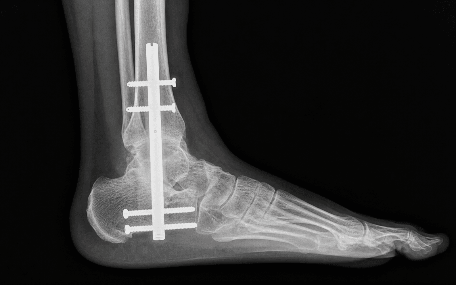
The trap: A plantar midline entry incision or medialised nail entry transits directly through (or immediately adjacent to) the lateral plantar nerve, artery and vein as they course from the medial heel pad distally toward the lateral midfoot.

The fix: Place the entry incision slightly LATERAL to the plantar midline, over the lateral half of the heel pad. Stay lateral to the medial band of the plantar fascia. Dissect bluntly through subcutaneous fat, identify the lateral plantar neurovascular bundle medially, retract medially, and enter the calcaneus under direct vision. The bundle should NEVER be the lateral-most structure in the wound.

Medial Plantar Nerve

Location: The medial plantar nerve runs along the medial border of the plantar fascia from the medial heel toward the great toe; it is generally spared by a lateralised entry portal but is at risk with extensive medial dissection or with correction of severe hindfoot valgus.

Risk: Injury produces numbness and painful neuroma of the medial sole and great toe β€” a significant functional disability. Identify and protect it during any medial dissection for tibial preparation or graft harvest.

Posterior Tibial Neurovascular Bundle

Location: The posterior tibial artery, tibial nerve and accompanying veins lie posterior to the medial malleolus and pass into the foot beneath the flexor retinaculum (tarsal tunnel).

Risk: In a posterior approach to the ankle and subtalar joint for joint preparation, retractors placed posteriorly can injure or stretch this bundle. Identify it before any posterior capsular or subtalar work, and keep retractors anterior to the bundle under direct vision.

Talar Avascular Necrosis β€” Nonunion Risk

The trap: In osteonecrosis of the talus (post-traumatic, post-TAR, idiopathic), the talar body is largely avascular. Standard TTC techniques that rely on tibiotalar coaptation will have a high rate of nonunion (up to 40-50%).

The fix: Recognise AVN preoperatively (MRI or CT with sclerosis/crescent sign). Plan for STRUCTURAL bone graft (tricortical iliac crest or femoral head allograft) bridging the tibial plafond to the calcaneus, or a hindfoot nail with tibiocalcaneal arthrodesis after talectomy. Warn the patient about the high nonunion rate and the potential need for revision.

Charcot Neuroarthropathy β€” Bone Quality and Alignment

Why different: Charcot feet have a hyperaemic, osteopenic, fragmented bone with poor screw purchase. The construct is load-bearing from day 1, and union rates are 50-70% in published series, lower than primary TTC.

Implications: Use a long hindfoot nail extending to the tibial metaphysis, supplement with autogenous bone graft (reamings from the tibia, iliac crest, or proximal tibia), consider adjunctive plate fixation of the tibiotalar or subtalar joints, and accept prolonged non-weight-bearing (often 3 months) until radiographic union. Correct the rocker-bottom deformity and restore a plantigrade foot.

Varus Malalignment β€” Worst Outcome

The trap: Leaving the hindfoot in varus at the end of TTC fusion produces a poor outcome β€” the patient loads the lateral border, develops fifth MT overload, lateral foot pain, and may ulcerate over the base of the fifth metatarsal.

The fix: Aim for a plantigrade foot with hindfoot valgus of 5 degrees, neutral forefoot, and tibial axis passing through the central hindfoot. If the foot is plantigrade but in varus, the outcome is worse than in valgus β€” small degrees of residual valgus are well tolerated, varus is not.

Mnemonic

P.L.A.N.T.A.RPLANTAR β€” Entry Portal Anatomy and Dangers

Mnemonic

H.I.N.D.F.O.O.THINDFOOT β€” Indications and Decision-Making

Mnemonic

N.A.I.LNAIL β€” Intraoperative Pearls

Surgical Indications

Primary Indications for TTC Arthrodesis

  • Combined tibiotalar AND subtalar arthritis β€” most common indication; isolated ankle or subtalar fusion will not address the patient's pain
  • Failed total ankle replacement (TAR) with subtalar arthritis, talar component subsidence, or aseptic loosening where revision TAR is not feasible
  • Charcot neuroarthropathy (Eichenholtz stage 2-3) of the hindfoot with instability, deformity, or ulceration β€” a load-sharing construct that bypasses both joints
  • Avascular necrosis of the talus with collapse β€” either preserve the talus with structural graft bridging to the calcaneus, or perform talectomy with tibiocalcaneal arthrodesis
  • Severe hindfoot deformity (post-traumatic varus, equinocavovarus, equinus contracture) that requires correction across both joints
  • Failed prior ankle or subtalar arthrodesis with nonunion, malunion, or symptomatic adjacent joint disease
  • Tumour resection of the talus requiring intercalary reconstruction

Relative Indications

  • Salvage after failed TAR in a lower-demand patient where conversion to TTC nail is the definitive solution
  • Talar body fracture with severe comminution not amenable to ORIF
  • Severe post-traumatic hindfoot bone loss requiring bridging graft

Contraindications

Absolute:

  • Active infection (osteomyelitis of the tibia, talus, or calcaneus) β€” must be cleared first with staged debridement
  • Uncorrectable vascular insufficiency β€” check ankle-brachial pressure index; revascularise first
  • Severe peripheral neuropathy with open ulceration through the planned incision β€” heal the wound first

Relative:

  • Young, high-demand patient β€” consider staged ankle and subtalar fusion to preserve whatever hindfoot motion is possible
  • Active Charcot with severe bone fragmentation (Brodsky 3B) β€” consider external fixation or staged management
  • Smoker with poor healing β€” counsel about nonunion risk (up to 2-3x higher); consider bone graft adjuncts
  • Severe osteoporosis (T-score less than minus 3) β€” consider plate-augmented construct or external fixation

Evidence for Operative Treatment

Outcomes of Primary TTC Arthrodesis

  • A systematic review of retrograde hindfoot nailing for combined hindfoot pathology reports union rates of 70-90% in primary cases with modern compression/angle-stable nail designs
  • Patient satisfaction exceeds 75% in most series, with substantial pain relief and improved function; gait analysis shows near-normal ankle power but loss of hindfoot inversion/eversion
  • AOFAS scores improve from a mean of 30-40 preoperatively to 70-80 postoperatively in most series
  • Time to union: 3-6 months in primary cases; up to 9-12 months in Charcot, AVN, and revision

Failed Total Ankle Replacement

  • A meta-analysis of TAR failures converted to TTC nail reports union rates of 75-85%, with patient satisfaction approaching that of primary TTC
  • Tibiotalocalcaneal nail is now considered the gold standard salvage for failed TAR with subtalar arthritis
  • Bone loss from the explanted talar component often requires structural graft or bone substitute augmentation

Charcot Neuroarthropathy

  • A multi-centre review of TTC nailing for Charcot reports union rates of 50-70%, with limb salvage rates exceeding 85% at 5 years
  • Rocker-bottom deformity correction with the nail permits ulcer healing in 70-90% of patients
  • Solid intramedullary constructs have largely replaced external fixation for Charcot hindfoot reconstruction
  • A long hindfoot nail extending to the tibial metaphysis is preferred for stable load-sharing

Avascular Necrosis of the Talus

  • AVN is a high-risk subset: union rates 50-70% in published series, lower than primary TTC
  • Structural bone graft (autogenous iliac crest tricortical or femoral head allograft) bridging the tibial plafond to the calcaneus is often required
  • Talectomy with tibiocalcaneal arthrodesis is a salvage option for severe AVN with collapse

Indication-Specific Outcomes and Technical Nuances


Key Evidence

Evidence

Risk factors for nonunion following tibiotalocalcaneal arthrodesis: a systematic review and meta-analysis

Level I
Patel S, Baker L, Perez J, Vulcano E, Kaplan J, Aiyer A β€’ Foot Ankle Surg
Clinical implication: Preoperative counselling must address modifiable risk factors (smoking cessation, glycaemic control) and set realistic expectations for Charcot and AVN patients where nonunion rates are highest.
Evidence

Tibiotalocalcaneal arthrodesis with a retrograde intramedullary nail: a prospective cohort study at a minimum five year follow-up

Level II
Perez-Aznar A, Gonzalez-Navarro B, Bello-Tejeda LL, Alonso-Montero C, Lizaur-Utrilla A, Lopez-Prats FA β€’ Int Orthop
Clinical implication: TTC arthrodesis with a retrograde nail provides durable pain relief and functional improvement at five years; use a long nail extending to the tibial metaphysis to reduce stress fracture risk.
Evidence

Tibiotalocalcaneal arthrodesis with structural allograft for management of large osseous defects of the hindfoot and ankle: a systematic review and meta-analysis

Level I
Cifaldi A, Thompson M, Abicht B β€’ J Foot Ankle Surg
Clinical implication: Structural allograft is effective for bridging large bone defects in TTC arthrodesis, particularly in failed TAR and talar AVN; counsel about lower union rates compared with primary cases.
Evidence

Comparison of dynamic versus static locked retrograde tibiotalocalcaneal arthrodesis with intramedullary nail fixation: evaluation of the RAIN database

Level III
Dujela MD, Berlet GC, Houng BE, Hyer CF β€’ J Foot Ankle Surg
Clinical implication: Both dynamic and static locking achieve reliable union in TTC arthrodesis; nail length to the tibial metaphysis is the more important technical variable than locking mode.
Evidence

Tibiotalocalcaneal arthrodesis using retrograde intramedullary nail fixation: comparison of patients with and without diabetes mellitus

Level III
Wukich DK, Mallory BR, Suder NC, Rosario BL β€’ J Foot Ankle Surg
Clinical implication: Diabetes is a significant risk factor for nonunion and complications after TTC arthrodesis; optimise glycaemic control preoperatively and counsel about higher risk.

Clinical Decision Scenarios

Practise clinical reasoning and management decisions out loud

Viva scenarioAdvanced
Clinical prompt

β€œA 62-year-old man with Type 2 diabetes and a 6-month history of midfoot and ankle swelling presents with a rocker-bottom deformity of the right foot. He has a non-healing plantar ulcer under the cuboid. Examination shows a warm, swollen, erythematous foot with intact sensation (protective sensation absent on 10 g monofilament testing). What is your assessment and management plan?”

Practical approach
This patient has Charcot neuroarthropathy of the hindfoot and midfoot with an associated plantar ulcer β€” Brodsky type 2 with extension to the midfoot. The rocker-bottom deformity has caused the cuboid to protrude plantarly, leading to a pressure ulcer. **Assessment**: - Confirm Charcot stage (Eichenholtz) β€” based on clinical and radiographic findings - Bilateral weight-bearing foot and ankle radiographs (AP, mortise, lateral) to assess bone fragmentation, subluxation, and deformity - MRI to assess the extent of bone oedema, fragmentation, and any occult infection - Inflammatory markers (CRP, ESR, WCC) and ulcer swab β€” important to exclude osteomyelitis, which would change management - HbA1c, fasting glucose, albumin, and lymphocyte count β€” optimise the patient before surgery - Vascular assessment: ankle-brachial pressure index, toe pressures, and consider vascular review if ABPI less than 0.8 **Staged management**: - Stage 1: Treat the ulcer. Offload the foot in a total contact cast or Charcot restraint orthotic walker (CROW) boot. Treat any superimposed infection with oral or IV antibiotics. Optimise diabetes (HbA1c less than 7.5%). Improve nutrition (albumin greater than 30 g/L). - Stage 2: Once the ulcer is healed and the soft tissues are quiescent, perform a tibiotalocalcaneal arthrodesis with a retrograde intramedullary nail. Use a long nail to the tibial metaphysis and adjunctive bone graft. Correct the rocker-bottom deformity (Achilles lengthening, plantar fascia release, bony realignment). - Stage 3: Strict non-weight-bearing in a cast for 3 months, then progressive weight-bearing in a CROW boot for a further 3-6 months, then long-term custom-molded foot orthosis. **Outcomes to counsel the patient about**: - Limb salvage rate 85-90% at 5 years with TTC nail - Union rate 50-70% in Charcot (lower than primary TTC) - Risk of infection, nonunion, malunion, and stress fracture at the nail tip - Long recovery (6-12 months) and the need for prolonged offloading - The patient should understand that this is a limb-salvage operation, not a cure β€” and the alternative is below-knee amputation **Key technical points**: - Use a long hindfoot nail to the tibial metaphysis (180-300 mm) - Adjunctive bone graft (autogenous proximal tibia or iliac crest) Β± BMP - Consider adjunctive plate fixation for severely comminuted joints - Achilles lengthening to achieve a plantigrade foot - Strict glycaemic control throughout
Viva scenarioAdvanced
Clinical prompt

β€œA 58-year-old woman underwent a total ankle replacement 8 years ago for post-traumatic ankle arthritis. She now presents with progressive hindfoot pain, swelling, and difficulty weight-bearing. Radiographs show talar component subsidence, polyethylene wear, and subtalar joint space narrowing. She has failed conservative management. What are the options and which do you recommend?”

Practical approach
This patient has a failed total ankle replacement (TAR) with talar component subsidence, polyethylene wear, and symptomatic subtalar arthritis β€” the classic scenario for tibiotalocalcaneal (TTC) arthrodesis as a salvage procedure. **Options**: 1. **Revision TAR**: Generally not feasible here because of talar bone loss, polyethylene wear, and the additional problem of subtalar arthritis. Revision TAR has higher failure rates than primary TAR and is not suitable for this patient. 2. **Isolated ankle arthrodesis (with subtalar fusion as a second stage)**: Would address the ankle but not the subtalar arthritis. The patient would still have hindfoot pain. Furthermore, ankle arthrodesis after TAR is technically demanding (loss of talar bone stock) and may be best combined with subtalar fusion. 3. **Tibiotalocalcaneal (TTC) arthrodesis with retrograde intramedullary nail**: The gold standard salvage for failed TAR with subtalar arthritis. The nail spans the entire hindfoot, bypassing both joints, and provides a stable plantigrade foot. 4. **Below-knee amputation**: The last resort if reconstruction fails or the patient is not fit for reconstruction. **My recommendation**: TTC arthrodesis with a retrograde intramedullary nail. **Preoperative workup**: - Weight-bearing AP, mortise, and lateral ankle radiographs; Saltzman hindfoot view - CT to assess talar bone loss and joint destruction - Inflammatory markers (CRP, ESR, WCC) to exclude low-grade infection - Aspiration of the ankle if infection is suspected - Bone quality assessment (DEXA scan if osteoporotic risk factors) **Operative plan**: - Explant the TAR components carefully, preserving as much host bone as possible - Assess the talar bone defect; if significant, use structural graft (femoral head allograft or tricortical iliac crest) to fill the defect - Use a LONG hindfoot nail (180-300 mm) extending to the tibial metaphysis - Pack cancellous autograft (tibial reamings) into the joints and around the graft - Achieve a plantigrade foot with 5 degrees of hindfoot valgus - Multiplanar locking screws in the calcaneus for rotational control - BMP adjunct in revision cases **Postoperative plan**: - Strict non-weight-bearing for 6-8 weeks, then progressive weight-bearing in a boot - CT at 6 months to confirm union - Counsel the patient that recovery is 6-12 months and the outcome is good but not equivalent to a primary TAR
Viva scenarioAdvanced
Clinical prompt

β€œA 45-year-old labourer presents 2 years after a talar neck fracture with collapse of the talar body and severe hindfoot pain. He is otherwise healthy and active. MRI shows extensive avascular necrosis of the talar body. What are the surgical options, and which do you recommend?”

Practical approach
This young, active patient has avascular necrosis (AVN) of the talar body with collapse β€” a high-risk situation for nonunion. The age and activity level must be factored into the decision. **Surgical options**: 1. **Tibiotalocalcaneal (TTC) arthrodesis with retrograde nail and structural bone graft** β€” bridges the tibial plafond to the calcaneus with a structural block of iliac crest or femoral head allograft, with the necrotic talus preserved as a scaffold. 2. **Talectomy with tibiocalcaneal arthrodesis** β€” the talar body is excised and the nail fuses the tibia directly to the calcaneus. Lower height of the hindfoot but higher union rate than preserved talus in severe AVN. 3. **Ankle arthrodesis alone** β€” would address the tibiotalar joint but not the often-concomitant subtalar arthritis from the original injury. Will likely fail in the long term as the subtalar joint deteriorates. 4. **Total talar replacement (3D-printed custom talus)** β€” emerging technology; long-term data limited; not yet standard of care in most centres. 5. **Below-knee amputation** β€” last resort; the patient is young and otherwise healthy so not appropriate as first-line. **My recommendation**: For this young, active patient, I would discuss both TTC arthrodesis with structural graft AND talectomy with tibiocalcaneal arthrodesis, with the understanding that the latter has a higher union rate but a lower hindfoot height. In a labourer, the priority is a durable, fused, plantigrade foot that will tolerate heavy activity. I would lean toward talectomy with tibiocalcaneal arthrodesis because the union rate is higher (around 70 percent in published series) and the labourer's foot will tolerate the lower hindfoot height with a custom-molded orthosis. **If the patient prefers a preserved hindfoot height**: TTC arthrodesis with structural iliac crest or femoral head allograft and the necrotic talus left in situ. Higher nonunion risk (around 30-40 percent) but preserved height. **Operative plan (for talectomy with tibiocalcaneal arthrodesis)**: - Lateral transfibular or extensile lateral approach for talar body excision - Plantar entry portal as described - Long hindfoot nail to tibial metaphysis - Structural bone graft to fill the dead space and promote union - Multiplanar locking screws - Aim for 5 degrees of hindfoot valgus and a plantigrade foot **Postoperative plan**: - Strict non-weight-bearing for 6-8 weeks - Progressive weight-bearing in a boot from 8-12 weeks - CT at 6 months to confirm union - Long-term custom-molded foot orthosis - Counsel about the possibility of revision (higher than primary TTC) and the realistic functional outcome (stiff but pain-free foot)
Exam day cheat sheet
Tibiotalocalcaneal Arthrodesis (Hindfoot Nail) β€” Exam Day Summary

References

Evidence

Mid-term follow-up of patients with hindfoot arthrodesis with retrograde compression intramedullary nail in Charcot neuroarthropathy of the hindfoot

Level III
Chraim M, Krenn S, Alrabai HM, Trnka HJ, Bock P β€’ Bone Joint J
Clinical implication: Charcot neuroarthropathy is a recognised indication for TTC nail with lower union rates than primary fusion; use long nails and supplementary bone graft to maximise the chance of successful limb salvage.
Evidence

Tibio-talo-calcaneal arthrodesis with retrograde compression intramedullary nail fixation for salvage of failed total ankle replacement: a systematic review

Level IV
Donnenwerth MP, Roukis TS β€’ Clin Podiatr Med Surg
Clinical implication: TTC nail is the gold standard salvage for failed TAR with subtalar arthritis or talar component subsidence where revision arthroplasty is not feasible; anticipate talar bone loss and plan for structural graft.
Evidence

Salvage of avascular necrosis of the talus by combined ankle and hindfoot arthrodesis without structural bone graft

Level IV
Tenenbaum S, Stockton KG, Bariteau JT, Brodsky JW β€’ Foot Ankle Int
Clinical implication: AVN of the talus is a high-risk indication for TTC arthrodesis; structural bone graft improves union rates and should be used when talar collapse is present. Talectomy with tibiocalcaneal arthrodesis is the salvage option for advanced cases.
Editorially reviewed β€” transparent references and correction processPublished by OrthoVellum Medical Education TeamEditorial boardMethodologyReview policy
Educational disclosure

Educational content is reviewed for source visibility, editorial coherence, and correction readiness.

No individual clinician credential is claimed unless a named person is shown.

Verify before clinical use; this is not medical advice or a substitute for local guidance.