Microvascular fibular transfer for segmental bone defects and osteonecrosis | advanced
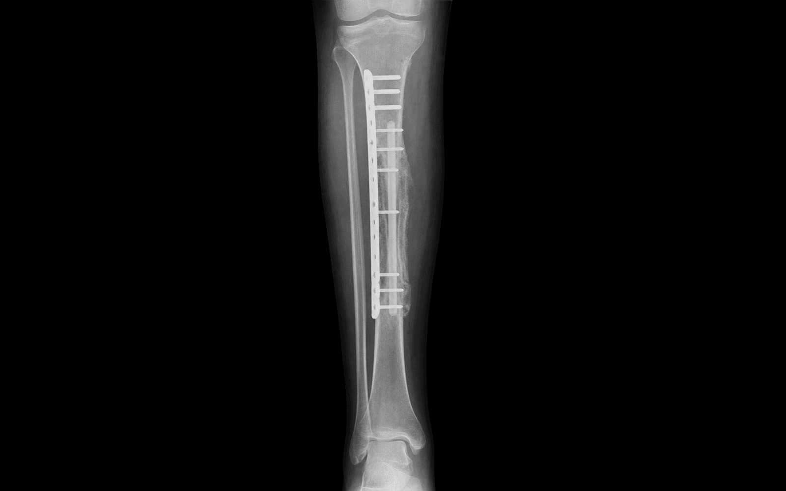
Surgical Imaging
Location: Wraps around the fibular neck proximally.
Risk: Resecting too far proximally endangers the nerve. Always preserve the proximal 6-8 cm of the fibula to protect the common peroneal nerve and lateral collateral ligament insertion.
Location: Distal tibiofibular joint.
Risk: Resecting the distal fibula destabilises the ankle mortise. You must preserve the distal 6-8 cm of the fibula to maintain the integrity of the syndesmosis and lateral malleolus.
Location: Runs closely along the posterior aspect of the fibula deep to the FHL.
Risk: Injury during osteotomy or deep dissection destroys the blood supply. Ensure osteotomies are performed carefully and the pedicle is identified and protected throughout harvest.
Why it happens: Due to tethering, denervation, or ischaemia of the Flexor Hallucis Longus (FHL) muscle belly, which is partially left on the graft to protect the pedicle.
Prevention: Meticulous dissection, careful haemostasis, and early active/passive range of motion exercises for the great toe post-operatively.
Risk: The fibular strut is initially mechanically weak compared to a femur or tibia and takes months to hypertrophy.
Management: Prolonged protected weight-bearing. Rigid initial fixation (often supplemented with an allograft shell in the Capanna technique) reduces the risk.
Timing: Highest risk in the first 72 hours post-op.
Implication: Leads to flap failure and graft necrosis. Requires meticulous anastomotic technique, appropriate vessel geometry without tension/kinking, and careful post-op monitoring using the skin paddle.
F.I.B.U.L.AFIBULA — Harvest Limits and Anatomy
G.R.A.F.TGRAFT — Indications for FVFG
Surgical Indications
Segmental Bone Defects
- Large defects: Typically indicated for long-bone defects greater than 6 cm.
- Oncology: Reconstruction after intercalary or intra-articular wide resection of primary bone sarcomas.
- Trauma: Severe open fractures with segmental bone loss where conventional grafting would fail.
- Infection: Reconstruction of defects following radical debridement for chronic osteomyelitis or infected nonunions.
- Congenital: Congenital pseudarthrosis of the tibia.
Femoral Head Osteonecrosis (AVN)
- Pre-collapse AVN: Ficat/Steinberg stage I or II, or early stage III with minimal collapse (less than 2 mm).
- Mechanism: The vascularised fibula provides structural support to the subchondral plate (preventing collapse) and brings a new blood supply to revascularise the necrotic segment.
- Patient selection: Most successful in younger patients (under 50 years) with symptomatic pre-collapse disease.
Advantages over Non-Vascularised Bone Graft
- Maintains osteocyte viability and heals by primary bone healing rather than creeping substitution.
- Undergoes rapid hypertrophy in response to mechanical stress (Wolff's law).
- Superior performance in poorly vascularised or irradiated recipient beds.
- Lower rate of late stress fracture and resorption.
Evidence Summary
Defect Reconstruction
- Vascularised fibular grafts achieve primary union rates of 70-90 percent in large defects.
- The Capanna technique (combining a vascularised fibula inside a massive structural allograft) provides immediate mechanical stability from the allograft and long-term biological viability from the fibula, significantly reducing the risk of stress fracture compared to FVFG alone.
Femoral Head AVN
- Multiple studies demonstrate that FVFG delays or prevents the need for total hip arthroplasty (THA) in young patients with early-stage osteonecrosis.
- Survival of the femoral head is closely correlated with the stage at the time of surgery; post-collapse heads (greater than 2 mm) have high failure rates.
Clinical Decision Scenarios
Practise clinical reasoning and management decisions out loud
“A 25-year-old man requires reconstruction of a 10 cm mid-diaphyseal tibial defect following resection of an adamantinoma. You plan a free vascularised fibular graft. What are the key anatomical landmarks and limitations you must respect during the fibular harvest?”
“You are treating a 30-year-old patient with pre-collapse osteonecrosis (Ficat Stage II) of the femoral head. You are discussing a free vascularised fibular graft. How does this procedure address the pathology, and what is the surgical technique?”