Rare Aggressive Vascular Malignancy | Poor Prognosis | Multimodal Treatment Required
- Extremely rare and highly aggressive - under 1% of sarcomas with 20-35% 5-year survival
- Three major associations: chronic lymphedema (Stewart-Treves), radiation therapy, and foreign bodies
- Multifocal disease common - 50% have multiple sites at presentation or develop additional lesions
- Early metastases - lung most common, occur early and frequently (50% of patients)
- Wide excision with negative margins mandatory - intralesional surgery leads to rapid recurrence
- “Stewart-Treves syndrome: angiosarcoma arising in chronic lymphedema (classically post-mastectomy)
- “Radiation-induced angiosarcoma has 5-10 year latency after breast cancer treatment
- “CD31 and CD34 positive on immunohistochemistry - confirms endothelial origin
- “Scalp angiosarcoma in elderly has worst prognosis - multifocal, infiltrative, high recurrence
The single most common cause of delayed angiosarcoma diagnosis is mistaking a bruise-like scalp lesion for benign ecchymosis. Any non-traumatic, persistent or enlarging purple scalp lesion in an older patient must be biopsied. By the time the diagnosis is clinically obvious, multifocal infiltrative disease has usually rendered cure impossible.
Overview and Epidemiology
Angiosarcoma is a malignant tumour of endothelial cells that can arise in skin, soft tissue, breast, liver, spleen, heart, and bone. It is among the most aggressive of all sarcomas, with a strong tendency to early haematogenous (especially pulmonary) metastasis, multifocal local disease, and high local recurrence even after complete excision.
- Accounts for under 1-2% of all soft tissue sarcomas
- Bimodal: young adults (deep soft tissue/visceral) and the elderly (cutaneous head and neck)
- The scalp and face are the most common cutaneous sites, typically in patients over 60
- Slight overall male predominance, although breast and Stewart-Treves disease occur in women
- Median 5-year survival across all sites is approximately 20-35%
- Therapeutic radiation (breast most commonly; 5-10 year latency)
- Chronic lymphedema (Stewart-Treves syndrome; usually over 10 year latency)
- Chemical carcinogens (vinyl chloride, arsenic, thorotrast - classically hepatic angiosarcoma)
- Chronic foreign bodies (vascular grafts, retained surgical material)
- Ultraviolet exposure (a driver of the high mutational burden in cutaneous head/neck disease)
Pathophysiology
Angiosarcoma arises from malignant transformation of vascular or lymphatic endothelial cells, producing irregular, anastomosing vascular channels lined by atypical endothelium that dissect through tissue planes. This infiltrative, channel-forming growth underlies two defining clinical problems: tumour extent is consistently underestimated at the margins, and disease is frequently multifocal ("skip" lesions), explaining the high recurrence rate even after apparently complete resection.
Molecular drivers differ by subtype, which is exam-relevant:
- Secondary angiosarcoma (post-radiation and lymphedema-associated) is characterised by high-level MYC (8q24) amplification in essentially 100% of cases, with FLT4 (VEGFR3) co-amplification in a subset. MYC amplification is absent in benign atypical vascular lesions, making it the key diagnostic discriminator in an irradiated field.
- Primary angiosarcoma generally lacks MYC amplification and shows other alterations (e.g. KDR/VEGFR2 mutations, PTPRB and PLCG1 mutations, and a high UV-signature mutational burden in cutaneous head/neck disease).
- Upregulation of vascular receptor tyrosine kinases (TIE1, KDR, TEK, FLT1) provides the rationale for anti-angiogenic and tyrosine-kinase-directed approaches.
The aggressive, vasoformative phenotype also accounts for the propensity to bleed and the characteristic bruise-like clinical appearance of cutaneous disease.
Classification
AJCC 8th Edition (Soft Tissue Sarcoma)
Angiosarcoma is staged using the AJCC soft tissue sarcoma staging system. However, most angiosarcomas are high-grade and present at advanced stage due to multifocal disease and early metastases.
- T
- T1 (under 5cm)
- Grade
- High (G3)
- Metastases
- M0
- 5-Year Survival
- 40-50%
- T
- T2-4 (over 5cm or deep)
- Grade
- High (G3)
- Metastases
- M0
- 5-Year Survival
- 30-40%
- T
- Any T
- Grade
- Any G
- Metastases
- M1 (metastatic)
- 5-Year Survival
- Under 10%
Most angiosarcomas present as Stage III or IV due to high grade and extent of disease.
Investigations and Staging
Diagnostic Imaging and Biopsy Protocol
Gold standard for local staging. Features: Heterogeneous signal, prominent enhancement (vascular tumor), infiltrative margins, multifocal areas.
Essential for metastatic workup. Lung metastases present in 30-40% at diagnosis. Also check liver, bone.
Mandatory for diagnosis. Request immunohistochemistry (CD31, CD34, Factor VIII, ERG). MYC amplification testing if radiation-associated.
Consider for extent of disease. Angiosarcoma is typically FDG-avid. Useful for detecting occult metastases and multifocal disease.
Histopathology
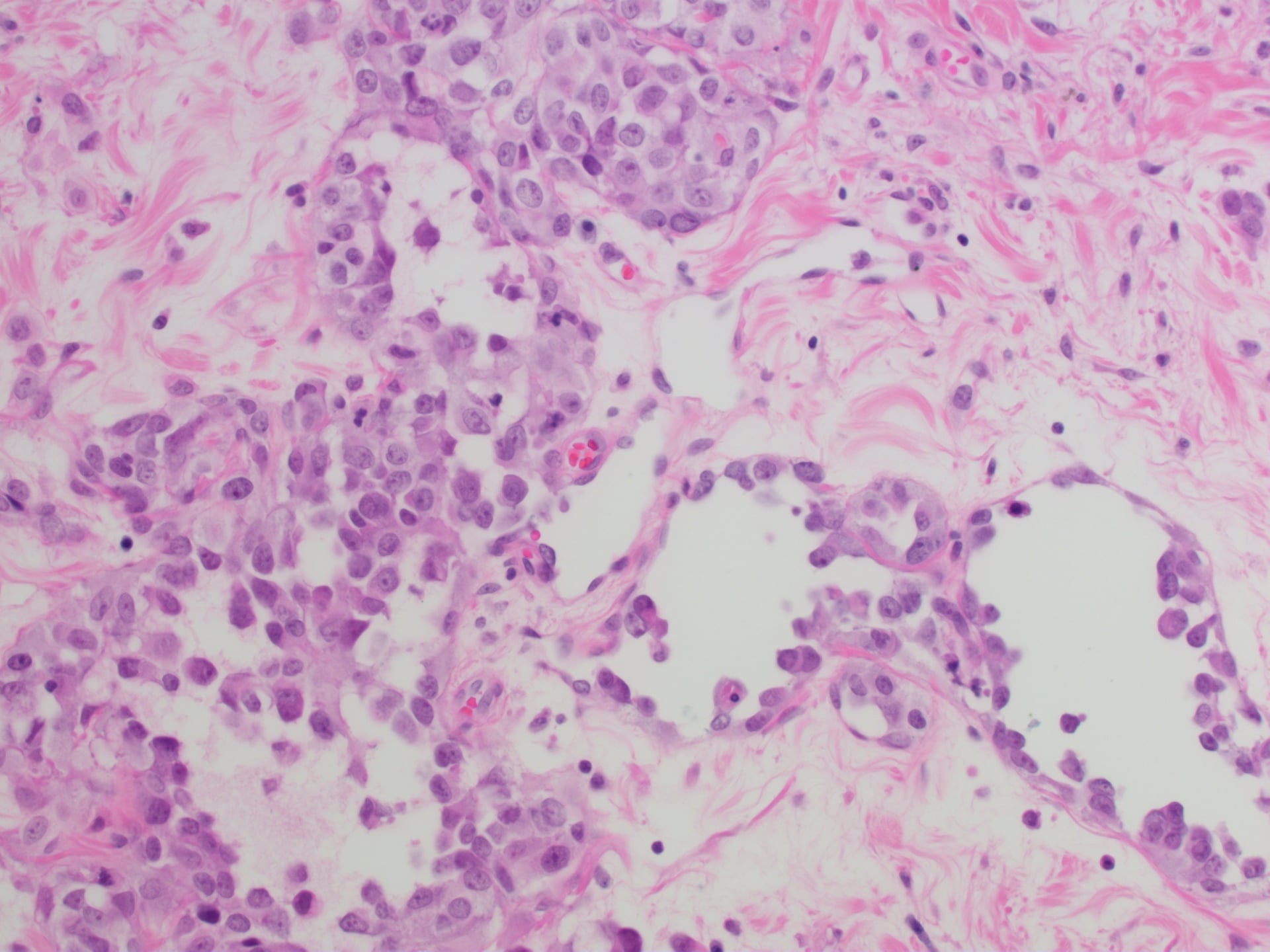
Morphology:
- Vasoformative: Blood-filled vascular channels
- Solid areas: Sheets of atypical endothelial cells
- High mitotic rate, nuclear atypia
- Variable differentiation (well-differentiated can appear bland)
Immunohistochemistry is essential:
- CD31: 90% positive (most sensitive)
- CD34: 80% positive
- Factor VIII: 60% positive
- ERG, FLI1: Positive nuclear staining
Differential Diagnosis
The most clinically dangerous error is mistaking a benign atypical vascular lesion (AVL) in an irradiated field for angiosarcoma, or vice versa. MYC amplification is the decisive discriminator.
- Key Distinguishing Feature
- Small, superficial, well-circumscribed; benign or pre-malignant
- Immunohistochemistry / Molecular
- MYC NOT amplified (negative) - key discriminator
- Key Distinguishing Feature
- Benign, lobular, no infiltration or marked atypia
- Immunohistochemistry / Molecular
- CD31 positive, MYC negative, low Ki-67
- Key Distinguishing Feature
- Spindle cells, slit-like vessels, plasma cells
- Immunohistochemistry / Molecular
- HHV-8 (LANA-1) positive - diagnostic
- Key Distinguishing Feature
- Epithelioid angiosarcoma can mimic carcinoma
- Immunohistochemistry / Molecular
- Cytokeratin positive AND vascular markers negative (angiosarcoma may be keratin-positive, so use CD31/ERG)
- Key Distinguishing Feature
- Pigmentation, prior naevus
- Immunohistochemistry / Molecular
- S100 / SOX10 / HMB-45 positive, CD31 negative
- Key Distinguishing Feature
- History of trauma, resolves over time
- Immunohistochemistry / Molecular
- No mass; biopsy any persistent non-traumatic scalp bruise
Clinical Presentation
Cutaneous Angiosarcoma (Head and Neck)
Classic presentation in elderly patients:
- Scalp lesion (most common site)
- Bruise-like appearance: Purple or red discoloration
- Rapid enlargement: Over weeks to months
- Multifocal: Multiple areas of involvement
- No trauma history: Unlike hematoma
Elderly patient with persistent scalp bruising without trauma should raise suspicion for cutaneous angiosarcoma. Biopsy is mandatory. Delay in diagnosis is common because lesion resembles benign ecchymosis.
Radiation-Induced Angiosarcoma
Post-breast cancer radiation:
- Latency: 5-10 years after radiotherapy
- Presentation: Skin changes on irradiated breast (redness, ecchymosis, palpable nodules)
- Location: Within radiation field
- Differential: Radiation dermatitis, inflammatory breast cancer
Stewart-Treves Syndrome
Post-mastectomy lymphedema:
- Latency: Usually greater than 10 years
- Presentation: Purple nodules or plaques in chronically edematous arm
- Rapidly progressive
- Often multifocal
Deep Soft Tissue Angiosarcoma
- Large mass in extremity or trunk
- Often painless until late
- Rapid growth
- May present with pathological fracture if bone involvement
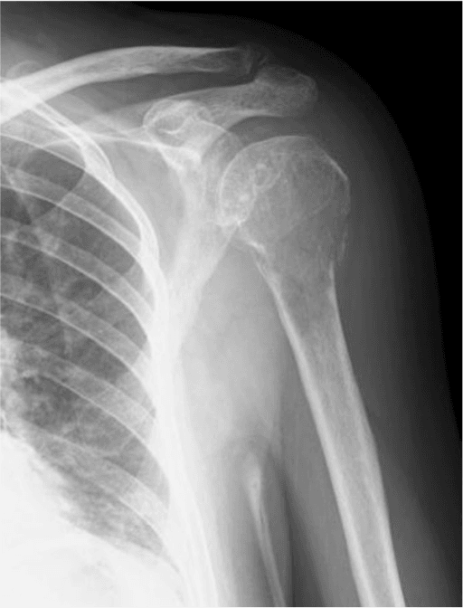
Primary Angiosarcoma of Bone (the orthopaedic-relevant form)
Primary angiosarcoma of bone is rare but is the form most likely to present to the orthopaedic surgeon:
- Multifocality is characteristic — it is classically multicentric, with several lesions within one bone or clustered in a single limb/anatomical region (a recognised diagnostic clue), although solitary lesions occur.
- Radiographically it is a permeative/lytic, destructive lesion with cortical breach and frequently a pathological fracture; matrix is absent (purely lytic).
- Histology is often epithelioid, which can mimic metastatic carcinoma — endothelial markers (CD31, ERG) are decisive.
- Differential of a destructive lytic bone lesion: epithelioid haemangioendothelioma (lower-grade vascular tumour, also can be multifocal), haemangioma, metastatic carcinoma, and myeloma — a multifocal lytic pattern in one region should prompt biopsy plus IHC rather than assumption of metastasis.
- Management mirrors high-grade bone sarcoma: biopsy and full staging (lung) before intervention, then wide resection where feasible ± radiotherapy/chemotherapy; high-grade disease carries a poor prognosis, and multifocality often precludes a curative resection.
Management
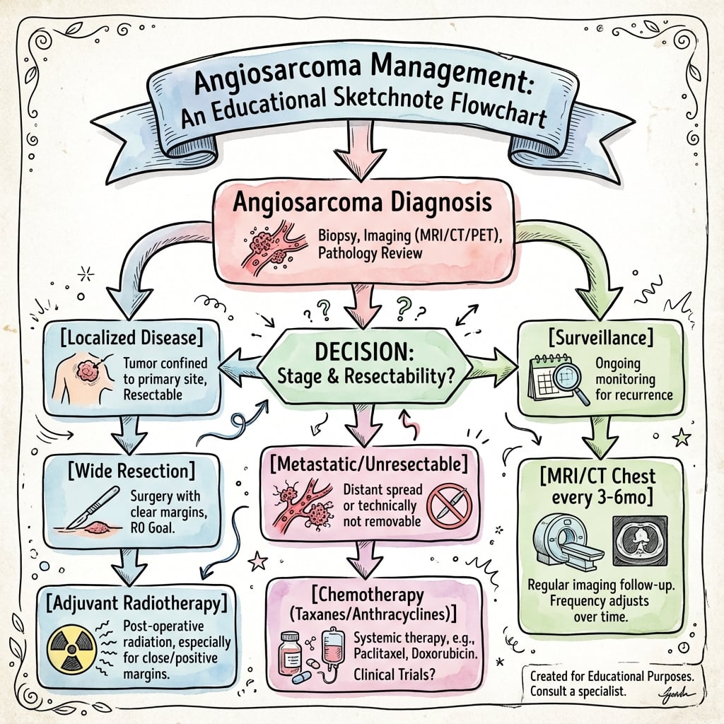
Treatment Algorithm
Surgery is the cornerstone of curative treatment. Wide excision with negative margins (R0 resection) is essential. However, achieving negative margins is challenging due to multifocal nature and infiltrative growth. Positive margins dramatically increase local recurrence (50-80% vs 20-30% with negative margins).
Surgical Resection Principles
Goals:
- Wide excision with 2-3cm margins (if feasible)
- En bloc resection of all involved areas
- Reconstruction as needed
Site-Specific Surgery:
- Surgical Approach
- Wide local excision, often requires skin grafting
- Challenges
- Multifocal disease makes complete excision difficult
- Surgical Approach
- Mastectomy + chest wall resection
- Challenges
- Extensive resection needed, often positive margins
- Surgical Approach
- Amputation (often required)
- Challenges
- Limb salvage rarely achievable due to extent
- Surgical Approach
- Wide excision + reconstruction
- Challenges
- Best chance of negative margins
Amputation indications:
- Stewart-Treves syndrome with extensive involvement
- Neurovascular encasement precluding limb salvage
- Recurrent disease after prior limb-sparing surgery
- Patient choice for local control
Amputation is often necessary for Stewart-Treves syndrome to achieve local control.
Multimodal Treatment by Subtype
- Surgery
- Wide excision (often incomplete)
- Radiation
- Adjuvant 60-66 Gy
- Chemotherapy
- Paclitaxel neoadjuvant or adjuvant
- Surgery
- Mastectomy + chest wall
- Radiation
- Consider (in previously irradiated field)
- Chemotherapy
- Adjuvant paclitaxel
- Surgery
- Amputation
- Radiation
- Adjuvant if residual limb
- Chemotherapy
- Palliative if metastatic
- Surgery
- Wide excision
- Radiation
- Adjuvant 60-66 Gy
- Chemotherapy
- Consider neoadjuvant + adjuvant
Complications
Disease-Related Complications
- Incidence
- 30-60%
- Management
- Re-excision, radiation, systemic therapy
- Incidence
- 50%
- Management
- Chemotherapy (paclitaxel), consider metastasectomy if oligometastatic
- Incidence
- 10-20% (vascular tumor)
- Management
- Embolization, surgical hemostasis
- Incidence
- Variable
- Management
- Compression, physiotherapy
Treatment-Related Complications
- Wound complications (15-25%): Infection, dehiscence, flap necrosis
- Radiation toxicity: Fibrosis, chronic pain (especially if re-irradiating)
- Chemotherapy toxicity: Neuropathy (paclitaxel), myelosuppression
Postoperative Care and Surveillance
Follow-Up Protocol
- Clinical examination
- CT chest every 3-4 months (high metastatic rate)
- MRI primary site every 4-6 months
- Clinical examination
- CT chest every 6 months
- MRI primary site every 6-12 months
- Clinical examination
- Imaging as clinically indicated
- Late recurrences and metastases possible
Surveillance continues indefinitely due to risk of late recurrence.
Prognosis and Outcomes
Prognostic Factors
- Favorable
- Deep soft tissue
- Unfavorable
- Head/neck, radiation-induced, lymphedema
- Favorable
- Under 5cm
- Unfavorable
- Over 10cm
- Favorable
- Negative (R0)
- Unfavorable
- Positive (R1/R2)
- Favorable
- Localized (M0)
- Unfavorable
- Metastatic (M1)
- Favorable
- Unifocal
- Unfavorable
- Multifocal
Cutaneous angiosarcoma of the scalp has the worst prognosis of all angiosarcoma subtypes (10-20% 5-year survival). Reasons: multifocal disease at presentation, infiltrative growth pattern, difficulty achieving negative margins, and elderly patient population. Even with aggressive multimodal therapy, outcomes remain poor.
Survival by Subtype
- 5-Year Survival
- 30-40%
- Key Factors
- Best prognosis, margins achievable
- 5-Year Survival
- 20-30%
- Key Factors
- Aggressive disease, prior radiation limits re-irradiation
- 5-Year Survival
- 10-20%
- Key Factors
- Worst prognosis, multifocal, elderly
- 5-Year Survival
- 15-25%
- Key Factors
- Extensive disease, requires amputation
Guidelines, Registries & Global Practice
Global Epidemiology
Angiosarcoma accounts for under 2% of all soft tissue sarcomas and roughly 1-2% of cutaneous sarcomas, making it one of the rarest yet most lethal sarcoma subtypes worldwide. It shows a slight male predominance overall, with a median age of 60-70 years for the common cutaneous head/neck form. Geographic and historical variation reflects exposure patterns: thorotrast-associated hepatic angiosarcoma (now historical), occupational vinyl chloride exposure (hepatic angiosarcoma, industrialised regions), and the rising incidence of radiation-associated breast angiosarcoma in high-income settings as breast-conserving therapy with radiotherapy has become standard.
Side-by-Side Guideline Comparison
- Region
- US
- Key Recommendation for Soft Tissue Sarcoma / Angiosarcoma
- Manage at sarcoma centres; core/incisional biopsy before resection; wide R0 excision; consider neoadjuvant/adjuvant chemo (paclitaxel or doxorubicin) and radiotherapy for high-grade disease
- Region
- Europe
- Key Recommendation for Soft Tissue Sarcoma / Angiosarcoma
- MDT at reference centre mandatory; biopsy planned so tract is excised; angiosarcoma treated as high-grade; taxanes recognised as active first-line, especially cutaneous/scalp
- Region
- UK
- Key Recommendation for Soft Tissue Sarcoma / Angiosarcoma
- Suspected sarcoma referred to specialist MDT before intervention; image-guided core biopsy; surgery plus radiotherapy for limb/trunk high-grade tumours
- Region
- Global pathology
- Key Recommendation for Soft Tissue Sarcoma / Angiosarcoma
- Diagnosis requires endothelial markers (CD31, ERG); MYC FISH/IHC to distinguish secondary angiosarcoma from atypical vascular lesions in irradiated fields
Population data sources:
- SEER (US) and national cancer registries provide the bulk of incidence and survival data given the rarity of the disease
- Rare-sarcoma networks (e.g. EURACAN, French Sarcoma Group, RARECARE) pool cases across centres
- No dedicated implant/arthroplasty registry applies; survival data derive from sarcoma-centre cohorts
Centralised reference-centre management is associated with better outcomes in soft tissue sarcoma generally.
Variation in care:
- High-resource: MRI staging, MYC molecular testing, MDT review, taxane/anthracycline access, reconstructive surgery and radiotherapy
- Limited-resource: Diagnosis often delayed; molecular testing unavailable; amputation or palliative care may be the only realistic options
- Universal principle: early biopsy of suspicious lesions and referral to the highest-capability centre available
Clinical trial enrolment is encouraged everywhere given the absence of curative systemic therapy in advanced disease.
Key documentation:
- Biopsy any persistent scalp bruising in elderly (delay common)
- Radiation-induced angiosarcoma: counsel breast cancer patients about late risk
- Stewart-Treves: document discussion of amputation vs palliative options
- Informed consent: include poor prognosis, high recurrence, multimodal treatment needs
- Surveillance protocol documented (intensive due to metastatic risk)
Controversies and Areas of Uncertainty
Angiosarcoma is rare enough that almost all evidence is retrospective; few questions are settled.
- Current Position
- Radical resection of the whole irradiated skin field improves survival (Li 2017)
- Uncertainty
- Multifocal field-effect biology means even R0 margins do not guarantee local control
- Current Position
- Weekly paclitaxel and doxorubicin appear equivalent (Penel 2012)
- Uncertainty
- No randomised head-to-head trial; choice driven by site, fitness and toxicity profile
- Current Position
- Can downstage locally advanced/unresectable disease (ANGIOTAX)
- Uncertainty
- No survival benefit proven; sequencing of surgery, radiotherapy and chemotherapy not standardised
- Current Position
- Used selectively for positive margins
- Uncertainty
- Cumulative toxicity in an already-irradiated field limits dose and benefit is unproven
- Current Position
- VEGF/VEGFR biology and high mutational burden (UV/cutaneous) are rationales
- Uncertainty
- Bevacizumab, pazopanib and checkpoint inhibitors remain investigational without level-1 evidence
Memory Aids
RALFCauses of Angiosarcoma
Hook:Three named clinical syndromes map onto these: radiation-induced breast AS, Stewart-Treves (lymphedema), and graft-associated AS.
VAMPCore Clinical Features
Hook:A vampire image fits a bruise-like, blood-filled, aggressive vascular tumour.
CEFEndothelial Confirmation on Biopsy
Hook:CD31 is the single most sensitive marker; reach for MYC FISH whenever the lesion sits in a previously irradiated field.
MCQ Practice Points
Q: What is the most common site for cutaneous angiosarcoma? A: Scalp - Cutaneous angiosarcoma has predilection for the scalp in elderly patients, presenting as bruise-like lesions without trauma. This subtype has the worst prognosis (10-20% 5-year survival) due to multifocal nature and infiltrative growth.
Q: What is Stewart-Treves syndrome? A: Angiosarcoma arising in chronic lymphedema, classically in the arm after mastectomy with axillary dissection. Latency is usually over 10 years. Presentation: violaceous nodules in lymphedematous limb. Prognosis extremely poor (15-25% 5-year survival). Treatment often requires amputation.
Q: What immunohistochemical markers confirm vascular origin in angiosarcoma? A: CD31 (90% positive - most sensitive), CD34 (80% positive), and Factor VIII/von Willebrand factor (60% positive). ERG and FLI1 show nuclear positivity. CD31 is the most sensitive endothelial marker.
Q: What is the preferred first-line chemotherapy for angiosarcoma? A: Weekly paclitaxel (80 mg/m²) is a standard first-line option, validated by the ANGIOTAX phase II study (non-progression 74% at 2 months). French Sarcoma Group data show doxorubicin-based regimens give comparable survival, so either is acceptable first-line; taxanes are particularly favoured for cutaneous/scalp disease.
Q: What is the 5-year survival for angiosarcoma overall? A: 20-35% for all sites combined. Prognosis varies by subtype: deep soft tissue (30-40%), radiation-induced (20-30%), scalp (10-20%), Stewart-Treves (15-25%). Poor prognosis due to early metastases, multifocal disease, and high local recurrence rates.
Exam Viva Scenarios
Practise clinical reasoning and management decisions out loud
“A 75-year-old man presents with a persistent purple discoloration on his scalp that has enlarged over 3 months. He denies trauma. On examination, there is a 5cm bruise-like lesion on the vertex. What is your differential diagnosis and management?”
“A 58-year-old woman treated with lumpectomy and radiation for breast cancer 8 years ago presents with skin changes on her treated breast - ecchymosis and a palpable 3cm mass. What is your assessment and management?”
“A 65-year-old woman with history of left mastectomy and axillary dissection 15 years ago presents with violaceous nodules on her chronically swollen left arm. Biopsy shows angiosarcoma. How do you manage this?”
Key Epidemiology
- Extremely rare: under 1% of soft tissue sarcomas
- Bimodal age: young adults (deep) and elderly (cutaneous)
- Scalp most common site for cutaneous type (40% of cases)
- Highly aggressive with poor prognosis (20-35% 5-year survival)
Classic Associations (RALF Mnemonic)
- Radiation therapy (5-10 year latency, 0.1-0.2% of irradiated patients)
- Arsenal chemicals (vinyl chloride, arsenic, thorotrast)
- Lymphedema chronic (Stewart-Treves syndrome)
- Foreign bodies (synthetic grafts, surgical materials)
Clinical Features (VAMP Mnemonic)
- Vascular markers positive (CD31 90%, CD34 80%, Factor VIII 60%)
- Aggressive behavior (early metastases, multifocal, rapid growth)
- Multifocal disease (50% at presentation or develop multiple lesions)
- Poor prognosis (20-35% overall, 10-20% scalp, 15-25% Stewart-Treves)
Diagnostic Workup
- Biopsy: Core needle with immunohistochemistry (CD31, CD34, Factor VIII)
- MRI primary site: Assess extent and multifocality
- CT chest/abdomen/pelvis: Metastases in 30-40% at diagnosis (lung most common)
- PET-CT: Consider for extent and occult disease (angiosarcoma FDG-avid)
Treatment Principles
- Surgery: Wide excision 2-3cm margins, R0 resection critical
- Radiation: Adjuvant 60-66 Gy (all high-grade cases)
- Chemotherapy: Paclitaxel first-line (40-50% response rate)
- Scalp: Often unresectable, multimodal therapy
- Stewart-Treves: Amputation often required for local control
Prognosis by Subtype
- Deep soft tissue: 30-40% 5-year survival (best of poor options)
- Radiation-induced breast: 20-30% 5-year survival
- Cutaneous scalp: 10-20% 5-year survival (worst prognosis)
- Stewart-Treves: 15-25% 5-year survival
- Metastatic disease: under 10% 5-year survival
Complications and Surveillance
- Local recurrence: 30-60% despite multimodal therapy
- Distant metastases: 50% (lung most common)
- Follow-up: Q2-3mo years 1-2, Q4-6mo years 3-5, then Q6-12mo
- CT chest Q3-4mo first 2 years (high metastatic rate)
- Late recurrences possible - indefinite surveillance
Evidence Base and Key Studies
Cutaneous Angiosarcoma of the Face and Scalp: Outcomes After Definitive Treatment
- 70 patients with non-metastatic face/scalp angiosarcoma treated curatively
- 5-year overall survival 43%, disease-specific survival 46%
- Tumour size over 5 cm and satellitosis predicted inferior survival
- Combined surgery plus radiotherapy improved local control and survival versus single-modality therapy
Cutaneous Angiosarcoma of the Face/Scalp: Reevaluating Surgery and Radiotherapy
- 143 patients treated 1962-2019; median follow-up 33 months
- 5-year local control 51%; 5-year disease-specific survival 56%
- Worse outcomes with tumours over 5 cm, multifocal disease, and single-modality therapy
- Combined surgery plus radiotherapy (S-XRT) improved local control and disease-specific survival on multivariable analysis
Weekly Paclitaxel for Unresectable Angiosarcoma: the ANGIOTAX Study
- Phase II trial of weekly paclitaxel 80 mg/m² in 30 metastatic/unresectable angiosarcoma patients
- Non-progression rates 74% at 2 months and 45% at 4 months
- Median time to progression 4 months; median overall survival 8 months
- 3 of 3 locally advanced breast angiosarcomas achieved partial response enabling curative-intent surgery
Metastatic Angiosarcomas: Doxorubicin, Weekly Paclitaxel and Metastasectomy
- French Sarcoma Group retrospective analysis of 149 metastatic angiosarcomas (1996-2009)
- Median overall survival 11 months; performance status the only independent prognostic factor (HR 2.49)
- Doxorubicin-based first-line (HR 0.38) and weekly paclitaxel (HR 0.36) both improved survival versus palliative care
- Metastasectomy independently improved outcome (HR 0.09) in selected patients
Radiation-Associated Breast Angiosarcoma: R0 Resection Still Carries Poor Prognosis
- Population-based cohort of 35 secondary breast angiosarcomas (31 underwent surgery)
- Median latency after radiotherapy 7 years (range 3-25 years)
- R0 resection achieved in 23 of 31, yet two-thirds developed local recurrence
- Median disease-specific survival just over 3 years (37 months)
Radical Resection Improves Survival in Radiation-Associated Breast Angiosarcoma
- 76 women with radiation-associated breast angiosarcoma; median latency 85 months
- Radical resection (all/nearly all irradiated skin) vs conservative resection compared
- 5-year disease-specific survival 86% (radical) vs 46% (conservative)
- 5-year local recurrence 23% (radical) vs 76% (conservative)
Consistent MYC Amplification Distinguishes Secondary (Radiation/Lymphedema) Angiosarcoma
- High-level MYC (8q24) amplification found in 100% of secondary angiosarcomas
- MYC amplification absent in atypical vascular lesions and other radiation-associated sarcomas
- FLT4 (VEGFR3) co-amplification in 25% of secondary angiosarcomas
- MYC amplification is an early, defining event distinguishing secondary from primary angiosarcoma
Stewart-Treves Syndrome: Pathogenesis and Management
- Lymphangiosarcoma arising in chronic lymphedema (classically post-radical mastectomy)
- Also reported in congenital and other causes of chronic secondary lymphedema
- Chemotherapy and radiotherapy have not significantly improved survival
- Early amputation or wide local excision offers the best chance of long-term survival; untreated survival is 5-8 months