Rare Benign Epiphyseal Cartilage Tumor | Skeletally Immature | Chicken-Wire Calcification
- Epiphyseal location in a skeletally immature patient is the characteristic pattern
- Chicken-wire calcification pattern on histology is diagnostic hallmark
- ABC formation occurs in a reported 15-35% of cases, creating a mixed cystic-solid appearance - a diagnostic trap, but it does NOT predict recurrence
- Painful symptoms with activity-related pain and joint effusion common
- Curettage with adjuvants is standard - but recurrence depends on WHO: 5.0% in a 199-case series that was 52% skeletally MATURE, versus 32% in an 87-case series with ALL physes OPEN (PMIDs 26041854, 19723993)
- “Think chondroblastoma for any epiphyseal lesion in adolescent with pain
- “Chicken-wire calcification refers to calcified lacunar rim around chondroblasts
- “Giant cells present but not as numerous as giant cell tumor
- “Rare benign metastasis to lungs can occur (not malignant transformation)
Overview and Epidemiology
Chondroblastoma is a rare benign cartilage tumour, under 1% of all bone tumours, and the only benign cartilage tumour that arises in the epiphysis; enchondroma and osteochondroma arise in the metaphysis or diaphysis. Benign is not the same as harmless. It causes significant pain, it recurs after curettage, and in rare cases it seeds the lungs.
Who. Peak age is 10-25 years (range 5-30), with a male predominance of 2:1. In the largest multicentre series, 199 extremity chondroblastomas, the physis was already closed in 52% at presentation.
Where. Ninety-five per cent arise at the ends of long bones, and the knee accounts for roughly half (proximal tibia plus distal femur was 46.7% in the modern series). The classic list and the largest modern series disagree on the order:
- Traditional teaching, from the older Mayo material: proximal humerus 20-25%, distal femur 15-20%, proximal tibia 15-20%, proximal femur 10-15%, talus 5-10%
- The 199-case multicentre series (PMID 26041854): proximal tibia 27.6%, proximal femur 26.1%, distal femur 19.1%, with the proximal humerus well down the list
- The 87-case paediatric open-physis series (PMID 19723993) agrees, naming the proximal tibia and the proximal femoral epiphysis as commonest
Do not spend long defending either ordering: the four sites are all common. What matters clinically is that the proximal humerus was the only significant predictor of local recurrence (p = 0.001) in the 199-case series, so remember it for its risk rather than its frequency.
Rare sites. Flat bones and the spine are rarely involved; vertebral involvement was 9 of 856 cases (1.4%) in the Mayo review. Calcaneus, talus and patella are rare sites, and the atypical sites are covered under presentation.
The physis narrows the differential. Chondroblastoma arises while the physis is open, may extend into the metaphysis after physeal closure, and in rare cases extends across an open physis, which is unusual among tumours. Giant cell tumour typically presents after physeal closure, at 20-40 years. The status of the growth plate is critical to narrowing the differential:
- Epiphyseal lesion with an open physis: think chondroblastoma (or infection)
- Epiphyseal lesion with a closed physis: think giant cell tumour (or clear cell chondrosarcoma)
Pathophysiology and Pathology
Histogenesis. Chondroblastoma arises from germinative cartilage cells in the epiphysis, the primitive chondroblasts that normally form secondary centres of ossification. The trigger for neoplastic transformation is unknown, but recurrent H3F3B K36M mutations are found in 95% of cases and appear to be the driver.
The mutation is somatic rather than germline, sits in the histone H3.3 variant and affects epigenetic regulation, and it can be detected with a mutation-specific antibody, which helps to separate chondroblastoma from its mimics.
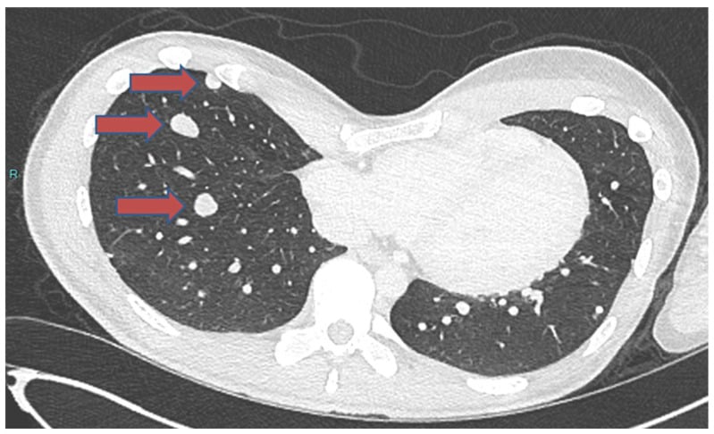
Natural history. Growth is slow and progressive. In 5-10% the tumour penetrates the articular cartilage into the joint. A secondary aneurysmal bone cyst develops in a reported 15-35%. In under 1%, viable tumour cells implant in the lungs without malignant transformation, the benign metastasising chondroblastoma.
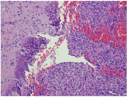
Gross pathology. The curettage specimen is friable red-grey tissue, gritty from its calcifications. The tumour is well circumscribed within the epiphysis, with cystic haemorrhagic areas where a secondary ABC is present.
Histology
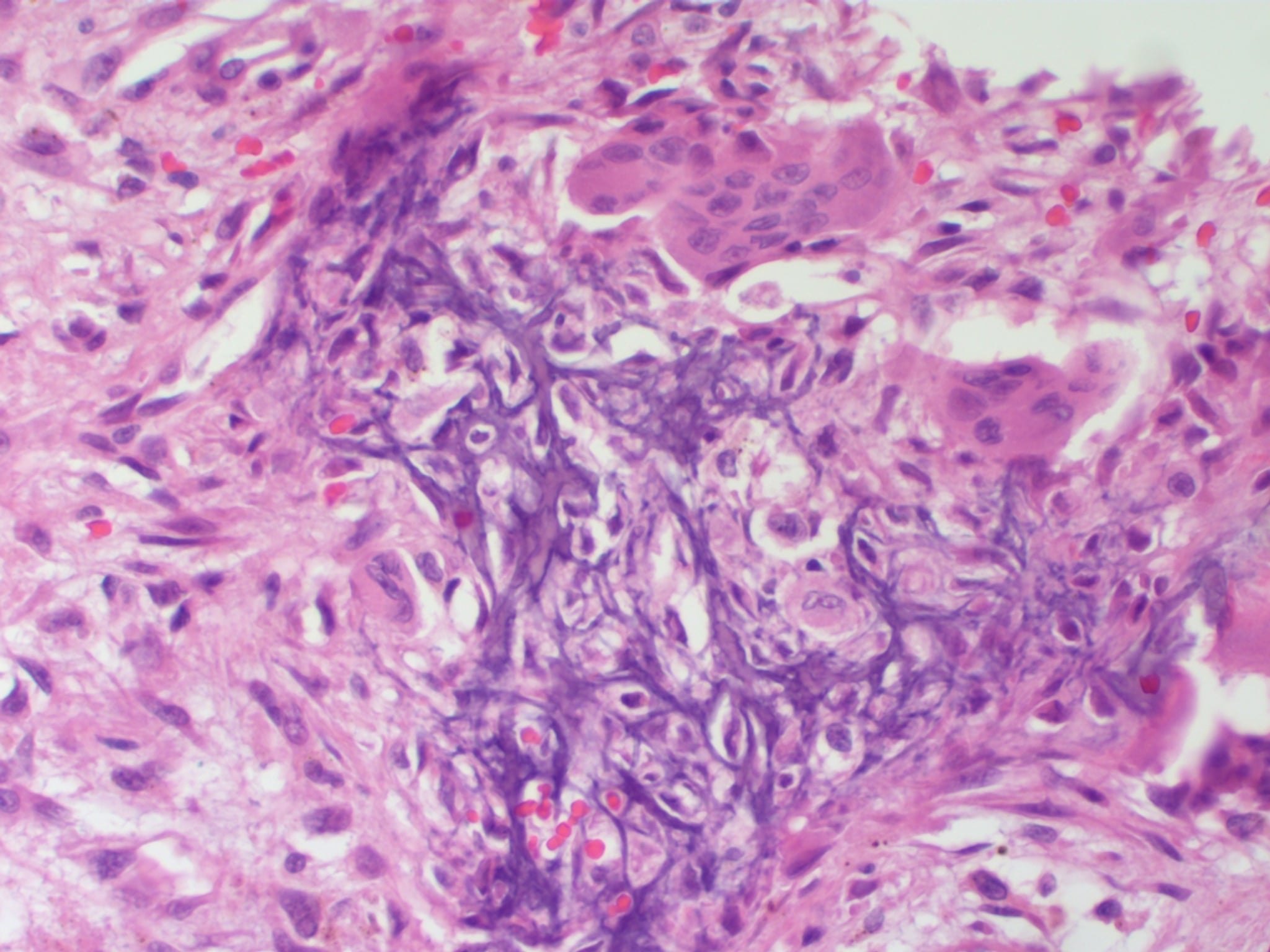
Chondroblasts, the neoplastic cell. Mononuclear round to polygonal cells with distinct cell borders, pink cytoplasm and oval nuclei carrying longitudinal grooves that give a coffee-bean appearance. These are the tumour cells; everything else on the slide is a reaction to them.
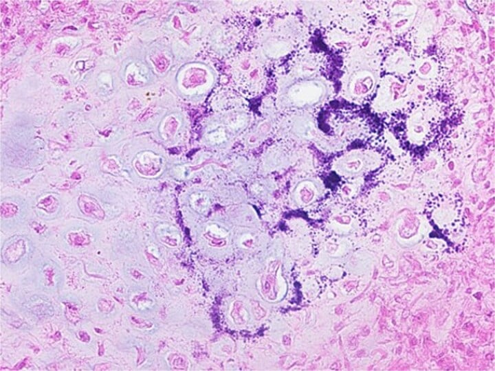
Chicken-wire calcification, the pathognomonic feature. Dystrophic calcification of the pericellular (lacunar) matrix forms a lacy network of calcified rims encircling individual chondroblasts. On haematoxylin and eosin it is a fine purple-blue net outlining cells, and at low magnification it resembles chicken-wire fencing. It is this rim around single cells, and not the giant cells, that separates chondroblastoma from giant cell tumour and from every other bone tumour.
Giant cells, reactive. Osteoclast-type multinucleated giant cells are scattered through the tumour, but fewer than in giant cell tumour. They are reactive, not neoplastic, which is why counting them is the classic route to the wrong diagnosis.
Chondroid matrix, variable. Cartilaginous matrix is present focally, with chondroblasts sitting in hyaline or myxochondroid ground substance that is often calcified. Its absence does not exclude the diagnosis.
Immunohistochemistry
- Staining
- Positive (strong)
- Interpretation
- Confirms chondroid differentiation
- Staining
- Positive
- Interpretation
- Specific for chondroblastoma
- Staining
- Positive (mutation-specific antibody)
- Interpretation
- Highly specific, aids diagnosis
- Staining
- Negative
- Interpretation
- Excludes epithelial tumors
Secondary Aneurysmal Bone Cyst
A minority, but far from a trivial one. The reported frequency is higher when histological rather than radiographic criteria are used, which is the main reason published figures disagree. The cyst creates blood-filled spaces within the tumour, and it matters because it can:
- Obscure the underlying chondroblastoma on imaging, so the lesion appears purely cystic
- Lead to rapid expansion and pathological fracture
- Make biopsy diagnosis difficult if the solid component is not sampled
What it does not do is predict recurrence. In the paediatric multicentre series, an aneurysmal bone-cyst component on histology had no significant influence on local recurrence, and neither did sex, radiographic aggressiveness or the surgical method used. The only factor that mattered was a strictly epiphyseal location. An ABC component is a diagnostic trap, not a prognostic one.
Classification
WHO classification (2020). Chondroblastoma is listed as a benign cartilage tumour with locally aggressive (intermediate) behaviour, ICD-O code 9230/0. Its defining characteristics are the epiphyseal location, the mononuclear chondroblasts, the chicken-wire calcification pattern and the H3K36M mutation.
Enneking staging. Most chondroblastomas present as stage 2 lesions:
- Stage 1 (latent): intracapsular and well demarcated, with minimal symptoms or an incidental finding; observation may be appropriate
- Stage 2 (active): the most common presentation; symptomatic with progressive growth, but contained by natural barriers; curettage with adjuvant therapy
- Stage 3 (aggressive): breaks through the cortex or physis with extracompartmental extension; may require en bloc resection
Clinical Presentation
Symptoms. A dull aching pain, worse with activity, in 90%, usually chronic over months to years. Reactive synovitis in the adjacent joint gives an effusion in 40%, with stiffness from pain and effusion, and a limp when the lower limb is involved.
Examination. Tenderness is localised to the epiphyseal region, there is an effusion from reactive synovitis, and range of motion is restricted by pain rather than a mechanical block. The joint may be warm but is not erythematous, and chronic knee pain leaves quadriceps wasting.
Typical scenarios. Each common site has a benign condition it is mistaken for:
- Proximal humerus: an adolescent athlete with shoulder pain worse on overhead activity and limited abduction and forward flexion, mistaken for rotator cuff tendinitis or impingement
- Distal femur: an adolescent with anterior knee pain and an effusion, worse running or jumping, mistaken for patellofemoral pain syndrome or Osgood-Schlatter disease
- Proximal tibia: an adolescent with knee pain localised to the proximal tibia and a common effusion, mistaken for a meniscal tear or tibial spine injury
- Proximal femur: a child or adolescent with hip pain and a limp, worse on internal rotation, mistaken for SCFE or Perthes disease
Think chondroblastoma when:
- An adolescent (10-25 years) has chronic epiphyseal pain
- Pain is worse with activity and not relieved by rest
- There is a joint effusion out of proportion to the trauma history
- The radiograph shows an epiphyseal lytic lesion with a sclerotic rim
- MRI shows bone marrow oedema extending beyond the lesion
Do not dismiss it as growing pains or an overuse injury: persistent epiphyseal pain in an adolescent warrants imaging.
Rare presentations. When the tumour has penetrated the articular cartilage it presents with mechanical symptoms, locking and catching, mimicking a loose body or a meniscal tear, and arthroscopy shows a cartilage defect with tumour tissue. Benign pulmonary metastasis is usually asymptomatic, discovered on routine chest radiograph or CT as multiple small pulmonary nodules months to years after treatment of the primary, and may regress spontaneously.
Atypical sites. The classic teaching is the long-bone epiphysis, but chondroblastoma also arises in epiphyseal-equivalent and atypical locations, and these behave or present differently:
- Apophyses, the greater trochanter, tibial tubercle, iliac crest and patella, share the biology of an epiphyseal lesion because apophyses are secondary ossification centres; keep chondroblastoma in the differential for a lytic apophyseal lesion in a young patient
- Tarsal bones, the talus and calcaneus, present late with vague hindfoot or ankle pain, often misdiagnosed as a sprain or a simple cyst, are technically difficult to access, and carry a higher recurrence rate than long-bone lesions
- Craniofacial skeleton, classically the temporal bone and skull base, occurs in older patients than long-bone disease and presents with hearing loss, temporomandibular symptoms or a skull-base mass
- Flat and irregular bones (rib, pelvis, scapula) are rare but reported
Investigations and Imaging
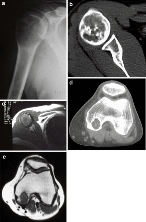
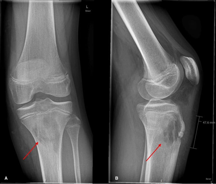
Radiographs. The first-line investigation, and often enough to suggest the diagnosis from location and appearance. The lesion is epiphyseal in a patient with an open or recently closed physis, eccentric and lytic with well-defined geographic borders, and carries a thin sclerotic rim of reactive bone in 50-70%. Stippled intralesional calcification may be visible; the chicken-wire pattern is a histological finding and is not seen on radiographs.
Size. Typically 2-5 cm: small lesions sit entirely within the epiphysis, larger ones expand it and may cross into the metaphysis.
Periosteal reaction. Usually absent. When present it generally reflects a secondary aneurysmal bone cyst or a pathological fracture, and a solid or aggressive periosteal reaction should prompt a rethink, since rare malignant behaviour is reported.
The sclerotic rim distinguishes chondroblastoma from infection, which has none. A rim that is very thick and irregular in an older patient should raise clear cell chondrosarcoma.
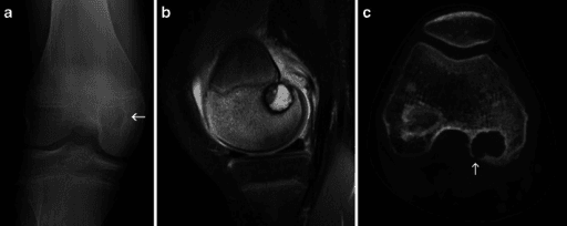
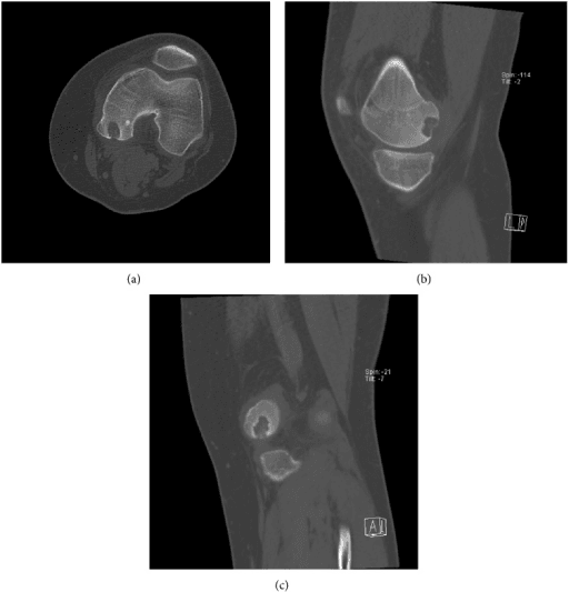
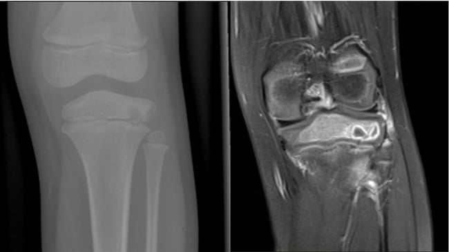
MRI. Mandatory for surgical planning. It shows the extent of marrow involvement, cartilage penetration into the joint, a secondary ABC component, and the relationship to the physis and neurovascular structures; it confirms the epiphyseal centre and the reactive joint effusion.
- T1: low to intermediate signal in the solid component
- T2: high signal in cartilage, very high signal in ABC cysts
- Enhancement: solid portions enhance; cystic ABC areas show fluid-fluid levels
Extensive bone marrow oedema on STIR sequences extends far beyond the tumour margins, through the epiphysis and into the metaphysis. It is a reactive inflammatory response, not tumour infiltration, and true tumour size is better judged on T1-weighted images, where only the solid lesion shows low signal. Curette the lytic cavity, not the oedema zone.
CT. Detects intralesional calcification as subtle stippled density, assesses cortical breakthrough where an ABC component has caused expansion, and plans the placement of the cortical window.
Biopsy. Required for a definitive diagnosis before surgery. Open biopsy is preferred to needle biopsy because it gives adequate tissue and avoids sampling only the ABC component, and because small epiphyseal lesions are difficult to target with a needle; pathological fracture through the biopsy tract is a risk.
When biopsying an epiphyseal lesion with ABC features, take multiple samples from solid areas. Sampling only the cystic component misses the chondroblastoma. Review the imaging to target solid nodules for needle placement.
- Indication
- Large accessible lesion
- Advantage
- Minimally invasive, outpatient
- Risk
- Sampling error if ABC component targeted
- Indication
- Small epiphyseal lesion
- Advantage
- Adequate tissue, direct visualization
- Risk
- Requires OR, pathological fracture risk
- Indication
- Expendable bone (fibular head)
- Advantage
- Diagnostic and therapeutic in one procedure
- Risk
- Not feasible for most locations
Differential Diagnosis
- Age and Physis
- 10-25y, open
- Location
- Epiphysis
- Imaging and Clinical
- Well-defined, sclerotic rim, matrix calcification
- Histology and Giant Cells
- Chicken-wire calcification, H3K36M positive; giant cells moderate
- Age and Physis
- 20-40y, closed
- Location
- Epiphysis-metaphysis
- Imaging and Clinical
- Eccentric, lytic, soap-bubble, no sclerotic rim, extends to subchondral bone
- Histology and Giant Cells
- Sheets of giant cells; numerous (predominant)
- Age and Physis
- Over 30y, closed
- Location
- Epiphysis
- Imaging and Clinical
- Lytic, may have calcification; larger, aggressive features
- Histology and Giant Cells
- Clear cytoplasm, low-grade; giant cells absent
- Age and Physis
- Any age, open or closed
- Location
- Epiphysis
- Imaging and Clinical
- Fever, elevated inflammatory markers
- Histology and Giant Cells
- Inflammatory cells, organisms; giant cells absent
- Age and Physis
- 5-15y
- Location
- Variable, usually metaphysis
- Imaging and Clinical
- Lytic, may be aggressive; systemic involvement possible
- Histology and Giant Cells
- Langerhans cells, eosinophils; giant cells absent
- Age and Physis
- Any age
- Location
- Epiphysis, subchondral
- Imaging and Clinical
- Well-defined, subchondral, communicates with the joint
- Histology and Giant Cells
- Degenerative
Against giant cell tumour. Both are epiphyseal lesions with giant cells, and the table carries most of the separation: age and physeal status, the sclerotic rim and calcification that giant cell tumour lacks, and the rim around single cells rather than sheets of giant cells on the slide. Two further points. Chondroblastoma is S100 positive and giant cell tumour is S100 negative, and giant cell tumour is locally aggressive, recurring in 20-50%.
Management Algorithm
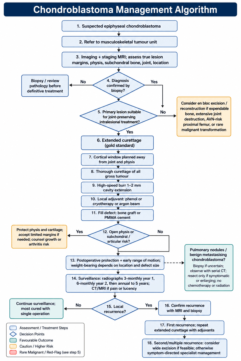
The decision. Extended curettage with adjuvants is the gold standard for the primary lesion, and the goal is to remove the tumour while preserving the joint and the growth plate. Wide en bloc excision is the exception, kept for:
- Recurrent chondroblastoma after failed curettage (two or more recurrences)
- Expendable bone (proximal fibula, fibular head)
- Extensive joint destruction precluding curettage
- Malignant transformation
- Proximal femur with AVN risk
Recurrence is not one number. Extended curettage with adjuvants is conventionally quoted as reducing recurrence from around 30% after simple curettage to roughly 10-20%. Treat that as received teaching rather than a proven effect: the comparison comes from historical and between-series data, never from a randomised trial.
The variable that separates the published series is skeletal maturity. In the 199-case multicentre series, 52% skeletally mature, recurrence was 5.0% after curettage and 0% after en bloc resection; in the 87-case paediatric series with every physis open it was 32%, strictly epiphyseal lesions did significantly worse (p = 0.004), and the surgical method used had no significant influence. A single-unit series of 53 sat between them at 13.2%.
Proximal fibula. The fibular head is expendable and not critical for knee stability, which makes it the ideal site for excision. En bloc resection with 5-10 mm margins, detaching biceps femoris and protecting the common peroneal nerve; no reconstruction is needed and the functional outcome is excellent.
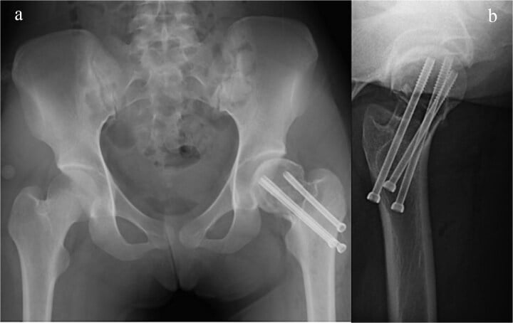
Proximal femur. Curettage here risks avascular necrosis if the blood supply is disrupted, so wide excision with femoral head replacement is considered for recurrent disease or in the older adolescent near skeletal maturity. Wide excision means resecting the femoral head and neck and reconstructing with a modular endoprosthesis or allograft, at significant morbidity and cost.
Other sites. The proximal humerus, distal femur and proximal tibia are hard to reconstruct, so curettage is preferred even for recurrences and wide excision is kept for multiple recurrences or malignant transformation.
Malignant transformation. Extremely rare, at under 1%, and when it occurs it is usually after multiple curettages with high-dose radiation. Rapid growth after years of stability, cortical destruction with a soft-tissue mass, and high-grade chondrosarcoma or dedifferentiated areas on histology are the features. Treatment is wide excision with margins and the prognosis is poor (high-grade sarcoma). Do not irradiate chondroblastoma: radiation increases the risk of malignant transformation.
Surgical Technique
Extended Curettage with Adjuvants
- Review the MRI to identify the true tumour margins, not the oedema
- Plan the cortical window away from the articular surface
- Protect the adjacent physis if it is still open
- Identify the neurovascular structures at risk
- Create a cortical window larger than the lesion diameter, using drill and osteotome
- Position it to reach all of the tumour while preserving subchondral bone
- Save the bone window for later replacement if possible
- Thorough curettage with sharp curettes of all sizes
- Remove all gross tumour tissue and membrane
- Extend to subchondral bone but do not penetrate the articular cartilage
- Inspect the cavity walls under direct vision
- High-speed burr: remove a further 1-2 mm of cavity wall
- Phenol: 5% phenol-soaked gauze for 2 minutes, then copious saline lavage
- or cryotherapy: 2-3 liquid nitrogen freeze-thaw cycles
- or argon beam: thermal ablation of the cavity surface
- Morselised allograft or autograft, or PMMA cement
- Fill the cavity completely to restore structural integrity
- Replace the cortical window, or use cement as the bone substitute
How adjuvants extend the margin. Curettes alone leave microscopic tumour on the cavity wall. The burr removes it mechanically, and a chemical or thermal adjuvant then kills cells a further millimetre or two beyond that, so the two together push the effective margin past the visible cavity in a lesion that cannot be resected because of where it sits.
Phenol, the most common adjuvant, cauterises chemically and penetrates 1-2 mm; cryotherapy ruptures cell membranes with freeze-thaw cycles; argon beam ablates the surface thermally; hydrogen peroxide is a further chemical option; and the exothermic polymerisation of PMMA reaches 60-80°C and kills cells.
Filling the void. Two options:
- PMMA cement gives mechanical support and early weight-bearing, its exothermic reaction adds an adjuvant effect, and recurrence is easier to see at the cement-bone interface
- Bone graft, autograft or allograft, is a biological reconstruction preferred in young patients, but may obscure recurrence on imaging
In a patient with an open growth plate:
- Stay within the epiphysis and do not cross the physis into the metaphysis
- Use imaging guidance or fluoroscopy to confirm the margins
- If the tumour extends across the physis (rare), accept some residual tumour to preserve growth
- Growth arrest from physeal damage causes limb length discrepancy; discuss the risk of growth disturbance with the family before surgery
Subchondral bone. Preserve a minimum of 5 mm of subchondral bone if possible. Where the subchondral bone is involved, curette carefully to avoid cartilage damage and consider supporting the cartilage with bone graft, accepting the risk of future osteoarthritis if aggressive curettage is required.
- Approach
- Deltopectoral or deltoid-splitting
- Technical Considerations
- Protect axillary nerve, rotator cuff
- Complications to Avoid
- Avoid damage to growth plate if open
- Approach
- Medial or lateral parapatellar
- Technical Considerations
- Subchondral bone preservation
- Complications to Avoid
- Articular cartilage damage, physeal injury
- Approach
- Anterior or medial approach
- Technical Considerations
- Protect patellar tendon, meniscus
- Complications to Avoid
- Tibial tubercle avulsion in young patients
- Approach
- Lateral (trochanteric region)
- Technical Considerations
- Hip arthroscopy may be adjunct
- Complications to Avoid
- AVN, femoral neck fracture
- Approach
- Anteromedial or anterolateral
- Technical Considerations
- Joint access may require osteotomy
- Complications to Avoid
- Limited access, high recurrence risk
Complications
- Incidence
- 10-20% with adjuvants
- Risk Factors
- Incomplete curettage, no adjuvant
- Management
- Repeat curettage or wide excision for multiple recurrences
- Incidence
- 2-5%
- Risk Factors
- Large subchondral defect, early weight-bearing
- Management
- Non-weight-bearing, internal fixation if displaced
- Incidence
- 5-10%
- Risk Factors
- Subchondral curettage too aggressive
- Management
- Cartilage repair, accept degenerative arthritis risk
- Incidence
- Under 5%
- Risk Factors
- Physeal injury during curettage in young patient
- Management
- Limb length monitoring, epiphysiodesis or lengthening if needed
- Incidence
- 10-15%
- Risk Factors
- Prolonged immobilization, adhesions
- Management
- Aggressive physiotherapy, manipulation under anesthesia
- Incidence
- 2-3%
- Risk Factors
- Standard surgical site infection risk
- Management
- Antibiotics, debridement if deep infection
Managing recurrence. The typical timeline is 1-3 years after surgery. Confirm it first with MRI and biopsy, since bone graft resorption can look the same, then escalate:
- First recurrence: repeat extended curettage with adjuvants, which succeeds in 70-80%
- Second recurrence: consider wide excision if feasible, in an expendable bone
- Multiple recurrences: en bloc resection, or accept the disease and manage the symptoms
Multiple recurrences do not indicate malignant transformation; chondroblastoma is benign but can be locally persistent, and radiotherapy is avoided because of the transformation risk.
Postoperative Care
The first fortnight. Hospital stay is usually 1-2 days. Analgesia is multimodal, moving to oral medication on day 1, and NSAIDs are avoided initially for a theoretical bone healing concern. Sutures or staples come out at 10-14 days; the wound is kept dry until healed and watched for haematoma and drainage.
Weight-bearing and activity. In the lower limb, touch weight-bearing for 4-6 weeks, progressing with bone healing to full weight-bearing typically by 8-12 weeks. In the upper limb, a sling for 2-4 weeks and then range-of-motion exercises. Impact activity is avoided for 3-6 months, and return to sport waits for imaging to confirm healing.
Surveillance. Recurrence typically occurs within 2 years, and most recurrences announce themselves with a return of pain before the imaging changes. Radiographs are taken every 3 months for the first year, every 6 months in the second, then annually to 5 years; CT or MRI follows any suspicious plain film.
- Assessment
- Wound healing, pain, ROM
- Imaging
- Radiograph (check bone healing)
- Action if Concern
- Early PT if stiff, check for infection
- Assessment
- Pain, function, range of motion
- Imaging
- Radiograph
- Action if Concern
- CT if suspicious lucency
- Assessment
- Symptom check, examination
- Imaging
- Radiograph
- Action if Concern
- MRI if soft tissue concern
- Assessment
- Full assessment
- Imaging
- Radiograph (± CT)
- Action if Concern
- Consider CT to confirm no recurrence
- Assessment
- 6-monthly clinical and radiographic
- Imaging
- Radiograph
- Action if Concern
- CT/MRI if symptoms recur
- Assessment
- Annual clinical and radiographic
- Imaging
- Radiograph
- Action if Concern
- CT/MRI if symptoms recur
Rehabilitation. Range of motion is the priority, then muscle strength, then proprioception and function. The details follow the site:
- Shoulder: early passive range of motion, no heavy overhead lifting for 3 months
- Knee: quadriceps strengthening, protection from impact
- Hip: protected weight-bearing and gait training
- Ankle and talus: gradual return to weight-bearing with orthotic support
Return to sport waits for all four: imaging confirming bone healing, full pain-free range of motion, strength within 90% of the contralateral limb, and completed sport-specific rehabilitation.
Outcomes and Prognosis
The long view is excellent. With appropriate treatment 80-90% are cured by a single curettage with adjuvants, recurrences are manageable with repeat curettage or excision, and functional outcomes are excellent in most cases. The risk of degenerative arthritis is low, under 10%, if the articular surface is preserved.
What to tell the family. Chondroblastoma is benign, not cancer. One operation is curative in most, repeat surgery is effective if it recurs, long-term function and a return to sport are expected, the lifetime risk of arthritis is low if the joint surface is protected, and there is no need for lifelong surveillance after 5 years disease-free.
Where its neighbours are. Chondroblastoma is defined by the company it keeps in the epiphysis: the lesion it is most often confused with is giant cell tumour of bone - which shares the epiphyseal location and, strikingly, a mutation in the same histone gene family (H3F3A in GCT, H3F3B here). The cystic component that obscures it is a secondary aneurysmal bone cyst, and in a skeletally immature epiphysis the other differential is chondromyxoid fibroma. The malignant cartilage lesion it must never be mistaken for is chondrosarcoma.
Guidelines, Registries & Global Practice
Global Epidemiology
Chondroblastoma is consistently reported as a rare benign bone tumour, accounting for under 1% of all primary bone tumours across published series. Demographics are stable across continents: a male predominance (roughly 2:1) and a young age at presentation. In the largest published multicentre extremity series (199 patients), the mean age was 18 years, with the proximal tibia (27.6%) and proximal femur (26.1%) the most common sites, and the physis open in 25.7%, closing in 22.2% and closed in 52.1% at presentation. In a paediatric-only multicentre cohort restricted to open physes (87 patients), the mean age was 12.5 years with the proximal tibia and proximal femoral epiphysis predominating.
- Consensus Position
- WHO 2020 lists chondroblastoma as a benign cartilage tumour (ICD-O 9230/0) defined by H3F3B K36M mutation
- Evidence Base / Source
- WHO Classification of Tumours: Soft Tissue and Bone, 5th ed (2020); Behjati 2013
- Consensus Position
- Mandatory biopsy of epiphyseal lesions; H3K36M IHC / H3F3B testing to confirm and exclude mimics
- Evidence Base / Source
- Behjati 2013 (PMID 24162739); Kervarrec 2017 (PMID 28059095)
- Consensus Position
- Intralesional (extended) curettage with grafting/cement is first-line worldwide; en bloc resection reserved for expendable/recurrent disease
- Evidence Base / Source
- Xu 2015 (PMID 26041854); Suneja 2005 (PMID 15972914)
- Consensus Position
- Avoided as primary therapy due to malignant transformation concern; not endorsed in benign-tumour guidance
- Evidence Base / Source
- Historical case-series consensus; sarcoma network guidance
Guideline & Network Positions
- Relevant Position
- Benign cartilage tumour, locally aggressive potential; molecularly defined entity
- Practice Implication
- Standardises diagnosis; H3K36M as defining marker
- Relevant Position
- Bone sarcoma guidelines mandate referral of bone tumours to specialist sarcoma centres before biopsy
- Practice Implication
- Biopsy and surgery in a multidisciplinary bone tumour unit
- Relevant Position
- Suspected primary bone tumours referred to a recognised bone tumour treatment centre
- Practice Implication
- Centralised care; planned biopsy through the operating unit
- Relevant Position
- Bone cancer pathway: image, then refer indeterminate aggressive lesions to a sarcoma centre for biopsy
- Practice Implication
- Avoids inappropriately placed biopsy tracts
Chondroblastoma is too rare for a dedicated implant registry; the evidence base is built from multicentre tumour-unit cohorts rather than arthroplasty-style registries. The recurring registry-grade message is that biopsy and definitive surgery should occur within a specialist musculoskeletal tumour unit, because an inappropriately placed biopsy tract can compromise later limb-salvage surgery. Reported local recurrence ranges widely by case-mix - approximately 5% in a mixed-maturity extremity cohort (Xu 2015) up to 32% in strictly open-physis paediatric lesions (Sailhan 2009) - reflecting that open-physis, strictly epiphyseal and proximal humeral/femoral lesions are the highest-risk groups.
Practice Variation
- Adjuvant choice varies by centre and region: high-speed burr is near-universal, with phenol, cryotherapy, argon-beam or PMMA cementation selected by surgeon preference and lesion site; no head-to-head trial establishes superiority.
- Void filling differs by maturity: bone graft is favoured in skeletally immature patients to preserve biology, whereas PMMA is often chosen in older patients for immediate stability and easier surveillance of the cement-bone interface.
- Image-guided ablation (radiofrequency / cryoablation) is an emerging option for small, surgically difficult lesions in some interventional-radiology-led centres but is not yet standard of care.
Documentation & Consent (Global Best Practice)
- Record growth-plate status (open vs closed) explicitly on imaging reports — it drives the differential
- Document that the differential includes chondroblastoma, giant cell tumour and infection, and that biopsy precedes any definitive surgery
- Consent for curettage should cover recurrence (approximately 10-20% with adjuvants, higher in open-physis lesions), physeal/growth-disturbance risk (under 5%), articular damage and arthritis risk (5-10%), and phenol-related soft-tissue injury
- Counsel that wide excision is reserved for recurrent disease or expendable bone (e.g. proximal fibula)
MCQ Practice Points
Q: What is the classic location and age distribution for chondroblastoma?
A: Chondroblastoma is an epiphyseal tumor (arises in secondary ossification center) occurring in skeletally immature patients (10-25 years, M greater than F). Most common locations: Proximal humerus, proximal tibia, distal femur, proximal femur. The epiphyseal location in a young patient with open/recently closed physes is pathognomonic. This contrasts with giant cell tumor (metaphyseal/epiphyseal in skeletally mature patients).
Q: What are the characteristic imaging features of chondroblastoma?
A: Radiographs: Well-defined, geographic lytic lesion in the epiphysis; Sclerotic rim (narrow zone of transition); Internal matrix calcification (40-60%, "chicken-wire" pattern); Size usually 3-6 cm. MRI: Low-intermediate signal T1, heterogeneous T2; Marked surrounding bone marrow edema (disproportionate to lesion size - classic feature); May extend across physis. CT best demonstrates matrix calcification.
Q: What is the histological hallmark of chondroblastoma?
A: Chondroblasts: Round/polygonal cells with well-defined cytoplasmic borders, grooved or "coffee bean" nuclei. Chicken-wire calcification: Fine calcification surrounding individual cells (pericellular). Chondroid matrix: Immature cartilaginous matrix between cells. Giant cells: Scattered osteoclast-like multinucleated giant cells present but not as prominent as in GCT. Immunohistochemistry: S-100 positive, SOX9 positive; H3K36M mutation present in 95% of cases.
Q: What is the standard treatment for chondroblastoma?
A: Standard treatment is intralesional curettage with extended techniques (high-speed burr, electrocautery, phenol or hydrogen peroxide) followed by bone grafting or cement. Local recurrence rate: 10-20% (higher in skeletally immature patients with open physes). Cryotherapy should be used cautiously near physis/articular cartilage. Radiofrequency ablation is an option for small lesions. Rare malignant transformation or pulmonary metastases can occur (1-2%).
Q: How do you differentiate chondroblastoma from other epiphyseal lesions?
A: Chondroblastoma: Young patient (open physes), epiphyseal, sclerotic margin, matrix calcification, extensive edema. Giant cell tumor (GCT): Skeletally mature patient, extends from metaphysis into epiphysis, no sclerotic margin, no matrix, less edema. Clear cell chondrosarcoma: Older patient (30-60 years), proximal femur common, may have aggressive features. Langerhans cell histiocytosis: Younger children, may be epiphyseal but often diaphyseal, no matrix. Infection (Brodie abscess): Metaphyseal location more common, periosteal reaction.
Exam Viva Scenarios
Practise clinical reasoning and management decisions out loud
“How do you differentiate chondroblastoma from giant cell tumor?”
“Describe your surgical technique for extended curettage of a chondroblastoma in the proximal tibia of a 16-year-old.”
“What is your follow-up protocol after curettage of a chondroblastoma, and how do you detect recurrence?”
Key Facts
- Rare benign epiphyseal cartilage tumor (under 1% of bone tumors)
- Peak age 10-25 years (open physis), male 2:1
- Commonest sites: proximal tibia 27.6%, proximal femur 26.1%, distal femur 19.1% (PMID 26041854); proximal humerus is the classic site but predicted RECURRENCE (p=0.001), not frequency
- Epiphyseal location before physeal closure is the CHARACTERISTIC pattern, not pathognomonic - GCT, ABC, infection and Langerhans cell histiocytosis all occur there; the H3F3B K36M mutation (95%) is the specific discriminator
Histology Triad
- Chondroblasts - mononuclear cells with oval grooved nuclei
- Giant cells - osteoclast-type, fewer than in GCT
- Chicken-wire calcification - calcified lacunar rims (pathognomonic)
- S100 positive, H3K36M mutation in 95%
Clinical Presentation
- Activity-related pain for months, joint effusion common
- Epiphyseal tenderness, restricted ROM from pain
- X-ray: eccentric lytic with thin sclerotic rim
- MRI: extensive marrow edema (overestimates tumor size)
Differential Diagnosis
- Giant cell tumor: age 20-40y, closed physis, sheets of giant cells
- Clear cell chondrosarcoma: age over 30y, malignant, no chicken-wire
- Infection: fever, elevated CRP/ESR, no calcification pattern
- Chondromyxoid fibroma: metaphyseal, lobulated architecture
Treatment
- Extended curettage with adjuvants (gold standard)
- Adjuvants: phenol, high-speed burr, cryotherapy, or PMMA
- Reduce recurrence from 30% to 10-20%
- Wide excision for recurrent or expendable bones (fibular head)
Complications and Outcomes
- Recurrence 10-20% with adjuvants (1-3 years post-op)
- Secondary ABC in 15-35% (makes curettage more difficult, but does not predict recurrence)
- Malignant transformation extremely rare (under 1%)
- Benign pulmonary metastasis (under 1%) - observe, often regresses
Surgical Pearls
- Protect physis in young patients (avoid growth arrest)
- Preserve subchondral bone (prevent articular collapse)
- Biopsy solid component if ABC present (avoid sampling error)
- PMMA for large subchondral defects (structural support plus thermal kill)
Evidence Base and Key Studies
Defining Histone Driver Mutations (Landmark Molecular Study)
- p.Lys36Met (K36M) alterations, predominantly in H3F3B, found in 73 of 77 chondroblastomas (95%)
- Giant cell tumour of bone instead carries H3F3A G34W/G34L mutations (49/53, 92%)
- Mutations restricted to the neoplastic mononuclear stromal cells, not osteoclasts
- Demonstrates exquisite tumour-type specificity of histone H3.3 driver alterations
- Detectable by mutation-specific (H3K36M) immunohistochemistry for routine diagnosis
H3F3 Mutation as a Diagnostic Marker (Validation Study)
- Multicentre series of 281 bone lesions tested by high-resolution melting plus pyrosequencing
- H3F3 mutations identified in 88% of chondroblastomas and 85% of giant cell tumours of bone
- Mutations were sensitive and specific markers separating these from giant-cell-rich mimics
- Rare H3F3 mutations found in dedifferentiated chondrosarcoma mimicking giant cell tumour
- Supports reclassification of diagnostically difficult giant-cell-rich lesions
Surgical Outcomes - Largest Multicentre Series
- Multicentre retrospective analysis of 199 extremity chondroblastomas (145 male, 54 female; mean age 18 years)
- Most common site was proximal tibia (27.6%), then proximal femur (26.1%) and distal femur (19.1%)
- Physis was open in 25.7%, closing in 22.2% and closed in 52.1% at presentation
- Local recurrence 5.0% after curettage and 0% after en bloc resection
- Proximal humeral location was the only significant predictor of recurrence (p=0.001)
Recurrence Risk in Skeletally Immature Patients
- Multicentre review of 87 chondroblastomas, all in patients with open physes (mean age 12.5 years)
- Most common sites were proximal tibia and proximal femoral epiphysis
- Overall local recurrence 32% at minimum 24-month follow-up
- Strictly epiphyseal lesions had significantly higher recurrence than other locations (p=0.004)
- Aneurysmal bone cyst component and surgical method did not independently predict recurrence
Long-Term Function After Intralesional Curettage
- Single-unit retrospective review of 53 histologically proven chondroblastomas treated by intralesional curettage (1974-2000)
- Local recurrence in 7 patients (13.2%) over follow-up of 2 to 27 years
- Three recurrences controlled by repeat curettage; two required endoprosthesis and two below-knee amputation
- One patient had rare malignant metastasising chondroblastoma and died
- Mean Musculoskeletal Tumour Society functional score 94.2% in survivors