Bado Classification of Monteggia Fracture-Dislocations
A Monteggia fracture-dislocation requires urgent recognition — the most common exam pitfall is missing the radial head dislocation, especially in children where the ulnar fracture may be a subtle greenstick or plastic bowing deformity. Always draw the radiocapitellar line on every elbow radiograph: a line through the centre of the radial shaft must intersect the capitellum on both AP and lateral views. If it does not, the radial head is dislocated. In adults, treat with plate fixation of the ulna, which restores radial head reduction in most cases. In children, fix the ulna anatomically (plate or flexible intramedullary nail) and the radial head usually reduces. Document neurovascular status before and after treatment — the posterior interosseous nerve is at greatest risk in Type II injuries.
The Bado Classification
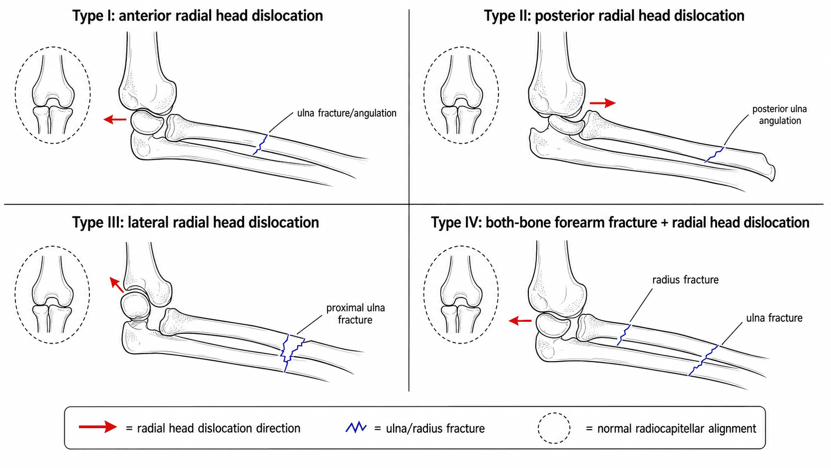
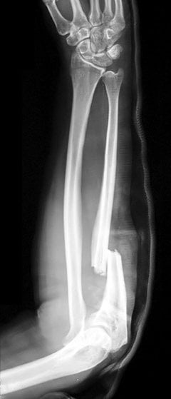
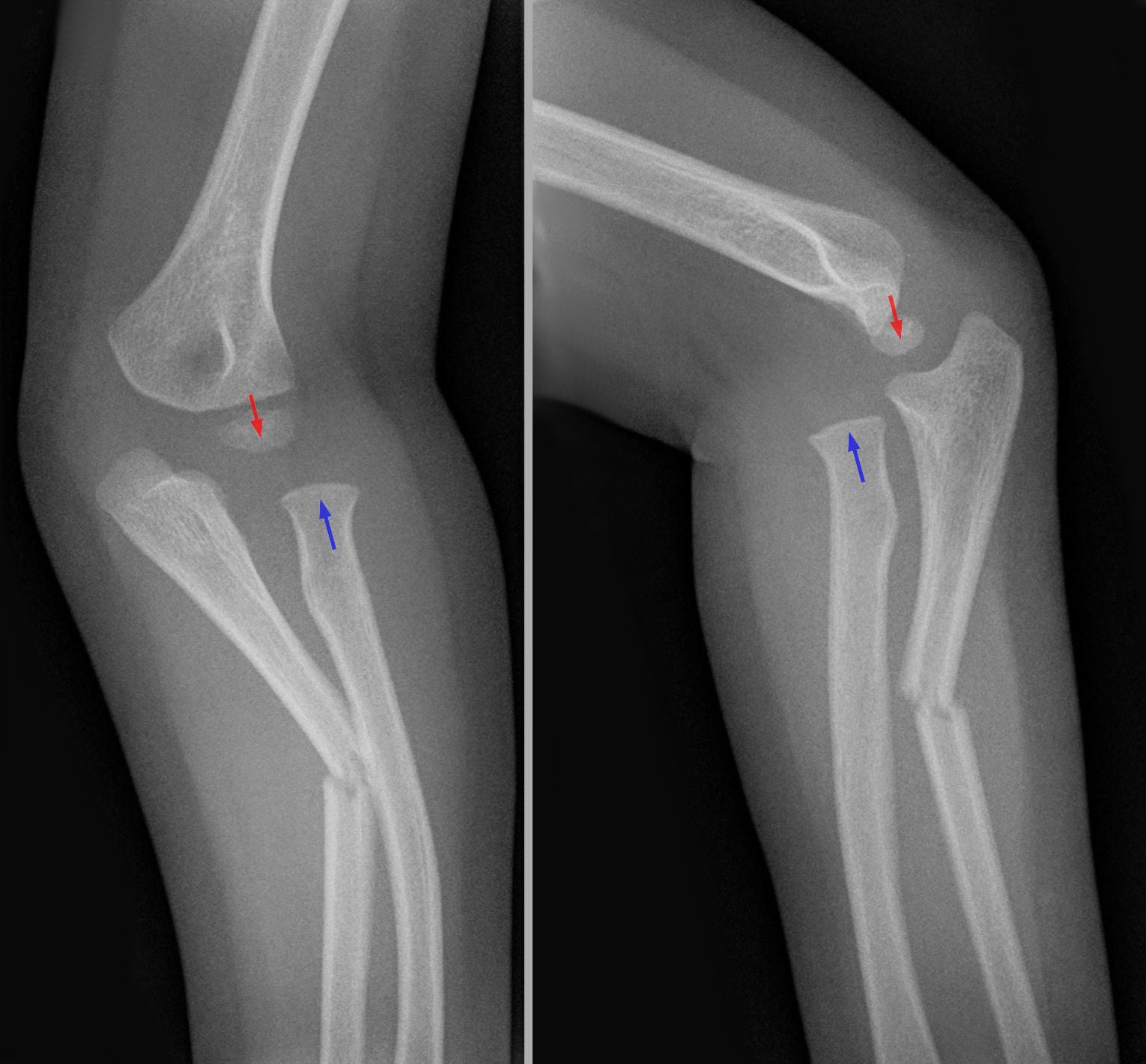
The Bado classification groups Monteggia fracture-dislocations into four types according to the direction of the radial head dislocation. Bado's classic English description (Clin Orthop 1967) remains the universal system and directly guides both diagnosis and treatment.
- Radial Head Dislocation
- Anterior
- Ulnar Fracture
- Diaphysis, any level
- Angulation
- Apex anterior
- Key Features
- Most common overall; the dominant paediatric pattern (up to 70 percent)
- Radial Head Dislocation
- Posterior / posterolateral
- Ulnar Fracture
- Diaphysis, any level
- Angulation
- Apex posterior
- Key Features
- Most common in adults; worse prognosis; comminuted ulna common
- Radial Head Dislocation
- Lateral / anterolateral
- Ulnar Fracture
- Metaphysis (coronoid region)
- Angulation
- Apex lateral
- Key Features
- Almost exclusively paediatric; ulna fracture near coronoid
- Radial Head Dislocation
- Anterior
- Ulnar Fracture
- Both radius and ulna at the same proximal level
- Angulation
- Apex anterior
- Key Features
- Both forearm bones fractured with radial head dislocation — equivalent to a Type I plus a radial fracture
APLB — Anterior · Posterior · Lateral · BothThe four Bado types by dislocation direction
Hook:Anterior–Posterior–Lateral–Both: the direction of the radial head dislocation defines the Bado type.
Bado's rule — the radial head dislocation is always at the same level as or proximal to the ulnar fracture — is the single most important diagnostic principle. On every elbow radiograph, trace the radial shaft proximally: if the line through the centre of the radius does not intersect the centre of the capitellum, the radial head is dislocated. In children, the ulnar fracture may be an occult greenstick or plastic bowing pattern, so the radiocapitellar line is often the only clue. Missing a Monteggia in a child is one of the most frequently cited paediatric orthopaedic pitfalls.
Bado also described "equivalent" lesions that behave like a Monteggia but do not show the classic ulna-fracture-plus-radial-head-dislocation, and examiners like the distinction:
- Type I equivalents (the commonly-taught group): isolated anterior dislocation of the radial head; an ulnar fracture with a radial NECK fracture (instead of a dislocation); an ulnar fracture with an anterior radial head dislocation plus a radial neck/proximal-radius fracture; and an isolated radial neck fracture.
- The practical rule: any isolated radial head dislocation or isolated radial neck fracture in a child should make you hunt for a subtle ulnar plastic-bowing injury and treat the whole thing as a Monteggia equivalent — correct the ulna and the radial head re-seats.
- The mimic to exclude — the transolecranon fracture-dislocation: here the trochlea is driven through the olecranon, but the proximal radioulnar joint stays INTACT (the radius and ulna move together, the radiocapitellar line is preserved). Because there is no proximal radioulnar dissociation, this is NOT a Monteggia — it is treated as an olecranon/elbow fracture-dislocation, and confusing the two changes the operation.
Management by Bado Type
- Adult Treatment
- ORIF ulna with plate; radial head usually reduces. Annular ligament repair if unstable.
- Paediatric Treatment
- Closed or open reduction of radial head; ulnar fixation with plate or flexible IM nail.
- Key Principle
- Restore ulnar length and alignment — the radial head follows.
- Adult Treatment
- ORIF ulna with plate; longer plates and often comminuted. Higher rate of radial head instability requiring annular ligament reconstruction.
- Paediatric Treatment
- ORIF ulna with plate; more challenging reduction. Annular ligament reconstruction if radial head subluxates.
- Key Principle
- More comminuted ulna in adults; worse outcomes than Type I.
- Adult Treatment
- Rare in adults; ORIF ulna with plate, annular ligament repair as needed.
- Paediatric Treatment
- Closed reduction often succeeds. If not, open reduction of radial head via lateral approach; repair annular ligament. Ulnar greenstick may need fixation.
- Key Principle
- The ulnar fracture is metaphyseal and may be greenstick — check stability after reduction.
- Adult Treatment
- ORIF both bones. Plate the ulna and either plate or fix the radius; annular ligament repair.
- Paediatric Treatment
- ORIF both bones. Plate the ulna, fix the radius; annular ligament repair if needed.
- Key Principle
- Both bones must be fixed anatomically to restore the radiocapitellar and distal radioulnar joints.
FARManagement principles for Monteggia lesions
Hook:Fix the ulna, Assess the radial head, Reconstruct if needed — the ulna drives the reduction.
Never accept a reduced Monteggia without confirming radial head stability. After anatomic ulnar fixation, image the elbow through a full pronation-supination arc and a full flexion-extension range on fluoroscopy. If the radial head subluxates or dislocates at any point, the annular ligament is incompetent and requires repair or reconstruction. Leaving an unstable radial head leads to progressive elbow dysfunction, pain, and early arthrosis. In children, chronic unreduced Monteggia lesions are far harder to treat than acute ones and may require ulnar osteotomy combined with annular ligament reconstruction.
The vivas raise three operative scenarios the body should answer:
- Annular ligament reconstruction (acute, radial head still unstable after ulnar fixation): the classic options are the Bell-Tawse reconstruction — a distally-based strip of triceps tendon/aponeurosis looped around the radial neck (often through a drill hole in the proximal ulna) to recreate the annular ligament — performed through a Boyd posterolateral approach that exposes the proximal radius and ulna. A temporary transcapitellar K-wire may protect the reduction but is used cautiously (wire-breakage risk).
- The irreducible acute radial head: if it will not reduce despite anatomic ulnar fixation, suspect soft-tissue interposition — the torn annular ligament infolded into the joint, or capsule/buttonholing. Do not force it: open the joint, extract the interposed tissue, reduce, and repair/reconstruct.
- Chronic / neglected Monteggia (the missed paediatric lesion): reconstruction centres on a proximal ulnar osteotomy to restore ulnar length and bow (typically an opening-wedge, slightly over-corrected to re-tension the soft tissues so the radial head re-seats), combined with open reduction of the radial head, with annular ligament reconstruction and/or temporary transcapitellar pinning added according to the rotational stability achieved. Outcomes are worse and less predictable than acute treatment, and a grossly dome-shaped/deformed radial head with a dysplastic capitellum may be irreducible — which is exactly why early recognition matters.
- Associated radial head fracture (Bado II, adults): ORIF if reconstructable, metal radial-head arthroplasty if not — avoid excision in the unstable Monteggia setting (the radial head resists proximal migration and valgus), and never excise the radial head in a child.
Paediatric Considerations and Pitfalls
- The ulnar fracture may be invisible. In children, the ulnar injury can be a greenstick fracture or plastic deformation (bowing) without a visible fracture line. The radial head dislocation is the clue — always draw the radiocapitellar line. Obtain true lateral views of the forearm (not just the elbow) to detect subtle ulnar bowing.
- Type I is overwhelmingly the dominant paediatric pattern — up to 70 percent of paediatric Monteggia lesions. The mechanism is a fall on an outstretched hand with the forearm in pronation, producing hyperpronation forces that drive the radial head anteriorly.
- Type III is almost exclusively seen in children. The ulnar fracture is at the metaphysis near the coronoid process, often a greenstick or buckle pattern. The radial head dislocates laterally. Closed reduction is usually successful; if not, open reduction through a lateral approach with annular ligament repair is needed.
- Chronic missed Monteggia is a preventable disaster. A missed Monteggia in a child presents weeks or months later with limited forearm rotation, elbow pain, and progressive deformity. Treatment of chronic cases requires ulnar osteotomy to restore length plus annular ligament reconstruction (Bell-Tawse or Boyd) — a far more complex procedure with worse outcomes than acute fixation. Document the radiocapitellar line on every paediatric elbow radiograph.
- Nerve injuries are more common than recognised. The posterior interosseous nerve (PIN) is at greatest risk in Type II (posterior) injuries because the radial head dislocates into or near the supinator muscle arcade. The anterior interosseous nerve (AIN) is at risk in Type I (anterior) injuries from the proximal fragment or reduction manoeuvres. Most are neurapraxias that recover, but they must be documented.
- Jupiter subtype classification refines the Bado types by dividing the ulnar fracture into proximal, middle, and distal thirds of the diaphysis (Types I-A, I-B, I-C and II-A, II-B, II-C). The subtype guides plate length and fixation strategy but does not change the fundamental treatment principle of restoring ulnar anatomy.
- Plastic bowing of the ulna is a paediatric-specific injury pattern where the ulna bends without a visible cortical break. It is a true fracture equivalent and will not remodel reliably without treatment. Recognise it by comparing with the contralateral forearm — any loss of ulnar straightness is significant.
PINNerve injuries in Monteggia lesions
Hook:Posterior type endangers the PIN; anterior type endangers the AIN. Always examine, always document.
Exam Viva
Practise clinical reasoning and management decisions out loud
“A 7-year-old girl fell on an outstretched hand and presents with a painful, swollen elbow. Radiographs show a greenstick fracture of the proximal ulnar diaphysis with apex anterior angulation. The radial head appears to sit in the centre of the capitellum on the AP view but you are not sure on the lateral. How do you proceed?”
“A 42-year-old man sustains a high-energy fall and radiographs show a comminuted fracture of the ulnar diaphysis with apex posterior angulation and a posteriorly dislocated radial head. After you plate the ulna anatomically, fluoroscopy shows the radial head remains dislocated through the pronation-supination arc. What is your next step and what are the surgical options?”
The four Bado types
- Type I: anterior radial head dislocation, ulnar diaphysis fracture, apex anterior — most common in children
- Type II: posterior/posterolateral radial head dislocation, ulnar diaphysis fracture, apex posterior — most common in adults
- Type III: lateral radial head dislocation, ulnar metaphyseal fracture near coronoid — paediatric only
- Type IV: anterior radial head dislocation, both radius and ulna fractured at the same proximal level
Diagnosis and radiographic rules
- Radiocapitellar line: must bisect the capitellum on every view — trace the radial shaft proximally
- Bado's rule: radial head dislocation is always at or proximal to the ulnar fracture level
- In children, the ulnar fracture may be greenstick or plastic bowing — the radiocapitellar line is the clue
- Obtain true lateral views of the entire forearm, not just the elbow
Management principles
- Restore ulnar anatomy: anatomic plate fixation in adults, plate or flexible IM nail in children
- Assess radial head stability fluoroscopically through full pronation-supination after ulnar fixation
- If the radial head remains unstable: open reduction and annular ligament reconstruction (Boyd or Bell-Tawse)
- Chronic missed Monteggia in children: ulnar osteotomy plus annular ligament reconstruction
Nerve injuries and complications
- Type II (posterior): PIN at greatest risk — radial head displaces near the supinator arcade
- Type I (anterior): AIN at risk — test FPL and index FDP specifically
- Most nerve injuries are neurapraxias recovering within 3 to 6 months — document at every visit
- Chronic unreduced Monteggia leads to progressive elbow stiffness, pain, and early arthrosis
Evidence Base
Every citation below has been checked against the source record in PubMed. The four-type framework comes from Bado's original 1967 description; the modern teaching that the character of the ulnar fracture — more than the Bado direction — guides treatment comes from Ring, Jupiter and Waters. Jupiter's posterior-Monteggia series remains the key adult evidence, and the Zhang review covers the neglected/chronic paediatric problem.
The Monteggia lesion
- Original four-type classification based on the direction of radial head dislocation relative to the capitellum
- Defined the essential lesion as fracture of the ulna combined with dislocation of the proximal radioulnar joint
- Established Bado's rule: the radial head dislocation is always at the same level as or proximal to the ulnar fracture
The anterior Monteggia fracture: observations on etiology and treatment
- Analysed the mechanism and treatment of anterior (Bado Type I) Monteggia fractures specifically
- Described the hyperpronation mechanism of injury driving the radial head anteriorly
- Recommended anatomic reduction and fixation of the ulna as the primary treatment, with annular ligament repair when the radial head remained unstable