Catterall Classification of Perthes Disease
- Grading outside the fragmentation phase. Catterall is only valid in the fragmentation (active) phase — a hip looks like Group I in the early avascular phase and "improves" in reossification. Stating a group on the wrong phase is a classic error.
- Quoting the group alone. Group is not sufficient: examiners want age at onset (the single most powerful factor) + Catterall group + head-at-risk signs + the Herring lateral pillar in the same breath.
- Missing a head-at-risk sign. It is two or more signs (or their persistence into reossification) — not a single transient sign — that changes management to containment.
- Confusing the classifications. Catterall (extent of head) ≠ Salter–Thompson (subchondral fracture extent, applied earlier) ≠ Herring (lateral pillar height) ≠ Stulberg (final head shape — an OUTCOME measure at maturity, not a staging tool).
- Treating containment as the goal in the wrong patient. Young child (under ~6), small group, no signs → observe (remodelling does the work). Surgery there is over-treatment.
Catterall vs the other Perthes classifications
A favourite viva probe is to compare the Perthes classifications — what each measures, when it is applied, and how reliable it is. Know that two are staging/extent systems (Catterall, Salter–Thompson), one is the modern prognostic system (Herring lateral pillar), and one is the outcome measure (Stulberg).
- What it measures
- Extent of femoral head epiphysis involved (4 groups, ¼ → whole) + 5 head-at-risk signs
- When applied
- Fragmentation phase
- Reliability / role
- Only moderate inter-observer agreement; historically important, exam-relevant
- What it measures
- Extent of the subchondral fracture line (crescent sign): Group A under half, Group B over half
- When applied
- Early — visible before full fragmentation
- Reliability / role
- Two groups → simpler/earlier than Catterall, but the crescent sign is not always seen
- What it measures
- Height of the LATERAL pillar of the epiphysis on AP (A intact, B over 50%, B/C border, C under 50%)
- When applied
- Early fragmentation (maximal collapse)
- Reliability / role
- The modern prognostic standard — best reliability; combined with age drives surgical decisions
- What it measures
- Sphericity/congruity of the head at SKELETAL MATURITY (classes I–V)
- When applied
- At maturity (healed)
- Reliability / role
- An OUTCOME measure — predicts long-term osteoarthritis; NOT a staging/decision tool
Catterall and Salter–Thompson describe how much head is involved (extent); Herring describes the lateral pillar and best predicts outcome during the disease; Stulberg describes the final shape and predicts arthritis after it heals. Age at onset overrides all of them as the single most powerful prognostic factor.
The topic says the Herring pillar "drives surgical decisions" — here is the actual decision rule from Herring's prospective multicentre study, which combines the pillar group with age:
- Pillar A (full lateral-pillar height preserved): good outcome regardless of treatment.
- Pillar B (over half the pillar height maintained): outcome depends on age — younger children do well whatever is done, but children older at onset (chronological age over 8 years) had better outcomes with operative containment (femoral or innominate osteotomy).
- B/C border: behaves like B — the older child benefits from surgery.
- Pillar C (under half the pillar height): poor outcome regardless of treatment.
- Bottom line: the group that benefits most from containment surgery is the older child (over 8) with a pillar B or B/C-border hip — A does well regardless and C does poorly regardless. This is exactly why age + lateral pillar (not the Catterall group alone) drive the operative decision.
Limitations & modern context
- Inter-observer reliability is only moderate, especially for Groups II and III — limiting the four-group system for research/outcome reporting.
- Grouping is only reliable during fragmentation — under-estimated early (avascular phase) and over-estimated once collapsed/reossified.
- It is purely radiographic and ignores age, range of motion and reossification pattern, all of which influence outcome.
- The Herring lateral pillar classification (height of the lateral pillar on the AP view) predicts outcome more reliably and has largely replaced Catterall for surgical decision-making.
- Catterall remains exam-relevant — name the four groups, the five head-at-risk signs and the age cut-off.
- The Stulberg classification of final head shape at maturity is the standard outcome measure (and predicts arthritis risk).
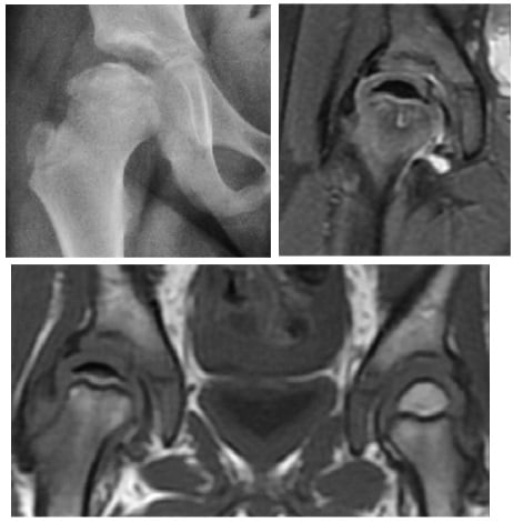
Group, head-at-risk signs & management
The aim of treatment is a spherical, well-contained femoral head at skeletal maturity. Decisions combine the Catterall group, the number of head-at-risk signs, the age at onset, and head containment.
- 1Stage and gradeGrade in the fragmentation phase (Catterall I–IV), count the head-at-risk signs, and state the Herring lateral pillar. Note the age at onset — the single most powerful prognostic factor.
- 2Observe the favourable hipGroup I–II with no/one head-at-risk sign, or any group in a child under ~6 with good containment and high remodelling potential — observation, NSAIDs, activity modification, physiotherapy to maintain range.
- 3Contain the at-risk hipGroup III–IV with two or more head-at-risk signs, or onset after ~8 years — containment surgery (femoral varus or Salter/pelvic osteotomy) to keep the head within the acetabulum during reossification.
- 4Salvage the deformed hipIf aspherical deformity is already established — shelf acetabuloplasty, cheilectomy or valgus/other osteotomy; the Stulberg class at maturity predicts long-term arthritis.
- Head-at-risk signs
- None or one
- Age at onset
- Any age
- Typical management
- Observation, NSAIDs, activity modification, physiotherapy
- Head-at-risk signs
- None or one, head contained
- Age at onset
- Under 6 years
- Typical management
- Observation; remodelling potential is high
- Head-at-risk signs
- Two or more signs
- Age at onset
- Any age
- Typical management
- Containment surgery — femoral or pelvic osteotomy
- Head-at-risk signs
- Any
- Age at onset
- Over 8 years
- Typical management
- Consider containment surgery; salvage (shelf, cheilectomy) if deformity established
A child presenting under 6 with any group has a substantially better outcome than a child presenting over 8 with the same group, because the remaining remodelling potential is greater. Always quote age, group and head-at-risk signs together — group alone is not enough, which is why the Herring lateral pillar classification is used alongside Catterall for modern decision-making.
Guidelines, registries & global practice
- British (BSCOS/BOA): Perthes is managed by paediatric orthopaedic specialists; Catterall III/IV with two or more head-at-risk signs, or onset after age eight, are referred early for consideration of containment in adequate-volume centres.
- AAOS: evidence-based guidance favours observation for young children with smaller groups and containment for older children with extensive involvement; age at onset is consistently the most powerful prognostic factor.
- EPOS (Europe): pairs Catterall grouping with the Herring lateral pillar and uses the head-at-risk signs as triggers for containment rather than as a standalone decision.
- Registries/audit: outcomes are reported by final Stulberg shape and reoperation rate rather than Catterall group alone, reflecting the modest reliability of the four-group system.
- Global variation: lower-resource settings rely more on plain radiographs and Catterall grouping; higher-resource centres increasingly use MRI to define head involvement and containment.
Mnemonics & memory aids
Quarter • Half • Three-Quarters • WholeGroups by quarter of head
Hook:The more head involved, the larger the sequestrum and the worse the eventual shape.
Some Girls Make Heads CrumbleThe five head-at-risk signs
Hook:Some Girls Make Heads Crumble = the 5 head-at-risk signs; two or more together → contain.
Old • Big • Risky → OperateWhen to contain
Hook:Old, Big and Risky → contain; young, small and no signs → observe (remodelling does the work).
The Catterall grouping system
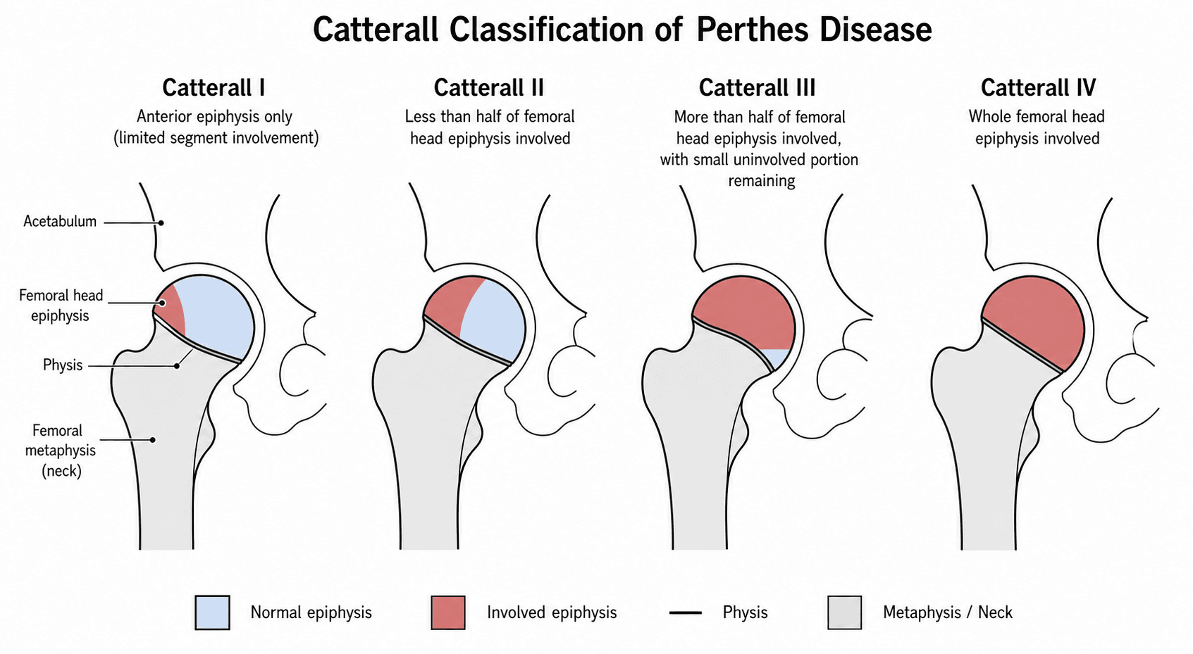
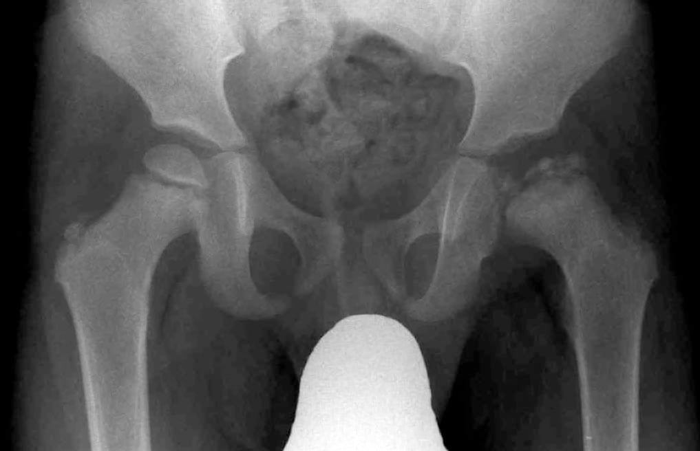
Introduced by Anthony Catterall in 1971, the classification grades the extent of femoral head involved by avascular necrosis on AP and frog-leg lateral radiographs, applied during the fragmentation phase when the sequestrum and new bone are visible.
- Extent of head involvement
- Less than one-quarter (anterior/central)
- Key radiographic features
- Small central or anterior sequestrum; no collapse; no subchondral fracture line
- Typical prognosis
- Good — full remodelling expected
- Extent of head involvement
- Up to one-half
- Key radiographic features
- Larger central sequestrum; anterolateral viable segment; viable medial and lateral columns
- Typical prognosis
- Good to fair — usually spherical
- Extent of head involvement
- About three-quarters
- Key radiographic features
- Large central sequestrum; thin viable posterior/medial sliver; subchondral fracture line often visible
- Typical prognosis
- Guarded — often aspherical
- Extent of head involvement
- Entire femoral head
- Key radiographic features
- Diffuse involvement; whole-head collapse; no preserved column; dense flattened head
- Typical prognosis
- Poor — aspheric head and early arthritis common
The grouping must be applied during the fragmentation (active) phase — not the early avascular phase (only a sclerotic head is seen) nor the late reossification phase (the head is healing). A hip may look like Group I early and reclassify to Group III once fragmentation reveals the true extent of the sequestrum.
The topic keeps referring to "the avascular / fragmentation / reossification phases" — these are the Waldenström radiographic stages of the disease course, and knowing them explains when each classification applies:
- 1. Initial / avascular (sclerosis): a smaller, dense, sclerotic epiphysis with a widened medial joint space; the subchondral fracture (crescent) line appears late (this is what Salter–Thompson grades). Roughly the first ~6 months.
- 2. Fragmentation: the necrotic bone resorbs and revascularises, giving a fragmented, "moth-eaten" epiphysis — this is the phase in which Catterall, Salter–Thompson and the Herring lateral pillar are applied, because the extent and the pillar height are only readable now.
- 3. Reossification (healing): new bone fills the epiphysis from the periphery inward — the final head shape is being set (so containment must be achieved before this completes).
- 4. Healed / remodelled (residual): the head shape is final — staged by Stulberg, which predicts long-term arthritis. This is why a hip under-reads in the avascular phase and "improves" in reossification: only the fragmentation phase reveals the true extent.
Head-at-risk signs
Catterall described five radiographic signs that mark a hip at risk of a poor outcome regardless of the group; two or more together carry a much worse prognosis.
- What it looks like
- Lateral displacement of the head with widening of the medial joint space (Waldenström)
- Why it matters
- Head is breaking out of acetabular containment; deformity will follow
- What it looks like
- Small radiolucent V-shaped wedge at the lateral margin of the epiphysis with speckled calcification
- Why it matters
- Lateral epiphyseal collapse; predicts aspherical healing
- What it looks like
- Wide, ill-defined lucency or cyst in the metaphysis adjacent to the physis
- Why it matters
- Severe physeal involvement; reduced remodelling potential
- What it looks like
- Physis lying horizontally rather than at the normal slope
- Why it matters
- Metaphyseal/physeal damage; disturbed further growth
- What it looks like
- Calcification lateral to the epiphysis and obliteration of the radiologic teardrop
- Why it matters
- Lateral buttress has failed; head uncovered and deforming
A single head-at-risk sign can be transient during the active phase. It is the combination of two or more signs, or their persistence into the reossification phase, that should change management. Document each sign explicitly and reassess on serial radiographs every 3–6 months while the disease is active.
Viva practice
Exam viva
Practise clinical reasoning and management decisions out loud
“A 7-year-old boy presents with a four-month history of a painless limp and limited internal rotation of the right hip. Radiographs in the fragmentation phase show involvement of about three-quarters of the femoral head, lateral subluxation, and a clear Gage sign. How would you classify the disease and what is your management plan?”
“A 5-year-old girl has a new diagnosis of Perthes disease in the early fragmentation phase. Radiographs show involvement of the anterior half of the head only, no head-at-risk signs, and good containment. How does the Catterall classification influence your management, and what would you tell the parents?”
Exam cheat sheet
- Four groups by head involvement: I <¼, II up to ½, III ~¾, IV whole head — grade in the fragmentation phase.
- Five head-at-risk signs: lateral subluxation, Gage sign, diffuse metaphyseal reaction, horizontal physis, lateral calcification/teardrop loss — ≥2 = poor prognosis → contain.
- Age at onset is the single most powerful prognostic factor (under 6 good, over 8 poor).
- Group I–II → observe; III–IV with ≥2 signs or age over 8 → containment surgery (femoral/pelvic osteotomy).
- State the Herring lateral pillar alongside Catterall (more reliable); Stulberg is the outcome measure.
- Catterall reliability is only moderate and valid only in the fragmentation phase.
The four groups
- Group I: less than one-quarter of the head (anterior/central)
- Group II: up to one-half, viable medial and lateral columns
- Group III: about three-quarters, large central sequestrum, subchondral fracture
- Group IV: the entire head with diffuse collapse
The five head-at-risk signs
- Lateral subluxation of the femoral head (Waldenström)
- Gage sign - radiolucent wedge at the lateral epiphysis
- Diffuse metaphyseal reaction (cysts/lucency in the metaphysis)
- Horizontal growth plate; lateral calcification with loss of the teardrop
Management decision rule
- Group I/II, no head-at-risk signs: observation, analgesia, physiotherapy
- Group III/IV, two or more head-at-risk signs: containment surgery (femoral/pelvic osteotomy)
- Age over 8 at onset: consider containment even with fewer signs
- State age, group and head-at-risk signs together in every answer
Limitations & modern context
- Inter-observer reliability of the four groups is only moderate
- Herring lateral pillar classification predicts outcome more reliably
- Stulberg classification is the standard long-term outcome measure
- Grouping is only reliable during the fragmentation phase
Evidence Base
Radiographic classifications in Perthes disease
- 5 observers classified 42 Perthes hips by the Catterall and the original/modified lateral pillar systems in 2 sessions.
- Catterall showed MODERATE inter-observer agreement (kappa 0.43) and a moderate association with 5-year femoral head sphericity (gamma 0.64); the ORIGINAL lateral pillar had the strongest association (gamma 0.75).
- Both Catterall and the original lateral pillar classification had sufficient reliability and prognostic value for clinical use; adding the borderline B/C group did not help.
Assessing the role of hip arthrography in the treatment decision making for children with Legg-Calvé-Perthes disease
- 47 operatively-treated LCPD children (mean age 7.5) staged by the Catterall and Herring classifications and head-at-risk signs; 10 surgeons assessed file sets with and without arthrography.
- Adding dynamic hip arthrography changed the chosen treatment strategy in 43.4% of cases - especially in advanced disease.
- Decision-making reliability was only moderate (inter-observer Gwet AC1 ~0.48; PA AC1 ~0.51).
According to PubMed, the inter-observer reliability (kappa 0.43) and prognostic value of the Catterall grouping versus the lateral pillar classification come from Huhnstock et al. 2017 (DOI), and the role of dynamic arthrography (changing the decision in 43% of cases) from Erkus et al. 2020 (DOI). The four groups, the five head-at-risk signs, the age-at-onset prognostic rule, and the Herring lateral pillar and Stulberg classifications are standard, well-established teaching.