Fixed Lateral Dislocation | Flexion Contracture | Extensor Realignment
- Patella dislocated laterally at birth with fixed flexion contracture and valgus
- Associated with external tibial torsion and quadriceps contracture
- Differs from habitual dislocation which reduces in extension
- Requires extensive lateral release, medial reefing, quadriceps lengthening and PHYSEAL-SPARING distal realignment (Roux-Goldthwait or Galeazzi) - not a tubercle osteotomy in a child
- Untreated leads to permanent deformity and early arthritis
- “Fixed lateral patella with absent medial retinaculum
- “Genu valgum and external tibial rotation are hallmarks
- “Differentiate fixed congenital from reducible habitual type
- “Early surgery before age 2 improves extensor mechanism balance
Overview and Epidemiology
Congenital dislocation of the patella is rare but important: the child is born with the patella sitting laterally, fixed, and it does not come back into the trochlea in any position.
It occupies the fixed end of the spectrum covered in paediatric patellar instability, and it should not be managed with the adult toolkit described in MPFL injury, most of which requires a closed physis.
What it is. Unlike habitual dislocation, which is reducible in extension, the congenital form remains dislocated in all positions and is associated with significant flexion contracture, valgus deformity and external tibial torsion. Early recognition and surgical realignment before age two allows trochlear remodelling and prevents lifelong extensor mechanism dysfunction and early osteoarthritis.
Who. The incidence is less than 1 in 100,000 live births, with a slight female predominance. Up to 40 percent of cases are bilateral. The recognised associations are nail-patella syndrome, trisomy 21 and Rubinstein-Taybi syndrome.
What it costs. The gait shows external rotation and a valgus thrust, and the knee has limited extension and instability. With growth the valgus and torsion progress, and untreated the end point is early patellofemoral arthritis.
Pathophysiology
Normal development. The patella develops within the quadriceps tendon and engages the trochlear groove by 12-16 weeks of gestation.
The primary failure. In congenital dislocation the quadriceps muscle mass fails to rotate internally during limb bud development, so the entire extensor apparatus is left rotated externally and the patella permanently lateral. It never enters the trochlear groove, and the medial retinaculum and medial capsular structures never develop beyond a rudiment.
The secondary changes. With the extensor mechanism lateral, the vastus lateralis and iliotibial band shorten and contract. The lateral femoral condyle remains hypoplastic, which deepens the lateral gutter and is the source of the genu valgum. The tibia develops external torsion as an adaptation to the laterally displaced, unbalanced extensor pull.
- Change
- Failed internal rotation of quadriceps
- Result
- Patella remains lateral
- Clinical Consequence
- Fixed dislocation at birth
- Change
- Lateral structures contract
- Result
- Flexion contracture develops
- Clinical Consequence
- Knee cannot extend fully
- Change
- Weight-bearing on valgus knee
- Result
- Progressive genu valgum
- Clinical Consequence
- Worsening mechanical axis
Classification and Types
Three patterns share the word "dislocation" and need three different operations, so the first job is to name the pattern. The congenital fixed type never reduces; the habitual type reduces in extension and dislocates on every flexion; the recurrent type dislocates episodically after trauma.
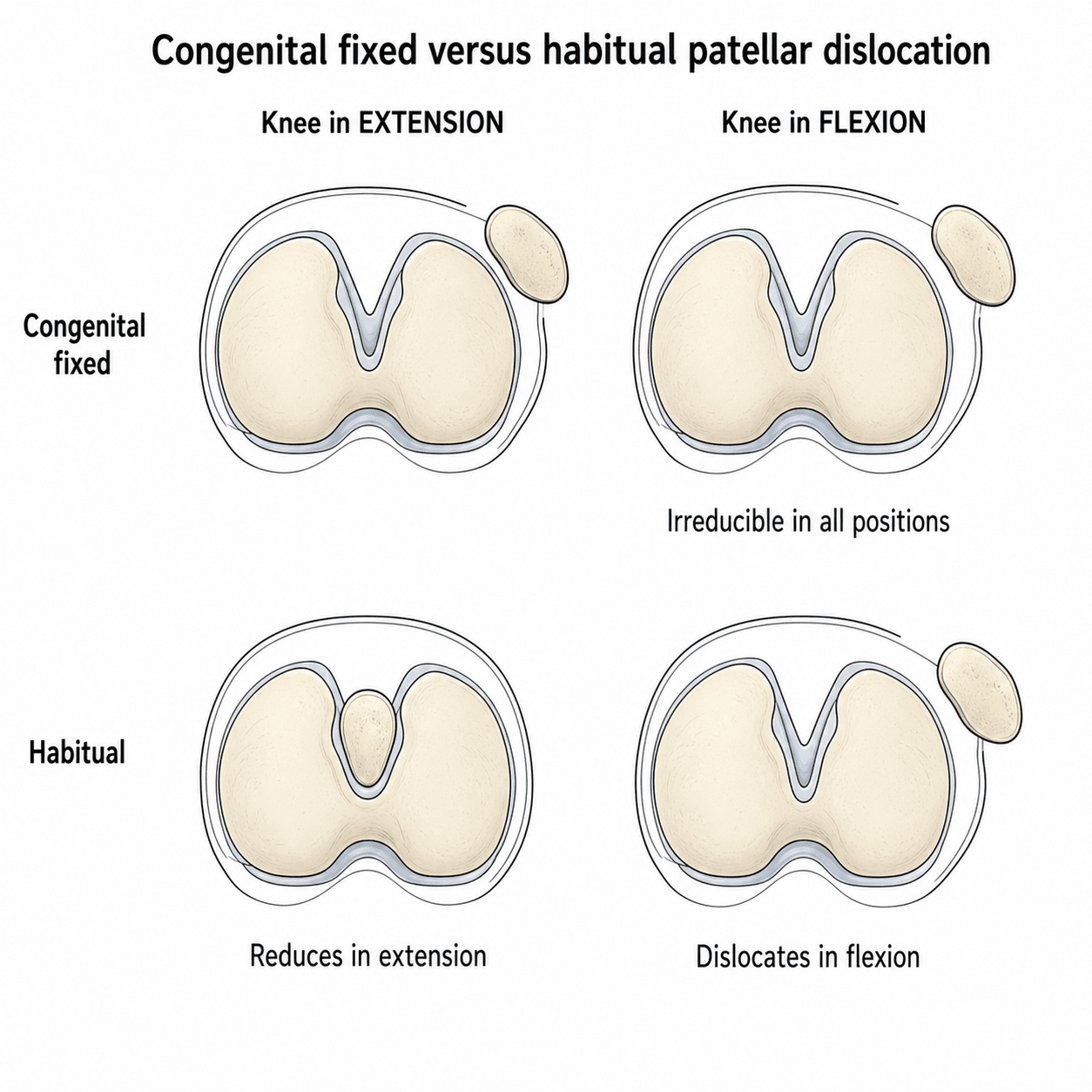
Congenital fixed. Present at birth and irreducible in any position, with a flexion contracture of 20-60 degrees, genu valgum and external tibial torsion; torsion greater than 30 degrees is common.
The quadriceps mechanism is contracted and laterally displaced, the medial retinaculum is absent or severely attenuated, the MPFL is deficient or absent, and the trochlea is flat or convex. Because every element of the extensor mechanism is malpositioned, this type needs a comprehensive realignment that addresses all of the deforming forces.
Habitual (obligatory). "Habitual" and "obligatory" are the same condition, and "intermittent" is the wrong word for it. The patella dislocates laterally every single time the knee flexes and relocates on extension, reproducibly, on every cycle; that is what separates it from recurrent instability, which is episodic. Get this wrong and you offer the wrong operation.
It is painless. Recurrent dislocation hurts and the child is apprehensive; obligatory dislocation usually does not, and the complaint is giving way or an odd-looking knee rather than pain. A child who has begun walking and whose kneecap visibly jumps laterally with every bend, without distress, has this diagnosis.
The lateral tissues are too tight; the medial ones have not failed. This is the mechanical inverse of recurrent dislocation. The contractures sit in the vastus lateralis, the iliotibial tract and the lateral fibres of rectus femoris.
A classical association is repeated intramuscular injections into the thigh in infancy: in one series of 26 knees in 25 children, every child had received them.
Flex the knee and let the patella do what it wants: full flexion is possible, but only because the patella is allowed to dislocate. Now hold the patella reduced and flex again: flexion is limited, in every case. The joint is buying its range by letting the patella escape laterally. That single manoeuvre demonstrates that the problem is a tight lateral extensor mechanism, not a lax medial one, and it is why lengthening, not medial reconstruction, is the operation.
Treatment is proximal quadriceps lengthening, and the word proximal is load-bearing. A review of 35 patients treated by quadricepsplasty concluded that quadriceps lengthening is essential and must be performed proximally, naming reformation of contractures and incomplete initial correction as the causes of failure.
Do not under-promise the difficulty: 12 of those 35 knees redislocated, though 79 percent were normal at a mean follow-up of nearly seven years. Medial plication is an adjunct in a minority, not the main event; avoid over-release, which causes medial instability; and MPFL reconstruction is the operation for the recurrent pattern, not this one.
- Congenital Fixed
- Birth, fixed
- Habitual
- Childhood, reducible
- Recurrent Traumatic
- After trauma
- Congenital Fixed
- Never reduces, even under anaesthesia
- Habitual
- Reduces in extension, every cycle
- Recurrent Traumatic
- Reduces with manipulation
- Congenital Fixed
- Always - never in joint
- Habitual
- EVERY flexion, obligatory
- Recurrent Traumatic
- Episodic, intermittent
- Congenital Fixed
- Usually painless; deformity is the complaint
- Habitual
- PAINLESS - a key discriminator
- Recurrent Traumatic
- Painful, often with apprehension
- Congenital Fixed
- Flexion + valgus fixed
- Habitual
- Lateral contracture IS the disease - vastus lateralis, ITB, lateral rectus femoris
- Recurrent Traumatic
- None or mild
- Congenital Fixed
- Whole extensor mechanism malpositioned
- Habitual
- LATERAL tissues too tight
- Recurrent Traumatic
- MEDIAL tissues too lax
- Congenital Fixed
- Extensive lateral release
- Habitual
- PROXIMAL quadriceps lengthening
- Recurrent Traumatic
- MPFL reconstruction
Clinical Assessment
History. The dislocation is noted at delivery or at the neonatal check. Ask about a family history of syndromic associations or other congenital anomalies, and about function: delayed walking, an external rotation gait and falls. Pain is usually minimal in infancy and appears with activity later.
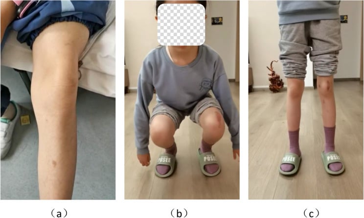
Examination. On inspection the patella is lateral, the knee flexed and in valgus, and the foot progression external. On palpation the patella is fixed laterally, the medial retinaculum is absent and the lateral condyle prominent. There is a fixed flexion contracture of 20-60 degrees with limited active extension. The apprehension test is negative, because the patella is already dislocated, and the Q-angle is increased.
The test that separates fixed from habitual. Attempt reduction with the knee in full extension, under anaesthesia if possible. In the congenital fixed type the patella remains lateral even in extension and cannot be reduced even under anaesthesia; in the habitual type it reduces fully in extension and dislocates only with flexion. This distinction determines the extent of surgery required.
- Onset
- Birth
- Reducibility
- Never reduces
- Associated Features
- Flexion contracture, valgus, ER torsion
- Key Test
- Fixed lateral on all views
- Onset
- Early childhood
- Reducibility
- Reduces in extension
- Associated Features
- Mild valgus, tight vastus lateralis
- Key Test
- Dislocates only in flexion
- Onset
- Variable
- Reducibility
- Variable
- Associated Features
- Nail-patella, trisomy 21, ligamentous laxity
- Key Test
- Systemic features
- Onset
- After injury
- Reducibility
- Reducible
- Associated Features
- Normal trochlea, MPFL tear
- Key Test
- Positive apprehension
Syndromes. Nail-patella syndrome features absent or hypoplastic patellae, nail dysplasia and iliac horns.
Trisomy 21 has ligamentous laxity and an increased risk of fixed dislocation. Always examine the hands and nails, and perform a karyotype if dysmorphic features are present; syndromic cases may require modified surgical goals.
Look beyond syndromes to limb deficiency. A fixed lateral patella can be one visible component of a postaxial limb deficiency: in the case reported on this page the same child had fibular hemimelia, proximal femoral focal deficiency and a tarsal coalition, with fibular hemimelia in the opposite leg too (PMID 35758357). Examine the fibula, the femoral length and the hindfoot, and image the other leg; the associated deformity may matter more for function than the patella does. The secondary genu valgum and the external tibial torsion are assessed as part of the paediatric rotational profile.
Investigations
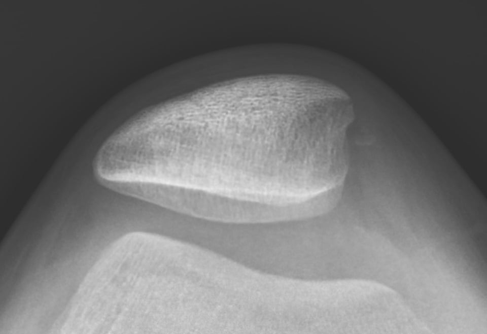
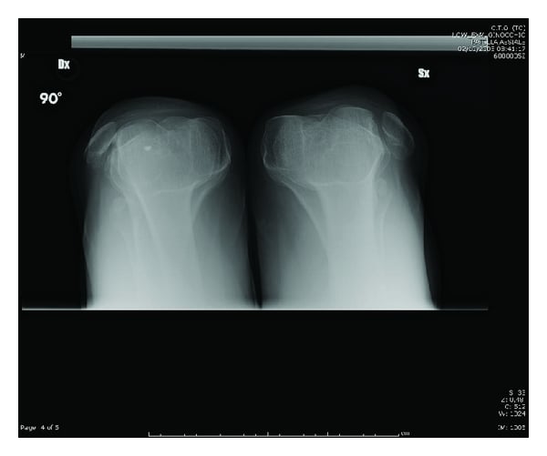
Radiographs. Take AP, lateral and skyline (Merchant or Laurin) views of both knees. The patella sits lateral to the trochlear groove on all views; look also for a hypoplastic lateral femoral condyle, a shallow or absent trochlea and patella alta. Standing alignment films demonstrate the genu valgum and external tibial torsion. Skyline views are essential but difficult in young children, and in infants the patella is cartilaginous and may not be visible on X-ray until 3-5 years.
Ultrasound. Where the patella has not ossified, ultrasound can confirm the cartilaginous patella lying lateral to the femur. Do not delay the diagnosis waiting for ossification: clinical examination and the attempted reduction are diagnostic.
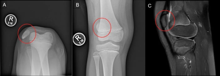
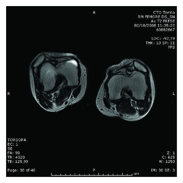
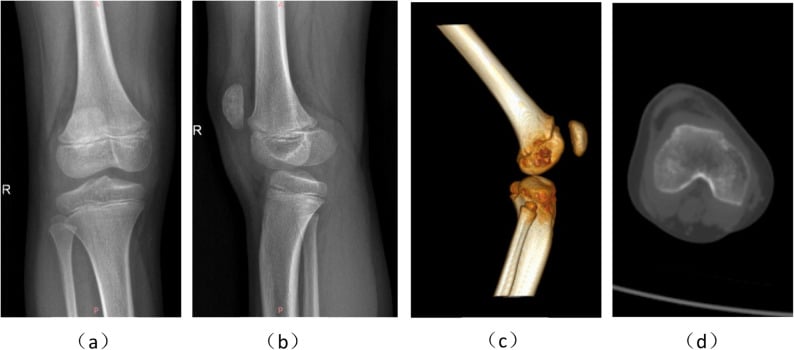
CT, for the older child. Before surgery in the older child, CT assesses trochlear morphology, the tibial tubercle-trochlear groove (TT-TG) distance, and femoral and tibial version. A TT-TG greater than 20 mm is the conventional trigger for distal realignment, but know where that number comes from before you quote it.
It is an absolute distance measured on adult CT, and an absolute distance scales with the size of the knee, so a 20 mm cut-off derived in adults cannot be transferred to a toddler whose distal femur is a fraction of adult width; a normal infant knee could never reach 20 mm. In the young child the decision to realign distally is made on the intra-operative tracking assessment, not on a CT number.
Do not order CT in an infant at all. The patella is unossified until roughly 3-5 years, so there is no ossified landmark to measure a TT-TG from, and it means irradiating a baby for a number you cannot use. CT belongs to the older child and adolescent, where it does real work quantifying external tibial torsion and femoral anteversion before a derotation.
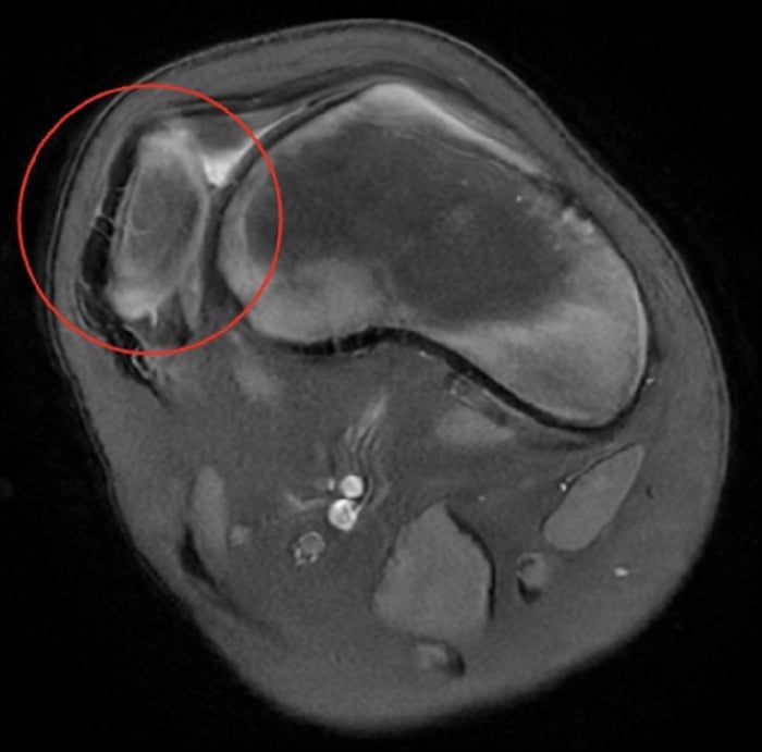
MRI, for complex cases. MRI evaluates cartilage status, the MPFL remnant, quadriceps muscle quality and associated ligamentous anomalies. The findings are an absent medial retinaculum, a contracted vastus lateralis and, in delayed presentation, chondral damage. It determines the need for MPFL reconstruction versus soft-tissue realignment only.
Management Algorithm
The goal. Achieve reduction and realignment before age two, so that the trochlea remodels, fixed contractures are prevented and the valgus and torsion do not progress; delay increases the risk of permanent valgus and torsion. The newborn with a fixed lateral patella is diagnosed clinically, treated in serial casts, and then operated on by 12-18 months.
Neonatal casting, 0-6 months. Weekly long-leg casts gradually correct the flexion contracture, with monitoring of skin integrity and neurovascular status. Know what casting can and cannot do: it treats the flexion contracture and does not reduce the patella, so it is preparation for surgery, not an alternative to it. The decision at 6 months is whether the contracture has been corrected enough to proceed; surgery is planned, not triggered by casting failure.
Infant surgery, 12-18 months. The operation has four components:
- Extensive lateral release of the iliotibial band, vastus lateralis and lateral capsule
- Medial reefing, a plication of the medial retinaculum
- Physeal-sparing distal realignment, a Roux-Goldthwait or Galeazzi procedure; never a tubercle osteotomy at this age
- Quadriceps lengthening, a V-Y quadricepsplasty, if the patella will not seat without tension
The knee goes into a long-leg cast in extension for 6 weeks. Follow-up is long term, watching for valgus recurrence and for the trochlea to develop.
The older child, over 4 years. A fixed deformity at this age is a missed congenital dislocation or a syndromic one. The same combination is used, lateral release, medial reefing, quadriceps lengthening and distal realignment, and the distal realignment is still a Roux-Goldthwait or Galeazzi while the apophysis is open. Possible additions are a femoral derotation, a derotation osteotomy if the torsion is severe, and a distal femoral osteotomy for severe valgus; the goal is correction of the mechanical axis and stable tracking.
Why early, and how sure we are. The mechanism is sound: the trochlea remodels only once the patella is reduced, so earlier reduction gives the groove more growth in which to deepen, and after roughly 5-6 years the trochlear shape is largely established.
But the teaching offers several thresholds, 6 months, 12-18 months and before age two, and the literature does not adjudicate between them; the best available review states in its own conclusion that further study is needed to define the optimal timing (PMID 34782069). So say "early, and for a mechanistic reason, before the trochlea sets", give 12-18 months as the conventional operating window, and acknowledge that the precise cut-off is unestablished. That is a stronger answer than defending a number the evidence does not support.
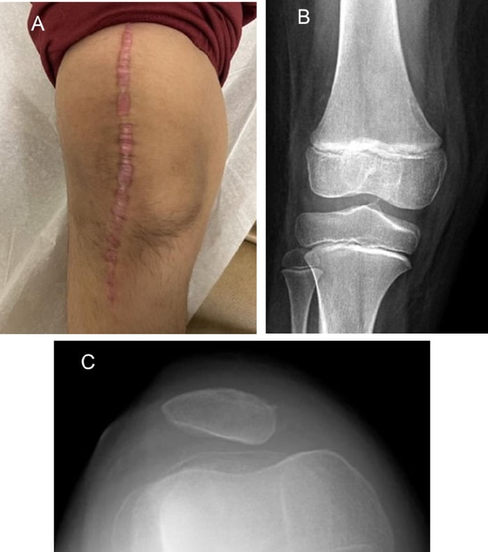
Late presentation is a worse starting position, not a closed door. A bilateral case diagnosed at eleven years and operated late had satisfactory results at 4-year follow-up (PMID 34327168), and an 18-year-old treated with trochleoplasty, quadriceps elongation and MPFL reconstruction had a centred, stable patella at 5 years (PMID 31834021). Telling the family of a 7-year-old that the window has shut is neither kind nor accurate.
And surgery is not automatic. Where knee function is only mildly impaired, observation alone has been suggested, with the decision resting on the degree of disability rather than on the radiograph (PMID 34327168).
Surgical Technique
Set-up. Supine with a tourniquet, the knee flexed 30 degrees initially. The approach is a midline or lateral parapatellar incision extending from mid-thigh to the tibial tubercle.
The steps.
- Release the iliotibial band from Gerdy's tubercle proximally to mid-thigh
- Divide vastus lateralis from the quadriceps tendon and the lateral intermuscular septum
- Perform a lateral retinacular release from the patella to the tibial tubercle
- Medialise the quadriceps tendon insertion with multiple drill holes or an anchor
- Plicate the medial retinaculum in pants-over-vest fashion
- If distal realignment is needed, use a physeal-sparing soft-tissue transfer, a Roux-Goldthwait or a Galeazzi semitendinosus tenodesis; do not perform a tibial tubercle osteotomy in a child with an open apophysis
- If the patella still will not reach the trochlea without tension after a complete lateral release, lengthen the quadriceps (V-Y quadricepsplasty) rather than force the reduction
- Check tracking through a full range of movement and ensure there is no over-medialisation
- Close in layers and apply a long-leg cast in full extension
At-risk structures. The common peroneal nerve during the iliotibial band release, and the medial collateral ligament during the medial reefing.
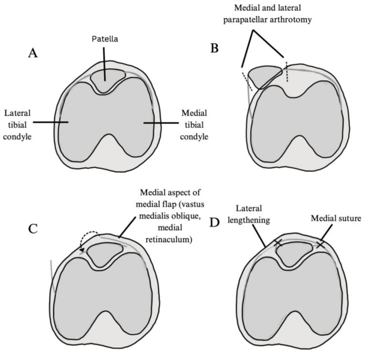
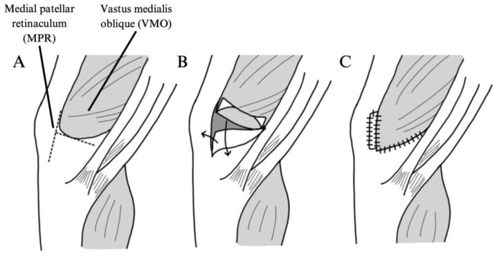
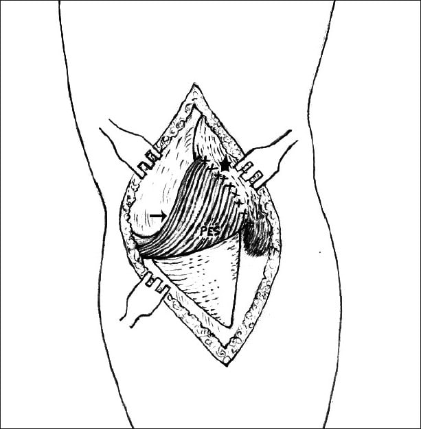
Distal Realignment in the Skeletally Immature Knee
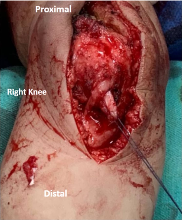
A true tibial tubercle osteotomy cannot be performed in the young child: the tubercle is an apophysis continuous with the proximal tibial physis, and disturbing it risks growth arrest and progressive genu recurvatum from an anterior tether.
In the immature knee, distal realignment of the extensor mechanism therefore relies on soft-tissue transfers rather than moving bone, combined with medial reefing or VMO advancement and the extensive lateral release.
- Roux-Goldthwait procedure: the patellar tendon is split longitudinally and the lateral half is detached distally and transferred medially, passed beneath the medial half and sutured to periosteum or pes, shifting the line of pull medially without touching the apophysis
- Galeazzi semitendinosus tenodesis: the semitendinosus, left attached distally, is rerouted and fixed to the patella as a medial check-rein, a physeal-sparing restraint against lateral dislocation
- A formal tibial tubercle (Elmslie-Trillat-type) osteotomy is reserved until the apophysis is closing or closed, in adolescence, when the TT-TG distance can be corrected directly
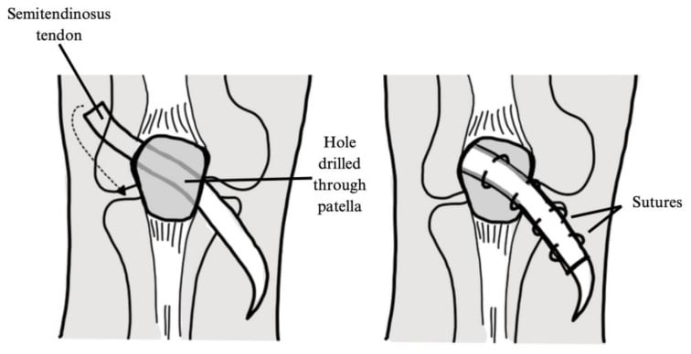
The Contracted Quadriceps: When Release Is Not Enough
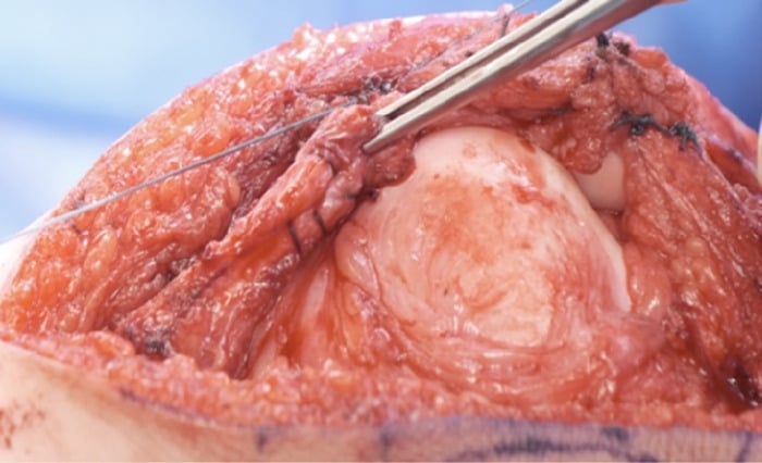
In long-standing or severe congenital dislocation the quadriceps, especially rectus femoris and vastus lateralis, is itself short and fibrotic, not merely laterally tethered.
Lateral release and medial reefing alone then cannot bring the patella distally into the trochlea without excessive tension, and forcing the reduction risks patella baja, skin necrosis or early recurrence.
The intra-operative rule. If, after a complete lateral release, the patella still will not reach the midline trochlea with the knee extended, the quadriceps is the remaining tether and must be lengthened rather than forced. Two techniques answer it:
- V-Y quadricepsplasty (quadriceps lengthening or Z-plasty): an inverted-V incision in the contracted quadriceps tendon and rectus, closed as a Y, that lengthens the extensor mechanism so the reduced patella seats in the groove while still allowing knee flexion. It is the key adjunct once a fixed flexion-extension mismatch persists after release
- Extensor-mechanism mobilisation (Stanisavljevic principle): subperiosteal release of the whole laterally displaced extensor apparatus off the lateral femur and proximal tibia so that it can be rotated medially as a unit
Complications
- Incidence
- 10-20 percent after surgery
- Risk Factors
- Inadequate lateral release, persistent valgus
- Management
- Revision realignment or trochleoplasty
- Incidence
- 5-10 percent
- Risk Factors
- Excessive tubercle transfer
- Management
- Lateral release revision
- Incidence
- 5-15 percent
- Risk Factors
- Over-release of lateral structures
- Management
- Anterior closing wedge osteotomy
- Incidence
- 15-25 percent
- Risk Factors
- Prolonged casting, scarring
- Management
- Manipulation under anaesthesia
- Incidence
- Rare
- Risk Factors
- Excessive medial reefing
- Management
- Patellar tendon lengthening
The most common cause of failure is inadequate release of the vastus lateralis and iliotibial band. The release must be extensive proximally to the mid-thigh. Incomplete release leaves a lateral tether that pulls the patella out again. Always confirm free medial translation of the patella to the midline after release before closing.
Outcomes and Prognosis
Read every percentage on this page as a conventional teaching estimate, not a measured rate. The entire published evidence base for this condition is narrative reviews and single case reports; the five papers cited below describe, between them, a handful of patients. No series exists that could generate a reoperation rate stratified by age band, and the reviews say so: "further study is needed to define the optimal timing and treatment strategy" (PMID 34782069) and "there is no agreement in literature on the ideal treatment" (PMID 34327168). The gradient, earlier does better because the trochlea remodels, is the teaching point and is mechanistically sound. The digits are not measurements.
- Expected Outcome
- Stable tracking, trochlear remodelling
- Reoperation Rate
- Less than 10 percent
- Long-term Function
- Near-normal gait and function
- Expected Outcome
- Good stability, mild residual valgus
- Reoperation Rate
- 15-20 percent
- Long-term Function
- Good for daily activities
- Expected Outcome
- Variable stability, persistent valgus
- Reoperation Rate
- 25-40 percent
- Long-term Function
- Functional limitation common
What predicts the result. The best prognosis follows surgery before age two, a complete lateral release, correction of the torsion and valgus, and compliant post-operative rehabilitation. The poor prognostic factors are delayed presentation after age five, syndromic laxity, incomplete release and persistent external torsion. In conventional teaching the key threshold is age two: outcomes decline sharply after this window because the trochlear remodelling potential is lost.
Guidelines, Registries & Global Practice
- Rare condition with incidence under 1 per 100000 births worldwide
- Bilateral cases reported consistently across all populations
- Syndromic associations (nail-patella, trisomy 21) recognised universally
- Missed diagnosis common in resource-limited settings without neonatal screening
- High-resource: Early ultrasound screening, CT planning, custom bracing post-op
- Limited-resource: Clinical diagnosis, serial casting with plaster, delayed surgery common
- Universal principle: Outcome depends on age at reduction more than technology
- Surgery: Extensive lateral release principles are the same globally
- Diagnosis emphasis
- Clinical differentiation of fixed vs habitual
- Surgical timing
- Surgery by 12-18 months for best remodelling
- Key technical points
- Extensive proximal lateral release mandatory
- Diagnosis emphasis
- Ultrasound confirmation in infants
- Surgical timing
- Serial casting first, surgery under age 2
- Key technical points
- Combined soft-tissue realignment; physeal-sparing distal transfer in the young child
- Diagnosis emphasis
- CT for TT-TG and version planning
- Surgical timing
- Individualised timing based on deformity
- Key technical points
- Address valgus and torsion in older children
No dedicated international registry exists for congenital patellar dislocation due to extreme rarity. Evidence is limited to small case series from tertiary paediatric centres. Principles are consistent across guidelines: differentiate fixed from habitual type, release lateral structures extensively, correct alignment early, and monitor for recurrence and arthritis long-term.
Controversies & Areas of Uncertainty
Some surgeons perform tibial tubercle osteotomy as early as age 4-5 years while others wait until physeal closure to avoid growth disturbance. Evidence is insufficient to define a safe lower age limit.
Trochleoplasty is rarely performed in young children because the trochlea remodels after reduction — and because a growing trochlea should not be cut. In older children with established dysplasia it remains controversial with limited paediatric data. In the skeletally mature knee the picture is different and this page's own evidence supports it: an 18-year-old treated with a subtraction trochleoplasty preserving the trochlear cartilage, plus quadriceps elongation, tubercle transfer and MPFL reconstruction, had a centred and stable patella at 5-year follow-up (PMID 31834021). The honest position is that trochleoplasty is contraindicated in the infant, doubtful in the child, and a reasonable component of a tailored adult reconstruction.
Over-plication risks patella baja and limited flexion. Under-plication risks recurrence. No validated intraoperative tensioning protocol exists.
Staged versus simultaneous surgery and the interval between sides are decided case-by-case. No comparative studies guide practice.
MCQ Practice Points
Q: What is the primary embryological cause of congenital patellar dislocation? A: Failure of internal rotation of the quadriceps muscle mass during limb bud development. This leaves the entire extensor mechanism laterally displaced, the patella never engages the trochlea, and the medial retinaculum fails to form.
Q: How do you differentiate congenital fixed dislocation from habitual dislocation on examination? A: Attempt reduction in full extension. In congenital fixed type the patella remains lateral in all positions including extension. In habitual type the patella reduces fully in extension and only dislocates with flexion beyond 30-40 degrees.
Q: What is the key surgical principle in congenital fixed dislocation? A: Extensive lateral release of the iliotibial band and vastus lateralis from mid-thigh to tubercle. Incomplete proximal release is the most common cause of recurrence. Medial reefing and tubercle transfer are adjuncts performed after adequate release.
Q: What is the ideal age for surgical realignment and why? A: 12-18 months. This window allows trochlear remodelling once the patella is reduced and prevents progressive valgus and external torsion. Surgery after age five has poorer outcomes because the trochlear shape is established and bony deformity is fixed.
Q: What torsional abnormality is commonly associated and how is it addressed? A: External tibial torsion greater than 30 degrees. It develops secondary to the laterally displaced extensor pull. If severe it is corrected with a tibial derotation osteotomy at the time of realignment surgery.
Exam Viva Scenarios
Practise clinical reasoning and management decisions out loud
“A 2-week-old infant is referred with a fixed lateral patellar dislocation noted at birth. The knee has a 40-degree flexion contracture and 15-degree valgus. Attempted reduction under anaesthesia fails. How do you manage this child?”
“A 7-year-old presents with longstanding external rotation gait and inability to fully extend the knee. Examination shows the patella fixed laterally with 30-degree valgus and 25-degree external tibial torsion. This was not treated in infancy. What is your surgical plan?”
Key Anatomy and Development
- Failure of quadriceps internal rotation leaves patella lateral from birth
- Medial retinaculum absent, lateral structures contracted
- Lateral femoral condyle hypoplastic, trochlea shallow
- External tibial torsion and genu valgum develop secondarily
Diagnosis
- Fixed lateral patella in all positions including extension
- Flexion contracture 20-60 degrees with valgus
- Differentiate from habitual (reduces in extension)
- Ultrasound in infants, skyline X-ray once ossified, CT for TT-TG
Treatment Algorithm
- Serial casting 0-6 months to correct contracture
- Surgery 12-18 months: extensive lateral release + medial reefing
- Distal realignment in the child = Roux-Goldthwait / Galeazzi (physeal-sparing); tubercle OSTEOTOMY only when the apophysis is closing
- TT-TG over 20 mm is an ADULT CT threshold - it does not scale to an infant knee, and the infant patella is unossified anyway
- Derotation or valgus osteotomy in older or missed cases
Surgical Pearls
- Release ITB and vastus lateralis proximally to mid-thigh
- Confirm free medial translation of patella before closing
- Avoid over-medialisation causing patella baja
- Long-leg cast in extension 6 weeks post-op
Complications and Prognosis
- Recurrence 10-20 percent if release incomplete
- Best outcomes with surgery before age 2
- Delayed surgery increases arthritis risk
- Monitor for over-medialisation and recurvatum
Evidence Base and Key Trials
The whole published evidence base is one review and four case reports. That is not a criticism of the authors — the condition is that rare — but it should change how you quote it. Read the set as answering five different questions, which is how they are grouped below.
Fixed (Congenital) Patellar Dislocation
- Lateral dislocation that is IRREDUCIBLE without surgical correction; no clear inheritance pattern, but associated with several congenital syndromes
- Typical findings: flexion contracture, loss of ACTIVE knee extension, increased tibial external rotation, and an absent patella in the trochlea
- Treatment has FOUR components: lateral release, medial stabilisation, QUADRICEPS LENGTHENING, and distal realignment - quadriceps lengthening is part of the operation, not an optional extra
- Results are generally favourable; PERSISTENT FLEXION CONTRACTURE and REDISLOCATION are the two most common complications
- The authors state explicitly that further study is needed to define the OPTIMAL TIMING and treatment strategy